
Principles of, incisions Flaps & Suturing
LS
Published · 58 slides · 0 views

1 / 1


Description
Principles of, incisions Flaps Suturing Dr.Mohammed Alaraji B.D.S.F.I.B.M.S Incision It is simply defined as a cut or wound made by cutting with a sharp instrument. The basic principles of incisions in oral surgery include: A blade number
Related Topics
Share
Embed code
Download this presentation From Below
"Principles of, incisions Flaps & Suturing" is the property of its rightful owner. Permission is granted to download and print the materials on this website for personal, non-commercial use only, and to display it on your personal computer provided you do not modify the materials and that you retain all copyright notices contained in the materials. By downloading content from our website, you accept the terms of this agreement.
Presentation Transcript
01
Principles of, incisions Flaps & Suturing Dr.Mohammed Alaraji
B.D.S.F.I.B.M.S<br>
B.D.S.F.I.B.M.S<br>
02
Incision It is simply defined as a cut or wound made by cutting with a sharp instrument.<br>
03
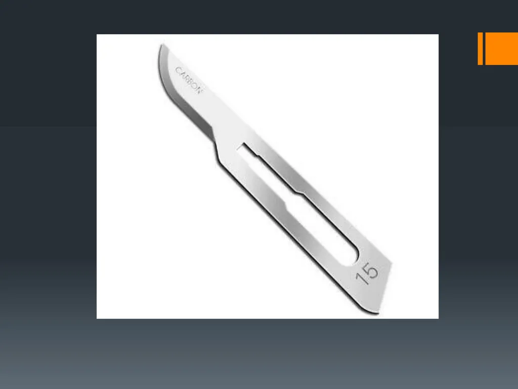
The basic principles of incisions in oral surgery include: A blade number 15 is suitable for most oral surgical procedures. Sometimes a blade number 12 is used.
A new and sterile blade should be used for each patient and it should be replaced with a new one intraoperatively if its cutting edge becomes blunted when necessary.
The scalpel blade is mounted on the scalpel-handle with the help of a needle holder, or hemostat, with which it slides into the slotted receiver with the beveled end parallel to that of the handle.
The scalpel is grasped in a pen grasp for maximum control and tactile sensitivity.<br>
A new and sterile blade should be used for each patient and it should be replaced with a new one intraoperatively if its cutting edge becomes blunted when necessary.
The scalpel blade is mounted on the scalpel-handle with the help of a needle holder, or hemostat, with which it slides into the slotted receiver with the beveled end parallel to that of the handle.
The scalpel is grasped in a pen grasp for maximum control and tactile sensitivity.<br>
04
The incision should be made at right angle to the underlying bone to ensure good healing when the tissues are re- apposed.
The scalpel should move at uniform speed and with sufficient firmness to cut through not only the mucosal surface but also the periosteum overlying the bone. It should be made, ideally, with a single movement, repeated strokes at the same place should be avoided as they may impair healing.<br>
The scalpel should move at uniform speed and with sufficient firmness to cut through not only the mucosal surface but also the periosteum overlying the bone. It should be made, ideally, with a single movement, repeated strokes at the same place should be avoided as they may impair healing.<br>
05
Flap is simply defined as a section of soft tissue that is outlined by surgical incisions, carries its own blood supply, allows surgical access to underlying tissues, can be replaced as required on its original position, maintained with sutures and is expected to heal.
Most of the oral surgical procedures require the reflection of a full mucoperiostial flap incorporating mucosa, submucosa and periosteum to gain access to the area that is the object of surgery.<br>
Most of the oral surgical procedures require the reflection of a full mucoperiostial flap incorporating mucosa, submucosa and periosteum to gain access to the area that is the object of surgery.<br>
06
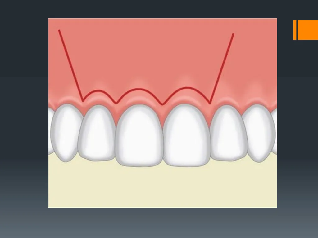
Flap design Flap design and incision should be carried out in such a way that injury of anatomic structures is avoided, such as: the mental neurovascular bundle, palatal vessels emerging from the greater palatine foramen and incisive foramen, lingual nerve, submandibular duct, facial artery and vein. So thorough knowledge of the anatomy of the orofacial region is essential.
The base of the flap should be wider than its apex (free gingival margin) to ensure adequate blood supply for better healing.<br>
The base of the flap should be wider than its apex (free gingival margin) to ensure adequate blood supply for better healing.<br>
07
The flap should be of adequate width for good visualization and accessibility of the operative field without subjecting the flap to tension and trauma during manipulation.
When planning the flap, the care should be given to the fact that the flap should be wider than the anticipated bony defect after completion of the procedure so that the flap margins, when sutured, should rest on intact and healthy bone to prevent wound dehiscence and poor healing.
Delicate handling of the flap during the surgical procedure without excessive tension of crushing in order not to compromise the blood supply which leads to delayed healing.<br>
When planning the flap, the care should be given to the fact that the flap should be wider than the anticipated bony defect after completion of the procedure so that the flap margins, when sutured, should rest on intact and healthy bone to prevent wound dehiscence and poor healing.
Delicate handling of the flap during the surgical procedure without excessive tension of crushing in order not to compromise the blood supply which leads to delayed healing.<br>
08
Vertical releasing incisions should start at the buccal vestibule and end at the interdental papilla which should either be excluded or included in the flap, the incision should always pass to the interdental papilla and not end at the labial or buccal surface of the tooth to ensure the integrity of the gingiva, but it should not pass through the papilla for accurate replacement of the flap.
Vertical releasing incision are contraindicated in certain sites in the oral cavity:
Lingual surface of the mandible: to avoid injury to the lingual nerve.
Canine eminence: because it increases the tension on the suture line which lead to wound dehiscence.
In the area of mental foramen, between mandibular first and second premolars: to avoid injury to the mental nerve.<br>
Vertical releasing incision are contraindicated in certain sites in the oral cavity:
Lingual surface of the mandible: to avoid injury to the lingual nerve.
Canine eminence: because it increases the tension on the suture line which lead to wound dehiscence.
In the area of mental foramen, between mandibular first and second premolars: to avoid injury to the mental nerve.<br>
09
Types of Mucoperiosteal Flaps<br>
10
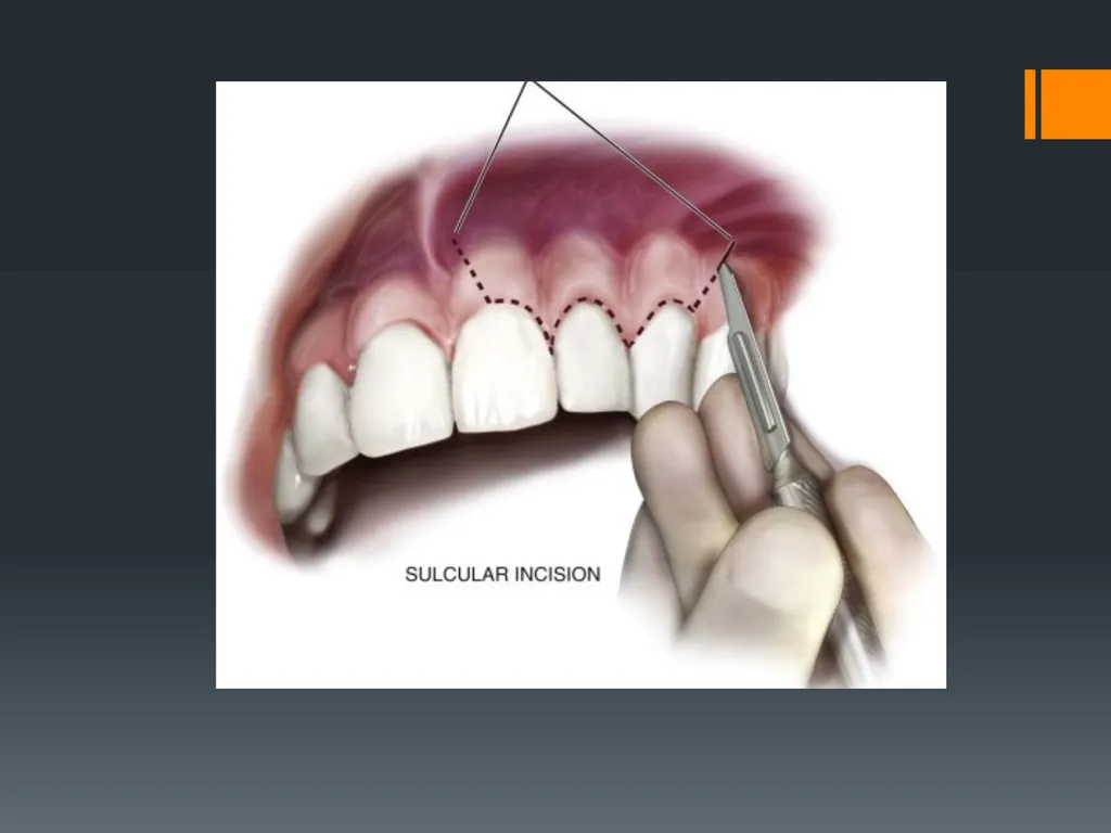
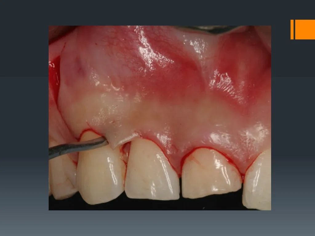
1- Envelope Flaps This type of flaps is made by a horizontal incision through gingival sulcus for the teeth or through the alveolar mucosa of the edentulous area with no vertical releasing incisions. The envelope flap is used for surgery of incisors, premolars and molars, on the labial or buccal and palatal or lingual surfaces.
The main indications of this type of flaps include: surgical extraction of impacted mandibular third molars, palatal approach to impacted maxillary canines or removal of mandibular tori.<br>
The main indications of this type of flaps include: surgical extraction of impacted mandibular third molars, palatal approach to impacted maxillary canines or removal of mandibular tori.<br>
11
The main advantages of this flap are; easy re-approximation to original position, good blood supply and it can easily modified to two-sided or three-sided flap by adding vertical releasing incisions to either ends of the flap when necessary.
Disadvantages
the limited accessibility and visualization.
difficulty in reflection with greater tension that can result in tearing at the ends of the flap.<br>
Disadvantages
the limited accessibility and visualization.
difficulty in reflection with greater tension that can result in tearing at the ends of the flap.<br>
12
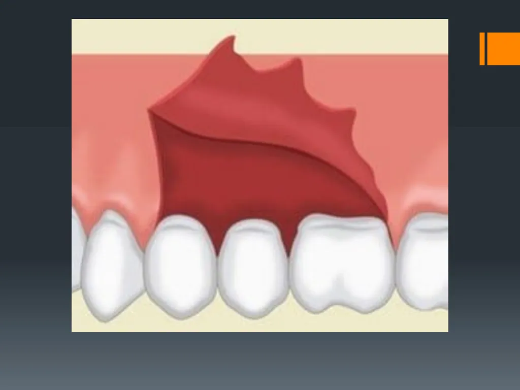
2- Two-sided Flap (Triangular Flap) This flap is the made with a horizontal incision along the gingival sulcus or alveolar ridge mucosa and a vertical releasing incision. The vertical incision begins approximately at the vestibular fold and extends to the interdental papilla of the gingiva. This flap is performed labially or buccally on both jaws .
indicated in the surgical removal of root tips, impacted teeth, small cysts, and apicectomies.<br>
indicated in the surgical removal of root tips, impacted teeth, small cysts, and apicectomies.<br>
13
Advantages are; it ensures an adequate blood supply, satisfactory visualization and accessibility, good re-approximation; it can be easily modified to a three-sided flap.
Disadvantages are; limited access, tension when flap is retracted and it may result in defect of attached gingiva.<br>
Disadvantages are; limited access, tension when flap is retracted and it may result in defect of attached gingiva.<br>
14
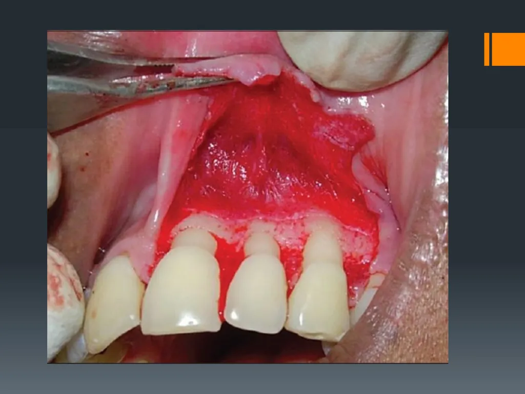
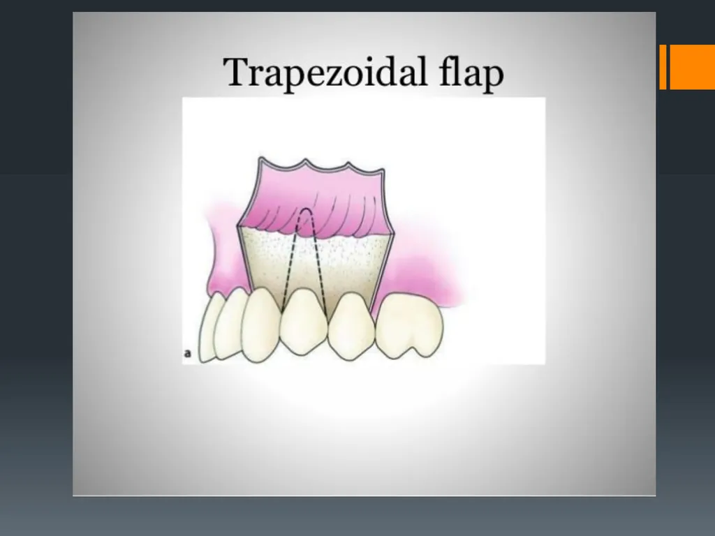
3- Three-sided Flap (Trapezoidal Flap) This flap consists of a horizontal incision along the gingival or alveolar ridge mucosa and 2 vertical releasing incisions, this flap is indicated when an extensive surgical field exposure is required especially when two-sided flap is inadequate.
The main advantages include; very good accessibility and visualization of the surgical field with minimal tension on the tissue, and good reapproximation of tissue to the original position.
The disadvantages are the possibility of producing an attached gingival defect. This flap cannot be lengthened or modified once reflected.<br>
The main advantages include; very good accessibility and visualization of the surgical field with minimal tension on the tissue, and good reapproximation of tissue to the original position.
The disadvantages are the possibility of producing an attached gingival defect. This flap cannot be lengthened or modified once reflected.<br>
15
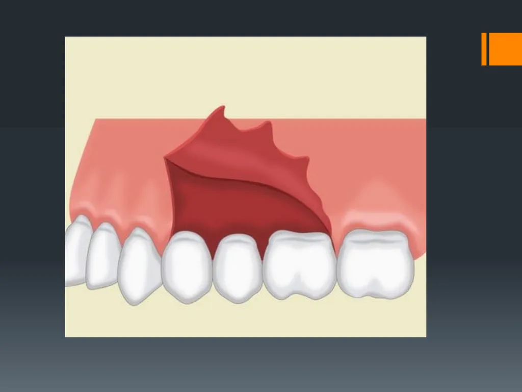
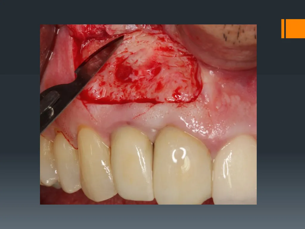
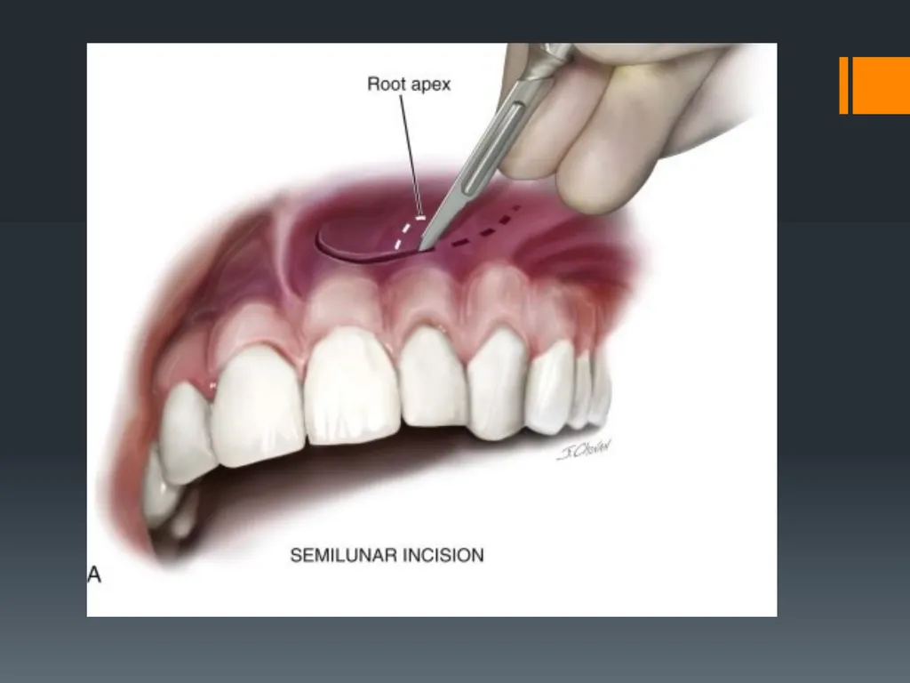
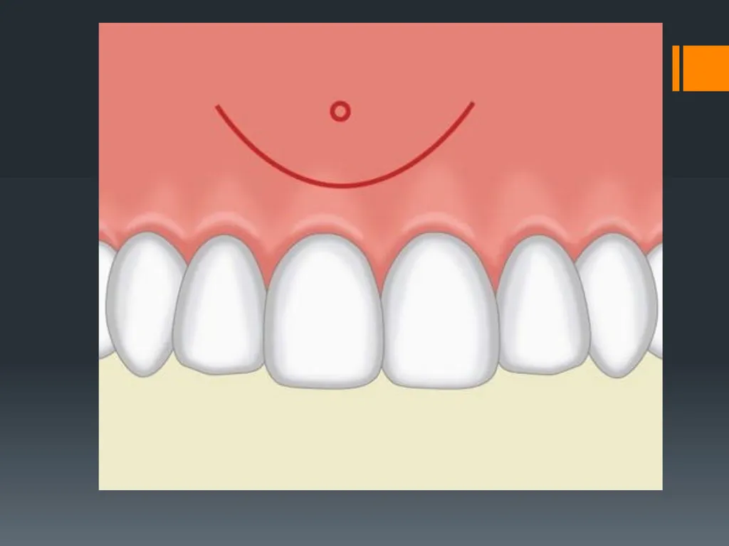
4-Semilunar Flap This flap is the result of a curved incision, which begins just beneath the vestibular fold and has a bow shaped course with the convex part towards the attached gingiva. The lowest point of the incision must be at least 0.5 cm from the gingival margin, so that the blood supply is not compromised. Each end of the incision must extend at least one tooth over on each side of the area of bone removal. The semilunar flap is used in apicoectomies and removal of small cysts and root tips.<br>
16
Advantages of this flap are small incision, easy reflection, no attached gingival defect especially around prosthetic appliances (crowns and bridges) and easy oral hygiene.
Disadvantages of this flap are limited accessibility and visualization of the surgical field, re-approximation may be difficult due to the absence of reference points, tendency to tear due to excessive tension on reflection and the possibility that the flap may made over defective bone as a result of inadequate planning or underestimation of the size of the bony defect so that the margins of the flap will not rest on intact bone leading to collapse of the flap and wound dehiscence.<br>
Disadvantages of this flap are limited accessibility and visualization of the surgical field, re-approximation may be difficult due to the absence of reference points, tendency to tear due to excessive tension on reflection and the possibility that the flap may made over defective bone as a result of inadequate planning or underestimation of the size of the bony defect so that the margins of the flap will not rest on intact bone leading to collapse of the flap and wound dehiscence.<br>
17
Other types of flaps V- Y-shaped incision. This flap used in surgical procedures of the palate, mainly for removal of exostoses (torus palatinus).
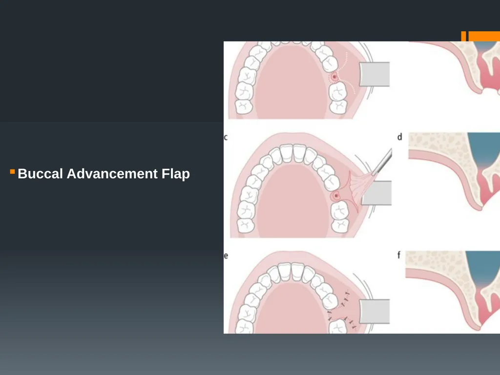
Buccal Advancement Flap Flaps that are used for closure of oroantral fistula or communication.
Palatal Transpositional Flap that incorporates the greater palatine vessel, it is rotated and sutured to the buccal tissues.<br>
Buccal Advancement Flap Flaps that are used for closure of oroantral fistula or communication.
Palatal Transpositional Flap that incorporates the greater palatine vessel, it is rotated and sutured to the buccal tissues.<br>
18
Buccal Advancement Flap<br>
19
Flap reflection The mucoperiosteal flap is reflected from the underlying bone using periosteal elevators. There are many types of mucoperiosteal elevators like Howarth& the no.9 Molt periosteal elevator . The elevators should be firmly pushed at approximately45° to the surface of the bone such that the periosteum is stripped from it. It is important to try to raise both mucosa and periosteum in one layer and this requires a considerable force to be applied.<br>
20
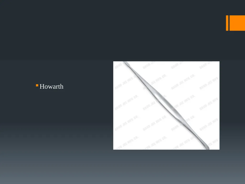
Howarth<br>
21
Suturing After completion of the surgical procedure, thorough irrigation of the surgical field using sterile normal saline follows. Then the flap is repositioned to its original position and held in place using sutures to protect the underlying tissues from infection and irritating factors and prevent postoperative hemorrhage. Sutures are also used to repair soft tissue lacerations, ligation of vessels and control of bleeding. The amount of suture material used should be kept to a minimum, particularly when braided, to reduce bacterial colonization. Suture material can be a nidus for infection, and knots can be the focus of a persistent and chronic inflammatory reaction (suture knot sinus).<br>
22
Suture Materials Suture materials are classified as either absorbable or non-absorbable material depending on whether the body tissues will degrade the suture material and absorb it over time. Absorption takes place either by Hydrolysis or by proteolytic enzymatic degradation depending on the material used. They can also be classified as monofilament or multifilament.<br>
23
A- Absorbable Sutures They are used in suturing of deep layers of wounds when multilayered suturing is required, they are also used in children, mentally handicapped patients and in patients who cannot return to the clinic to have their sutures removed.<br>
24
1- Plain Catgut it is made from collagen derived from healthy sheep or cattle intestine. its absorption is through phagocytosis and enzymatic degradation which occurs within 7-10 days .It is used for suturing subcutaneous tissues that do not require prolonged support. It is not suitable for suturing in oral surgery.<br>
25
2-Chromic Catgut it is made from collagen derived from healthy sheep or cattle intestine tanned with Chromium salts to facilitate handling and resist tissue degradation. It tensile strength is lost within18-2ldays, its absorption is like that of the plain Catgut but it takes longer time and with moderate tissue reaction. It has the same indication as for the plain Catgut and it is not suitable in oral surgery.<br>
26
3-Polyglactin (Vicryl) Synthetic suture made of copolymer of lactide and glycolide coated with polyglactin and calcium stearate.
It is braided multifilament Its absorption is through hydrolysis with complete absorption taking place within 60-90 days.<br>
It is braided multifilament Its absorption is through hydrolysis with complete absorption taking place within 60-90 days.<br>
27
4- Polydioxanone (PDS) Supplied as monofilament dyed or undyed, it is made of polyester polymer. Absorption occurs through hydrolysis which is complete in about 180 days.<br>
28
B-Non-absorbable sutures These sutures remain in the tissues and are not absorbed, but have to be cut and removed about 7 days after their placement.<br>
29
1- Silk it is made of raw silk from silkworms, and it is supplied as braided or twisted, dyed or undyed, coated with wax or silicon or uncoated. 80%-100% of its tensile strength is lost within 6 months. Fibrous encapsulation occurs in the body within 2-3 weeks, it causes moderate to high tissue reaction. It is used in ligation and suturing when long term tissue support is needed. Silk sutures are the easiest to use and the most economical, and have a satisfactory ability to make a secure knot. Sutures that are holding mucosa together usually stay no longer than 5 to 7 days.<br>
30
2- Nylon It is made of polyamide polymer and it is supplied usually as monofilament. It causes mild tissue reaction and it is used mainly for skin, in plastic surgery, neurosurgery, and ophthalmic surgery.<br>
31
Needles Needles are usually made of stainless steel which is strong and flexible material. There are different shapes, sizes and cross sections of needles. Needles of 18-26 mm in length are suitable for use in oral surgery.<br>
32
There are two basic needle types: "eyed."
Those that have the hole at the suture side of the needle and that need to be threaded with suture
eyeless" or "swaged."
those that have the suture attached to the needle .
As compared to a regular circle, needles are either; 1/4 circle, 1/2 circle, ¾ circle, 3/8 circle, or 1/8 circle or they can have different shapes like straight needles, J needles, or compound curve needles.<br>
Those that have the hole at the suture side of the needle and that need to be threaded with suture
eyeless" or "swaged."
those that have the suture attached to the needle .
As compared to a regular circle, needles are either; 1/4 circle, 1/2 circle, ¾ circle, 3/8 circle, or 1/8 circle or they can have different shapes like straight needles, J needles, or compound curve needles.<br>
33
According to the cross section of the needles, there are: ü Needles with round or oval cross section which are considered atraumatic and are mainly used for suturing thin mucosa. They are used in oral surgery especially in areas of thin mucosa they are also used in suturing of peritoneum, bowel, muscles and fat.
Needles with triangular cross section; these are either cutting or reverse cutting needles. The difference is that in addition to the two cutting edges of the triangle, cutting needles have a third cutting edge on the inside of the curvature while the reverse cutting needles the third cutting edge is on the outer convex curvature of the needle. These designs allow minimal soft tissue trauma during needle insertion as they cut a path through the soft tissues and do not therefore require excessive force on the part of the operator.<br>
Needles with triangular cross section; these are either cutting or reverse cutting needles. The difference is that in addition to the two cutting edges of the triangle, cutting needles have a third cutting edge on the inside of the curvature while the reverse cutting needles the third cutting edge is on the outer convex curvature of the needle. These designs allow minimal soft tissue trauma during needle insertion as they cut a path through the soft tissues and do not therefore require excessive force on the part of the operator.<br>
34
Instrument for Suturing includes: Needle Holder
Tissue Forceps
Suture Scissor<br>
Tissue Forceps
Suture Scissor<br>
35
Principles of suturing Suturing should be undertaken using a no-touch technique to reduce the risk of a needle-stick injury and the fewer the number of sutures used to produce the desired result. Insertion of too many sutures tears the tissue unnecessarily, and the resulting tangle of suture thread tends to accumulate plaque and promote inflammation.
Before the sutures are inserted the non-flap side of the incision should be undermined to facilitate the insertion of the needle.<br>
Before the sutures are inserted the non-flap side of the incision should be undermined to facilitate the insertion of the needle.<br>
36
When re-approximating the flap, the suture is passed first through the mobile (usually facial) tissue, the needle is re-grasped with the needle holder and is passed through the attached tissue of the lingual papilla. But if the two margins of the wound are close together, the surgeon may be able to insert the needle through both sides of the wound in a single pass. However, for better precision it is better to use two passes in most situations.<br>
37
v The tissue of the flap should be held firmly by the tissue forceps and the needle passed through the mucoperiosteum about 3 mm from the margin, more if the flap is friable because of chronic infection. The needle is then pushed through the corresponding tissue on the other side of the incision, again about 3-5 mm from the margin. The needle should enter the surface of mucosa at right angle.<br>
38
After the needle passes through both wound edges, the suture is pulled, so that the needle-bearing end is longer. Afterwards, the long end of the suture is wrapped around the handle of the needle holder twice. The short end of the suture is grasped by the needle holder and pulled through the loops. The suture is then tightened by way of its two ends, thus creating the first double-wrapped knot. Then a single-wrap knot is created, in the counterclockwise direction, which is named a safety knot<br>
39
Where possible, the knots should be drawn to lie to one or other side of the line of incision. Over-tightening of the suture, manifested by blanching of tissue, must also be avoided, it runs the risk of tissue necrosis and wound dehiscence. Overlapping of wound edges when positioning the knot should also be avoided.
Sutures placed intraorally are normally removed 5-7 days postoperatively. In the removal of sutures, normal dental tweezers should grasp the free ends of the thread and the suture should be cut by sharp scissors. The suture should then be pulled though in its entirety. The suture is better cut just as it enters the tissue to avoid pulling a contaminated suture through the tissue.<br>
Sutures placed intraorally are normally removed 5-7 days postoperatively. In the removal of sutures, normal dental tweezers should grasp the free ends of the thread and the suture should be cut by sharp scissors. The suture should then be pulled though in its entirety. The suture is better cut just as it enters the tissue to avoid pulling a contaminated suture through the tissue.<br>
40
Suturing Techniques<br>
41
1- Simple Interrupted Suture: This is the simplest and most frequently used type, and may be used in all surgical procedures of the mouth. The needle enters from the margin of the flap (mobile tissue) and exits at the same distance on the opposite side. The two ends of the suture are then tied in a knot. The advantage of the interrupted suture is that it is simple to execute and when sutures are placed in a row, inadvertent loosening of one or even losing one will not influence the rest.<br>
42
2-Continuous Suture This is usually used for the suturing of wounds that are long, e.g., for re-contouring of the alveolar ridge in the maxilla and mandible<br>
43
types: continuous simple (or nonlocking) suture is as follows: after passing the needle through both flap margins, an initial knot is made just as in the interrupted suture but only the free end of the suture is cut off. The needle-bearing suture is then used to create successive continuous sutures at the wound margins. The last suture is not tightened, but the loop created actually serves as the free end of the suture that is used to tie the knot<br>
44
continuous locking suture is a variation of the continuous simple suture. This type of suture is created exactly as described above, except that the needle passes through every loop before passing through the tissues, which secures the suture after tightening. Suturing continues with the creation of such loops, which make up parts of a chain along the incision. These loops are positioned on the buccal side of the wound, after being tightened.<br>
45
Mattress Suture This is a special type of suture and is described as horizontal and vertical. It is indicated in cases where strong and secure re-approximation of wound margins is required. The main indication for use of vertical mattress sutures is to evert the skin edges, the technique permits greater closure strength and better distribution of wound tension. The horizontal suture also allows eversion of the wound edges and is used in cases which require limiting or closure of soft tissues over osseous cavities, e.g., post-extraction tooth sockets. In the mattress suture the needle passes through the wound margins at a right angle, and the needle always enters and exits the tissues on the same side.<br>
46
Figure of Eight suturing Figure-of-eight suture occasionally placed over top of socket to aid in hemostasis, it is usually performed to help in maintaining a piece of oxidized cellulose in tooth socket after tooth extraction.<br>
47
Thank you<br>