
Radiation Safety for X-ray Diffraction Overview of
LD
Published · 20 slides · 0 views
1 / 1

Description
Radiation Safety for X-ray Diffraction Overview of Issue: Exposure types Short-term high-dose Long-term low-dose Invisible, odorless colorless; most exposures undetectable Lab users must understand radiation safety issues and pass an exam
Related Topics
Share
Embed code
Download this presentation From Below
"Radiation Safety for X-ray Diffraction Overview of" is the property of its rightful owner. Permission is granted to download and print the materials on this website for personal, non-commercial use only, and to display it on your personal computer provided you do not modify the materials and that you retain all copyright notices contained in the materials. By downloading content from our website, you accept the terms of this agreement.
Presentation Transcript
01
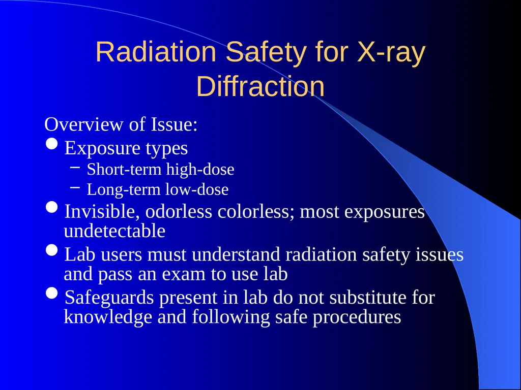
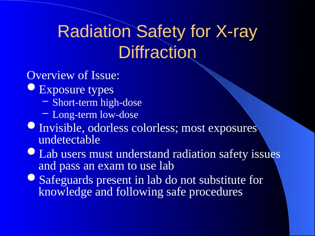
Radiation Safety for X-ray Diffraction Overview of Issue:
Exposure types
Short-term high-dose
Long-term low-dose
Invisible, odorless colorless; most exposures undetectable
Lab users must understand radiation safety issues and pass an exam to use lab
Safeguards present in lab do not substitute for knowledge and following safe procedures<br>
Exposure types
Short-term high-dose
Long-term low-dose
Invisible, odorless colorless; most exposures undetectable
Lab users must understand radiation safety issues and pass an exam to use lab
Safeguards present in lab do not substitute for knowledge and following safe procedures<br>
02
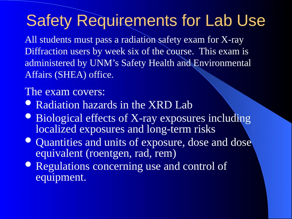
Safety Requirements for Lab Use The exam covers:
Radiation hazards in the XRD Lab
Biological effects of X-ray exposures including localized exposures and long-term risks
Quantities and units of exposure, dose and dose equivalent (roentgen, rad, rem)
Regulations concerning use and control of equipment. All students must pass a radiation safety exam for X-ray Diffraction users by week six of the course. This exam is administered by UNM’s Safety Health and Environmental Affairs (SHEA) office.<br>
Radiation hazards in the XRD Lab
Biological effects of X-ray exposures including localized exposures and long-term risks
Quantities and units of exposure, dose and dose equivalent (roentgen, rad, rem)
Regulations concerning use and control of equipment. All students must pass a radiation safety exam for X-ray Diffraction users by week six of the course. This exam is administered by UNM’s Safety Health and Environmental Affairs (SHEA) office.<br>
03
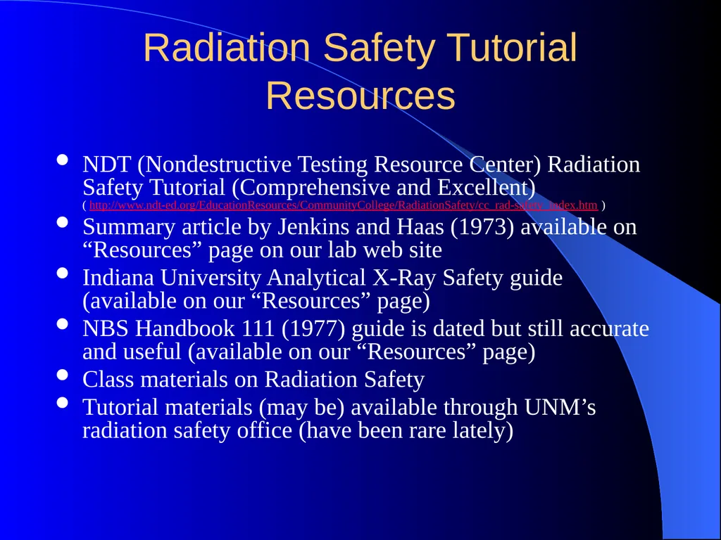
Radiation Safety Tutorial Resources NDT (Nondestructive Testing Resource Center) Radiation Safety Tutorial (Comprehensive and Excellent)
( http://www.ndt-ed.org/EducationResources/CommunityCollege/RadiationSafety/cc_rad-safety_index.htm )
Summary article by Jenkins and Haas (1973) available on “Resources” page on our lab web site
Indiana University Analytical X-Ray Safety guide (available on our “Resources” page)
NBS Handbook 111 (1977) guide is dated but still accurate and useful (available on our “Resources” page)
Class materials on Radiation Safety
Tutorial materials (may be) available through UNM’s radiation safety office (have been rare lately)<br>
Summary article by Jenkins and Haas (1973) available on “Resources” page on our lab web site
Indiana University Analytical X-Ray Safety guide (available on our “Resources” page)
NBS Handbook 111 (1977) guide is dated but still accurate and useful (available on our “Resources” page)
Class materials on Radiation Safety
Tutorial materials (may be) available through UNM’s radiation safety office (have been rare lately)<br>
04
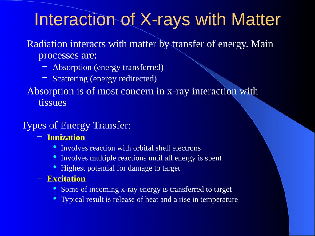
Interaction of X-rays with Matter Radiation interacts with matter by transfer of energy. Main processes are:
Absorption (energy transferred)
Scattering (energy redirected)
Absorption is of most concern in x-ray interaction with tissues Types of Energy Transfer:
Ionization
Involves reaction with orbital shell electrons
Involves multiple reactions until all energy is spent
Highest potential for damage to target.
Excitation
Some of incoming x-ray energy is transferred to target
Typical result is release of heat and a rise in temperature<br>
Absorption (energy transferred)
Scattering (energy redirected)
Absorption is of most concern in x-ray interaction with tissues Types of Energy Transfer:
Ionization
Involves reaction with orbital shell electrons
Involves multiple reactions until all energy is spent
Highest potential for damage to target.
Excitation
Some of incoming x-ray energy is transferred to target
Typical result is release of heat and a rise in temperature<br>
05
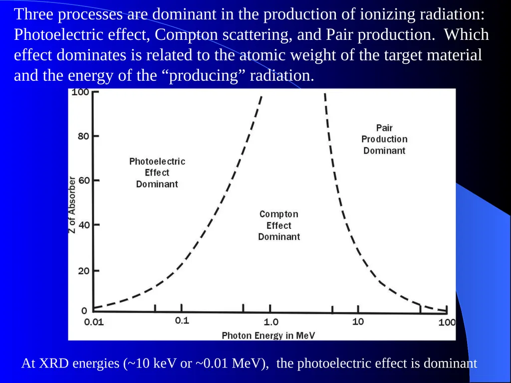
Three processes are dominant in the production of ionizing radiation: Photoelectric effect, Compton scattering, and Pair production. Which effect dominates is related to the atomic weight of the target material and the energy of the “producing” radiation. At XRD energies (~10 keV or ~0.01 MeV), the photoelectric effect is dominant<br>
06
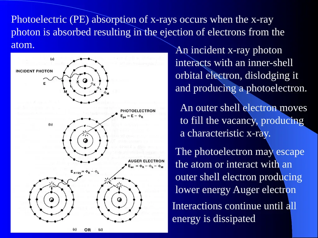
Photoelectric (PE) absorption of x-rays occurs when the x-ray photon is absorbed resulting in the ejection of electrons from the atom. An incident x-ray photon interacts with an inner-shell orbital electron, dislodging it and producing a photoelectron. An outer shell electron moves to fill the vacancy, producing a characteristic x-ray. The photoelectron may escape the atom or interact with an outer shell electron producing lower energy Auger electron Interactions continue until all energy is dissipated<br>
07
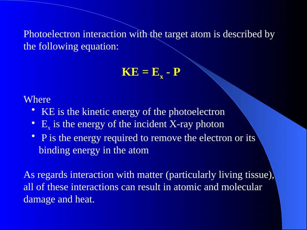
Photoelectron interaction with the target atom is described by the following equation:
KE = Ex - P
Where
KE is the kinetic energy of the photoelectron
Ex is the energy of the incident X-ray photon
P is the energy required to remove the electron or its binding energy in the atom
As regards interaction with matter (particularly living tissue), all of these interactions can result in atomic and molecular damage and heat.<br>
KE = Ex - P
Where
KE is the kinetic energy of the photoelectron
Ex is the energy of the incident X-ray photon
P is the energy required to remove the electron or its binding energy in the atom
As regards interaction with matter (particularly living tissue), all of these interactions can result in atomic and molecular damage and heat.<br>
08
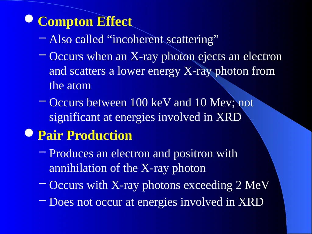
Compton Effect
Also called “incoherent scattering”
Occurs when an X-ray photon ejects an electron and scatters a lower energy X-ray photon from the atom
Occurs between 100 keV and 10 Mev; not significant at energies involved in XRD
Pair Production
Produces an electron and positron with annihilation of the X-ray photon
Occurs with X-ray photons exceeding 2 MeV
Does not occur at energies involved in XRD<br>
Also called “incoherent scattering”
Occurs when an X-ray photon ejects an electron and scatters a lower energy X-ray photon from the atom
Occurs between 100 keV and 10 Mev; not significant at energies involved in XRD
Pair Production
Produces an electron and positron with annihilation of the X-ray photon
Occurs with X-ray photons exceeding 2 MeV
Does not occur at energies involved in XRD<br>
09
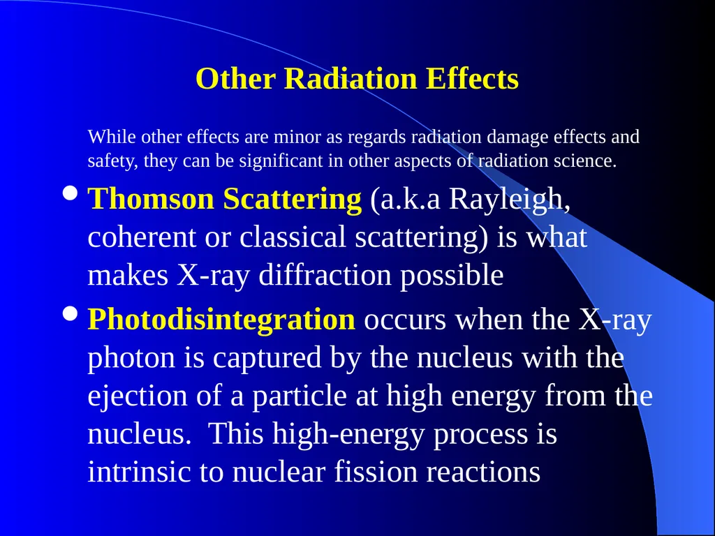
Other Radiation Effects
While other effects are minor as regards radiation damage effects and safety, they can be significant in other aspects of radiation science.
Thomson Scattering (a.k.a Rayleigh, coherent or classical scattering) is what makes X-ray diffraction possible
Photodisintegration occurs when the X-ray photon is captured by the nucleus with the ejection of a particle at high energy from the nucleus. This high-energy process is intrinsic to nuclear fission reactions<br>
While other effects are minor as regards radiation damage effects and safety, they can be significant in other aspects of radiation science.
Thomson Scattering (a.k.a Rayleigh, coherent or classical scattering) is what makes X-ray diffraction possible
Photodisintegration occurs when the X-ray photon is captured by the nucleus with the ejection of a particle at high energy from the nucleus. This high-energy process is intrinsic to nuclear fission reactions<br>
10
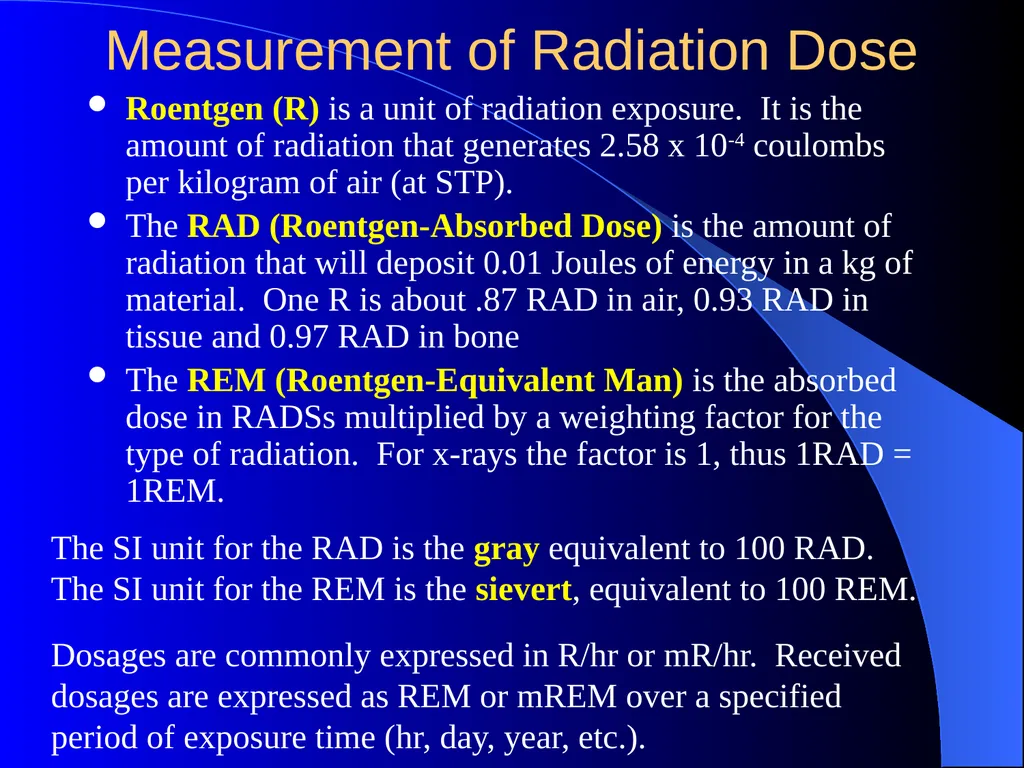
Measurement of Radiation Dose Roentgen (R) is a unit of radiation exposure. It is the amount of radiation that generates 2.58 x 10⁻⁴ coulombs per kilogram of air (at STP).
The RAD (Roentgen-Absorbed Dose) is the amount of radiation that will deposit 0.01 Joules of energy in a kg of material. One R is about .87 RAD in air, 0.93 RAD in tissue and 0.97 RAD in bone
The REM (Roentgen-Equivalent Man) is the absorbed dose in RADSs multiplied by a weighting factor for the type of radiation. For x-rays the factor is 1, thus 1RAD = 1REM. The SI unit for the RAD is the gray equivalent to 100 RAD. The SI unit for the REM is the sievert, equivalent to 100 REM. Dosages are commonly expressed in R/hr or mR/hr. Received dosages are expressed as REM or mREM over a specified period of exposure time (hr, day, year, etc.).<br>
The RAD (Roentgen-Absorbed Dose) is the amount of radiation that will deposit 0.01 Joules of energy in a kg of material. One R is about .87 RAD in air, 0.93 RAD in tissue and 0.97 RAD in bone
The REM (Roentgen-Equivalent Man) is the absorbed dose in RADSs multiplied by a weighting factor for the type of radiation. For x-rays the factor is 1, thus 1RAD = 1REM. The SI unit for the RAD is the gray equivalent to 100 RAD. The SI unit for the REM is the sievert, equivalent to 100 REM. Dosages are commonly expressed in R/hr or mR/hr. Received dosages are expressed as REM or mREM over a specified period of exposure time (hr, day, year, etc.).<br>
11
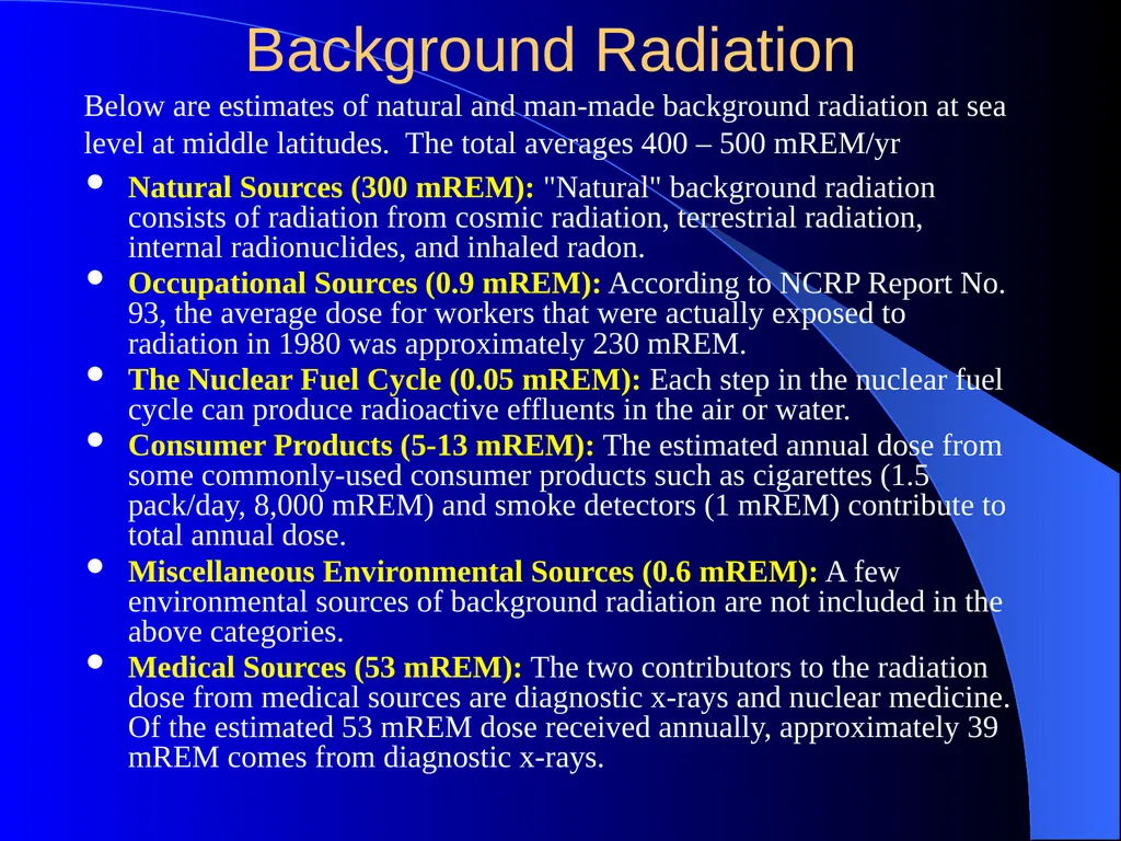
Background Radiation Natural Sources (300 mREM): "Natural" background radiation consists of radiation from cosmic radiation, terrestrial radiation, internal radionuclides, and inhaled radon.
Occupational Sources (0.9 mREM): According to NCRP Report No. 93, the average dose for workers that were actually exposed to radiation in 1980 was approximately 230 mREM.
The Nuclear Fuel Cycle (0.05 mREM): Each step in the nuclear fuel cycle can produce radioactive effluents in the air or water.
Consumer Products (5-13 mREM): The estimated annual dose from some commonly-used consumer products such as cigarettes (1.5 pack/day, 8,000 mREM) and smoke detectors (1 mREM) contribute to total annual dose.
Miscellaneous Environmental Sources (0.6 mREM): A few environmental sources of background radiation are not included in the above categories.
Medical Sources (53 mREM): The two contributors to the radiation dose from medical sources are diagnostic x-rays and nuclear medicine. Of the estimated 53 mREM dose received annually, approximately 39 mREM comes from diagnostic x-rays. Below are estimates of natural and man-made background radiation at sea level at middle latitudes. The total averages 400 – 500 mREM/yr<br>
Occupational Sources (0.9 mREM): According to NCRP Report No. 93, the average dose for workers that were actually exposed to radiation in 1980 was approximately 230 mREM.
The Nuclear Fuel Cycle (0.05 mREM): Each step in the nuclear fuel cycle can produce radioactive effluents in the air or water.
Consumer Products (5-13 mREM): The estimated annual dose from some commonly-used consumer products such as cigarettes (1.5 pack/day, 8,000 mREM) and smoke detectors (1 mREM) contribute to total annual dose.
Miscellaneous Environmental Sources (0.6 mREM): A few environmental sources of background radiation are not included in the above categories.
Medical Sources (53 mREM): The two contributors to the radiation dose from medical sources are diagnostic x-rays and nuclear medicine. Of the estimated 53 mREM dose received annually, approximately 39 mREM comes from diagnostic x-rays. Below are estimates of natural and man-made background radiation at sea level at middle latitudes. The total averages 400 – 500 mREM/yr<br>
12
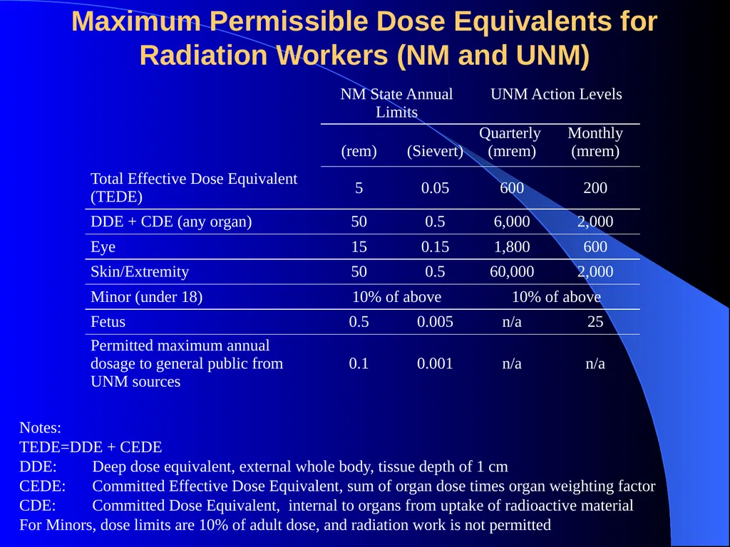
Maximum Permissible Dose Equivalents for Radiation Workers (NM and UNM) Notes:
TEDE=DDE + CEDE
DDE: Deep dose equivalent, external whole body, tissue depth of 1 cm
CEDE: Committed Effective Dose Equivalent, sum of organ dose times organ weighting factor
CDE: Committed Dose Equivalent, internal to organs from uptake of radioactive material
For Minors, dose limits are 10% of adult dose, and radiation work is not permitted<br>
DDE: Deep dose equivalent, external whole body, tissue depth of 1 cm
CEDE: Committed Effective Dose Equivalent, sum of organ dose times organ weighting factor
CDE: Committed Dose Equivalent, internal to organs from uptake of radioactive material
For Minors, dose limits are 10% of adult dose, and radiation work is not permitted<br>
13
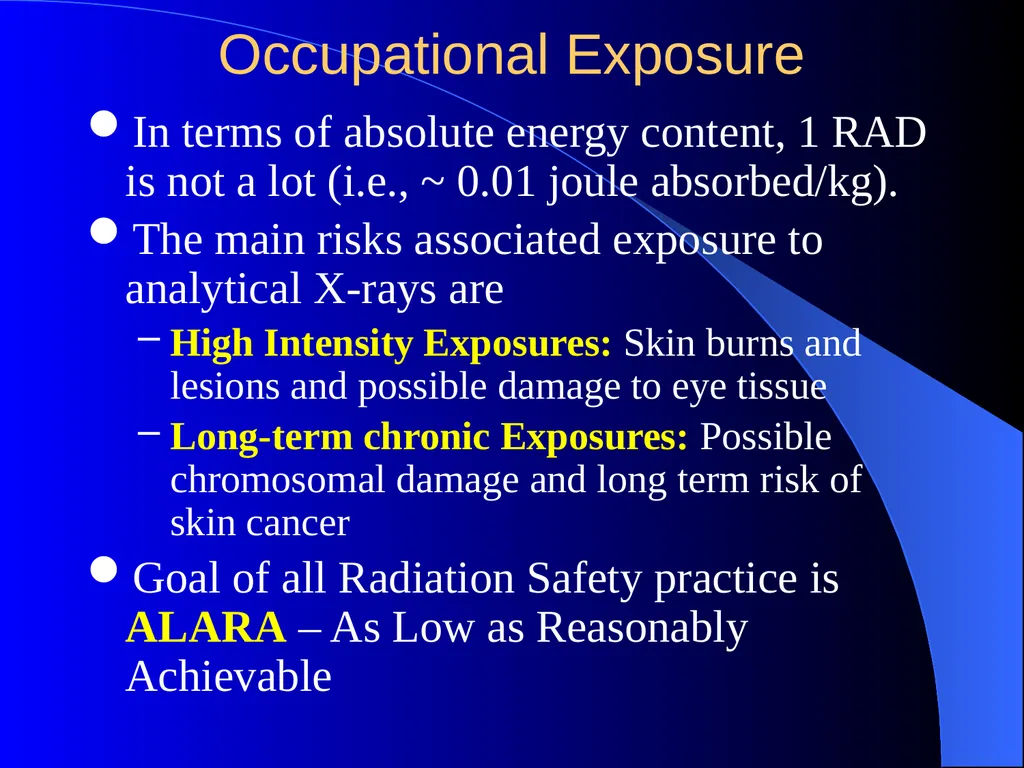
Occupational Exposure In terms of absolute energy content, 1 RAD is not a lot (i.e., ~ 0.01 joule absorbed/kg).
The main risks associated exposure to analytical X-rays are
High Intensity Exposures: Skin burns and lesions and possible damage to eye tissue
Long-term chronic Exposures: Possible chromosomal damage and long term risk of skin cancer
Goal of all Radiation Safety practice is ALARA – As Low as Reasonably Achievable<br>
The main risks associated exposure to analytical X-rays are
High Intensity Exposures: Skin burns and lesions and possible damage to eye tissue
Long-term chronic Exposures: Possible chromosomal damage and long term risk of skin cancer
Goal of all Radiation Safety practice is ALARA – As Low as Reasonably Achievable<br>
14
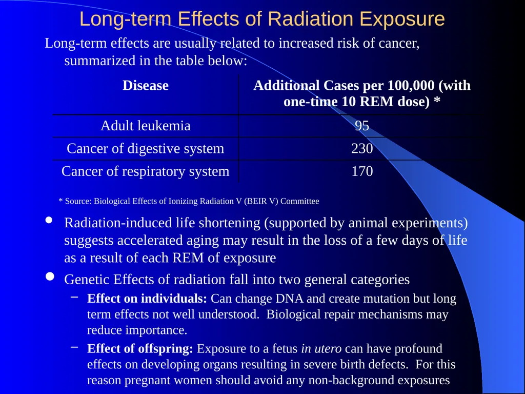
Long-term Effects of Radiation Exposure Long-term effects are usually related to increased risk of cancer, summarized in the table below: * Source: Biological Effects of Ionizing Radiation V (BEIR V) Committee Radiation-induced life shortening (supported by animal experiments) suggests accelerated aging may result in the loss of a few days of life as a result of each REM of exposure Genetic Effects of radiation fall into two general categories
Effect on individuals: Can change DNA and create mutation but long term effects not well understood. Biological repair mechanisms may reduce importance.
Effect of offspring: Exposure to a fetus in utero can have profound effects on developing organs resulting in severe birth defects. For this reason pregnant women should avoid any non-background exposures<br>
Effect on individuals: Can change DNA and create mutation but long term effects not well understood. Biological repair mechanisms may reduce importance.
Effect of offspring: Exposure to a fetus in utero can have profound effects on developing organs resulting in severe birth defects. For this reason pregnant women should avoid any non-background exposures<br>
15
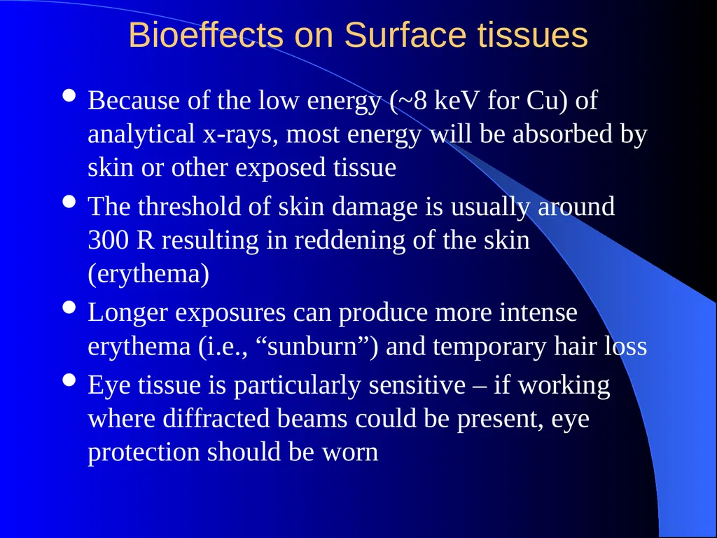
Bioeffects on Surface tissues Because of the low energy (~8 keV for Cu) of analytical x-rays, most energy will be absorbed by skin or other exposed tissue
The threshold of skin damage is usually around 300 R resulting in reddening of the skin (erythema)
Longer exposures can produce more intense erythema (i.e., “sunburn”) and temporary hair loss
Eye tissue is particularly sensitive – if working where diffracted beams could be present, eye protection should be worn<br>
The threshold of skin damage is usually around 300 R resulting in reddening of the skin (erythema)
Longer exposures can produce more intense erythema (i.e., “sunburn”) and temporary hair loss
Eye tissue is particularly sensitive – if working where diffracted beams could be present, eye protection should be worn<br>
16
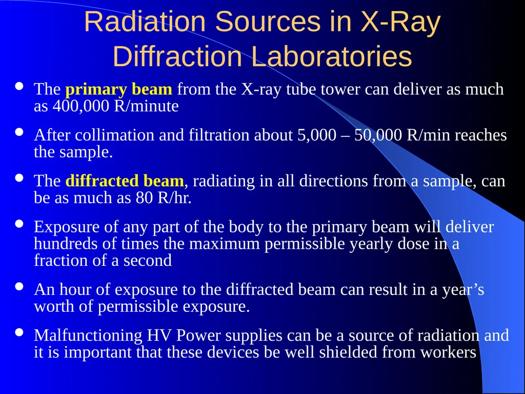
Radiation Sources in X-Ray Diffraction Laboratories The primary beam from the X-ray tube tower can deliver as much as 400,000 R/minute
After collimation and filtration about 5,000 – 50,000 R/min reaches the sample.
The diffracted beam, radiating in all directions from a sample, can be as much as 80 R/hr.
Exposure of any part of the body to the primary beam will deliver hundreds of times the maximum permissible yearly dose in a fraction of a second
An hour of exposure to the diffracted beam can result in a year’s worth of permissible exposure.
Malfunctioning HV Power supplies can be a source of radiation and it is important that these devices be well shielded from workers<br>
After collimation and filtration about 5,000 – 50,000 R/min reaches the sample.
The diffracted beam, radiating in all directions from a sample, can be as much as 80 R/hr.
Exposure of any part of the body to the primary beam will deliver hundreds of times the maximum permissible yearly dose in a fraction of a second
An hour of exposure to the diffracted beam can result in a year’s worth of permissible exposure.
Malfunctioning HV Power supplies can be a source of radiation and it is important that these devices be well shielded from workers<br>
17
Measures taken to Reduce Risk of Exposure in the Laboratory Complete enclosure of source and diffractometer whenever possible.
Spring-loaded fail safe shutters on the X-ray primary beam.
Fail-safe interlocks installed on the housing.
A fail-safe indicator light in the shutter-opening circuit.
Seal all openings in the housing with lead tape.
Periodic checks of the system for leakage at normal operating conditions using properly calibrated survey equipment. There is no required interval for this, but it must be requested if the following conditions exist:
Prior to the receipt of new equipment
Prior to a change in the arrangement, number , or type of local components in the system
Prior to any maintenance requiring the disassembly or removal of a local component in the system
Anytime a visual inspection of the system reveals an abnormal condition.
High-voltage power supplies, if not functioning properly, can be the source of X-rays. It is important that the HV voltage multipliers and other circuitry be properly shielded to eliminate this as a possible radiation source.<br>
Spring-loaded fail safe shutters on the X-ray primary beam.
Fail-safe interlocks installed on the housing.
A fail-safe indicator light in the shutter-opening circuit.
Seal all openings in the housing with lead tape.
Periodic checks of the system for leakage at normal operating conditions using properly calibrated survey equipment. There is no required interval for this, but it must be requested if the following conditions exist:
Prior to the receipt of new equipment
Prior to a change in the arrangement, number , or type of local components in the system
Prior to any maintenance requiring the disassembly or removal of a local component in the system
Anytime a visual inspection of the system reveals an abnormal condition.
High-voltage power supplies, if not functioning properly, can be the source of X-rays. It is important that the HV voltage multipliers and other circuitry be properly shielded to eliminate this as a possible radiation source.<br>
18
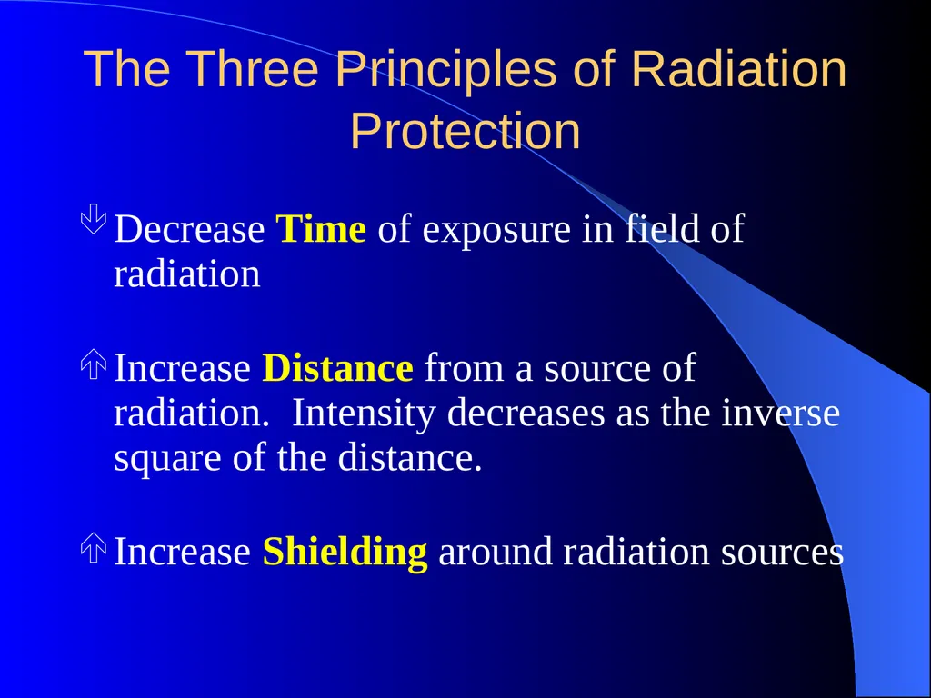
The Three Principles of Radiation Protection Decrease Time of exposure in field of radiation
Increase Distance from a source of radiation. Intensity decreases as the inverse square of the distance.
Increase Shielding around radiation sources<br>
Increase Distance from a source of radiation. Intensity decreases as the inverse square of the distance.
Increase Shielding around radiation sources<br>
19
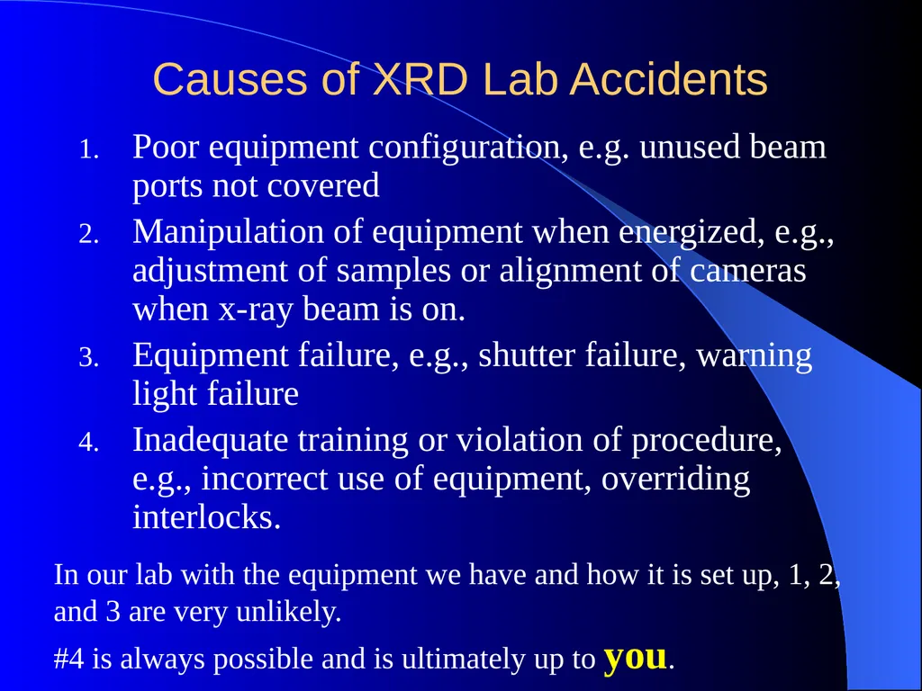
Causes of XRD Lab Accidents Poor equipment configuration, e.g. unused beam ports not covered
Manipulation of equipment when energized, e.g., adjustment of samples or alignment of cameras when x-ray beam is on.
Equipment failure, e.g., shutter failure, warning light failure
Inadequate training or violation of procedure, e.g., incorrect use of equipment, overriding interlocks. In our lab with the equipment we have and how it is set up, 1, 2, and 3 are very unlikely.
#4 is always possible and is ultimately up to you.<br>
Manipulation of equipment when energized, e.g., adjustment of samples or alignment of cameras when x-ray beam is on.
Equipment failure, e.g., shutter failure, warning light failure
Inadequate training or violation of procedure, e.g., incorrect use of equipment, overriding interlocks. In our lab with the equipment we have and how it is set up, 1, 2, and 3 are very unlikely.
#4 is always possible and is ultimately up to you.<br>
20
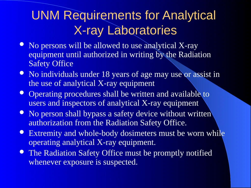
UNM Requirements for Analytical
X-ray Laboratories No persons will be allowed to use analytical X-ray equipment until authorized in writing by the Radiation Safety Office
No individuals under 18 years of age may use or assist in the use of analytical X-ray equipment
Operating procedures shall be written and available to users and inspectors of analytical X-ray equipment
No person shall bypass a safety device without written authorization from the Radiation Safety Office.
Extremity and whole-body dosimeters must be worn while operating analytical X-ray equipment.
The Radiation Safety Office must be promptly notified whenever exposure is suspected.<br>
No individuals under 18 years of age may use or assist in the use of analytical X-ray equipment
Operating procedures shall be written and available to users and inspectors of analytical X-ray equipment
No person shall bypass a safety device without written authorization from the Radiation Safety Office.
Extremity and whole-body dosimeters must be worn while operating analytical X-ray equipment.
The Radiation Safety Office must be promptly notified whenever exposure is suspected.<br>