
Respiratory Emergencies CHAPTER 17: Focused
TF
Published · 106 slides · 0 views

1 / 1












Description
Respiratory Emergencies CHAPTER 17: Focused Lecture National EMS Education Standard Competencies (1 of 5) Medicine Applies fundamental knowledge to provide basic and selected advanced emergency care and transportation based on assessment
Related Topics
Share
Embed code
Download this presentation From Below
"Respiratory Emergencies CHAPTER 17: Focused" is the property of its rightful owner. Permission is granted to download and print the materials on this website for personal, non-commercial use only, and to display it on your personal computer provided you do not modify the materials and that you retain all copyright notices contained in the materials. By downloading content from our website, you accept the terms of this agreement.
Presentation Transcript
01
Respiratory Emergencies CHAPTER 17: Focused Lecture<br>
02
National EMS Education Standard Competencies (1 of 5) Medicine
Applies fundamental knowledge to provide basic and selected advanced emergency care and transportation based on assessment findings for an acutely ill patient.<br>
Applies fundamental knowledge to provide basic and selected advanced emergency care and transportation based on assessment findings for an acutely ill patient.<br>
03
National EMS Education Standard Competencies (2 of 5) Respiratory
Anatomy, signs, symptoms, and management of respiratory emergencies including those that affect the
Upper airway
Lower airway<br>
Anatomy, signs, symptoms, and management of respiratory emergencies including those that affect the
Upper airway
Lower airway<br>
04
National EMS Education Standard Competencies (3 of 5) Anatomy, physiology, pathophysiology, assessment, and management of
Epiglottitis
Spontaneous pneumothorax
Pulmonary edema
Asthma<br>
Epiglottitis
Spontaneous pneumothorax
Pulmonary edema
Asthma<br>
05
National EMS Education Standard Competencies (4 of 5) Anatomy, physiology, pathophysiology, assessment, and management of
Chronic obstructive pulmonary disease
Environmental/industrial exposure
Toxic gas
Pertussis
Cystic fibrosis<br>
Chronic obstructive pulmonary disease
Environmental/industrial exposure
Toxic gas
Pertussis
Cystic fibrosis<br>
06
National EMS Education Standard Competencies (5 of 5) Anatomy, physiology, pathophysiology, assessment, and management of
Pulmonary embolism
Pneumonia
Viral respiratory infections
Obstructive/restrictive disease<br>
Pulmonary embolism
Pneumonia
Viral respiratory infections
Obstructive/restrictive disease<br>
07
Introduction Dyspnea is difficulty breathing.
Common complaint
Symptom of many conditions
Keep medical problems in mind as you obtain history and perform assessment.
Treat the symptoms, underlying problem, and anxiety.<br>
Common complaint
Symptom of many conditions
Keep medical problems in mind as you obtain history and perform assessment.
Treat the symptoms, underlying problem, and anxiety.<br>
08
Anatomy and Physiology Review (1 of 3) Upper airway: All airway structures above level of the vocal cords
Gas exchange: Blood in the pulmonary circulation picks up oxygen and releases carbon dioxide. © Jones & Bartlett Learning.<br>
Gas exchange: Blood in the pulmonary circulation picks up oxygen and releases carbon dioxide. © Jones & Bartlett Learning.<br>
09
Anatomy and Physiology Review (2 of 3) © Jones & Bartlett Learning.<br>
10
Anatomy and Physiology Review (3 of 3) © Jones & Bartlett Learning. © Jones & Bartlett Learning.<br>
11
Respiration (1 of 2) Two processes occur during respiration:
Inspiration
Expiration
Ventilation
Process of moving air into and out of the lungs
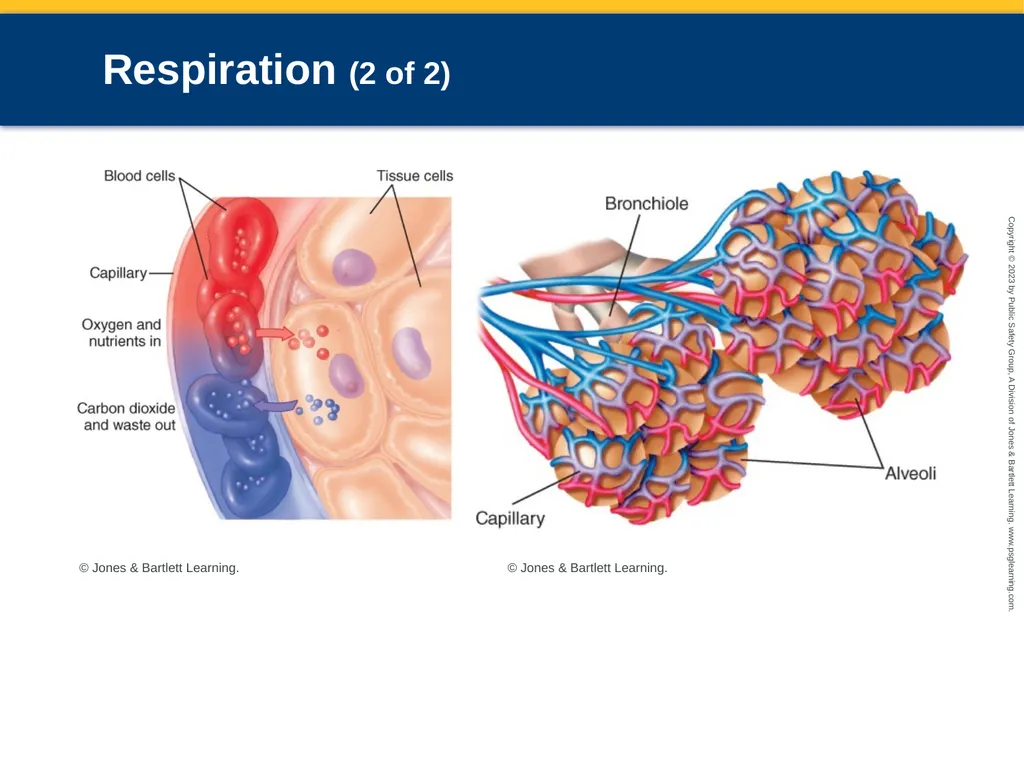
Heart pumps oxygenated blood throughout the body.
Carbon dioxide is exchanged for oxygen.<br>
Inspiration
Expiration
Ventilation
Process of moving air into and out of the lungs
Heart pumps oxygenated blood throughout the body.
Carbon dioxide is exchanged for oxygen.<br>
12
Respiration (2 of 2) © Jones & Bartlett Learning. © Jones & Bartlett Learning.<br>
13
Inspiration Stimulus to breathe comes from respiratory center in the medulla.
Diaphragm and intercostal muscles contract.
Thorax enlarges and intrapulmonary pressure fall below atmospheric pressure.
Air fills the lungs.
When pressure outside the thorax equals the pressure inside the thorax, inhalation stops.<br>
Diaphragm and intercostal muscles contract.
Thorax enlarges and intrapulmonary pressure fall below atmospheric pressure.
Air fills the lungs.
When pressure outside the thorax equals the pressure inside the thorax, inhalation stops.<br>
14
Expiration As the chest expands, stretch receptors send a signal to the apneustic center to inhibit the inspiratory center, and expiration occurs.
Prevents overexpansion of the lungs
Expiration lasts twice as long as inspiration.
Inspiratory/expiratory ratio<br>
Prevents overexpansion of the lungs
Expiration lasts twice as long as inspiration.
Inspiratory/expiratory ratio<br>
15
Pathophysiology Adequate ventilation, diffusion, and perfusion must occur for body to receive nutrients and oxygen.
Complications interfering with oxygen intake
Upper airway obstruction
Lower airway obstruction
Chest wall impairment
Neurologic control problems<br>
Complications interfering with oxygen intake
Upper airway obstruction
Lower airway obstruction
Chest wall impairment
Neurologic control problems<br>
16
Gas Exchange Interface (1 of 2) Alveoli are made up of two types of cells
Type I: Allow for better gas exchange
Type II: Make new type I cells and produce surfactant
Alveoli function best when partially inflated.
Surfactant reduces surface tension of the alveoli, making it easier for them to expand.<br>
Type I: Allow for better gas exchange
Type II: Make new type I cells and produce surfactant
Alveoli function best when partially inflated.
Surfactant reduces surface tension of the alveoli, making it easier for them to expand.<br>
17
Gas Exchange Interface (2 of 2) Conditions related to ventilation and/or perfusion can prevent oxygen from reaching bloodstream.
People with chronic lung disease make a surplus of red blood cells making blood thicker (polycythemia).
Cor pulmonale: Right-side heart failure because of chronic lung disease<br>
People with chronic lung disease make a surplus of red blood cells making blood thicker (polycythemia).
Cor pulmonale: Right-side heart failure because of chronic lung disease<br>
18
Carbon Dioxide Retention and Hypoxic Drive Rise in arterial carbon dioxide levels leads to less efficient respiratory centers.
Result of chronic carbon dioxide retention
Hypoxic drive
Backup system used to control breathing based on low levels of oxygen, rather than high levels of carbon dioxide
Stimulus to breathe is detection of low blood oxygen levels.<br>
Result of chronic carbon dioxide retention
Hypoxic drive
Backup system used to control breathing based on low levels of oxygen, rather than high levels of carbon dioxide
Stimulus to breathe is detection of low blood oxygen levels.<br>
19
Hypoventilation Excess carbon dioxide in blood results in respiratory acidosis.
Can occur if hypoventilation is not recognized
Carbon dioxide level is directly related to pH (acid-base balance).
Causes of hypoventilation:
Impaired lung function
Impaired mechanics of breathing
Impaired neuromuscular apparatus
Reduced respiratory drive<br>
Can occur if hypoventilation is not recognized
Carbon dioxide level is directly related to pH (acid-base balance).
Causes of hypoventilation:
Impaired lung function
Impaired mechanics of breathing
Impaired neuromuscular apparatus
Reduced respiratory drive<br>
20
Hyperventilation Arterial carbon dioxide falls below normal
May be an indicator of life-threatening illness
Tachypnea without physiologic demand for increased oxygen causes respiratory alkalosis.
Alkalosis causes symptoms of hyperventilation syndrome.
Can be body’s response to illness and a buildup of acids<br>
May be an indicator of life-threatening illness
Tachypnea without physiologic demand for increased oxygen causes respiratory alkalosis.
Alkalosis causes symptoms of hyperventilation syndrome.
Can be body’s response to illness and a buildup of acids<br>
21
Causes of Dyspnea (1 of 2) Dyspnea
Shortness of breath or difficulty breathing
One or more of these situations most likely exists:
Atelectasis
Damaged alveoli
Obstructed air passages, blood clots
Pleural space filled with air or excess fluid<br>
Shortness of breath or difficulty breathing
One or more of these situations most likely exists:
Atelectasis
Damaged alveoli
Obstructed air passages, blood clots
Pleural space filled with air or excess fluid<br>
22
Causes of Dyspnea (2 of 2) Patient with dyspnea may also report:
Air hunger
Chest tightness
Dyspnea is a common complaint in patients with cardiopulmonary disease.
Severe pain can cause a patient to experience rapid, shallow breathing without pulmonary dysfunction.<br>
Air hunger
Chest tightness
Dyspnea is a common complaint in patients with cardiopulmonary disease.
Severe pain can cause a patient to experience rapid, shallow breathing without pulmonary dysfunction.<br>
23
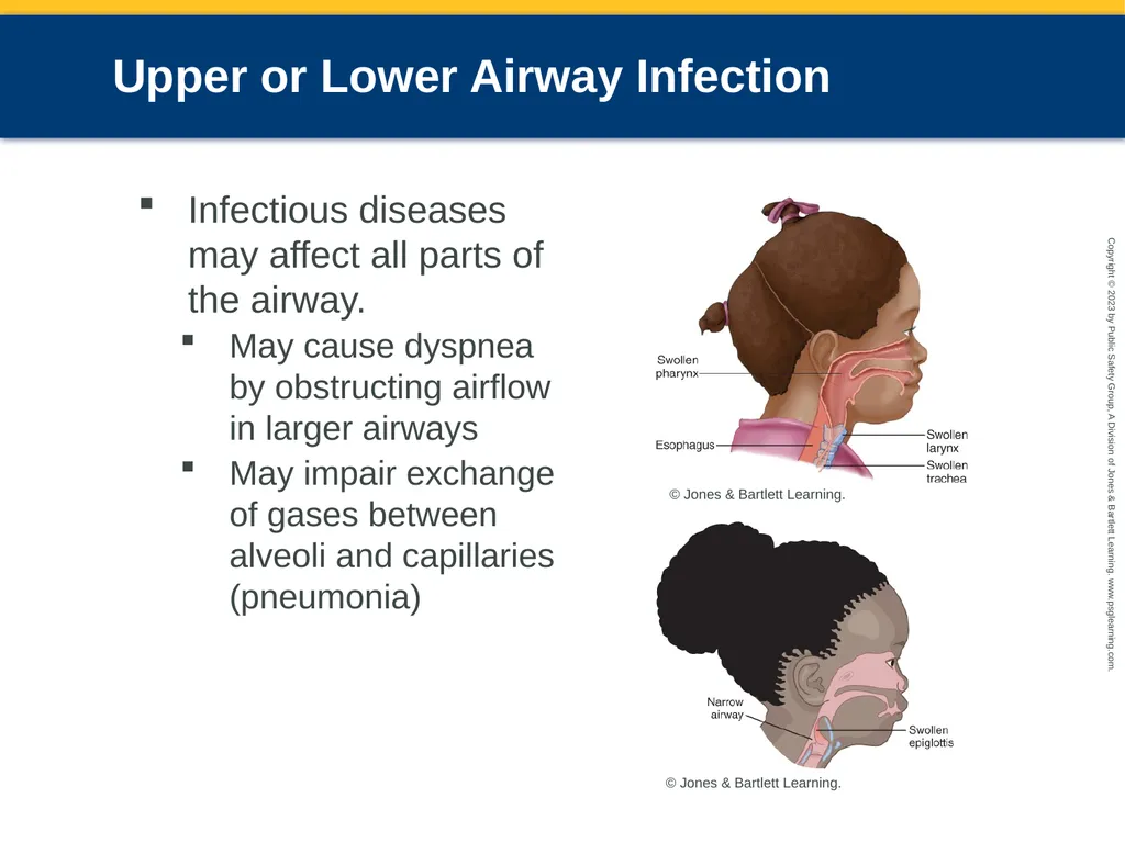
Upper or Lower Airway Infection Infectious diseases may affect all parts of the airway.
May cause dyspnea by obstructing airflow in larger airways
May impair exchange of gases between alveoli and capillaries (pneumonia) © Jones & Bartlett Learning. © Jones & Bartlett Learning.<br>
May cause dyspnea by obstructing airflow in larger airways
May impair exchange of gases between alveoli and capillaries (pneumonia) © Jones & Bartlett Learning. © Jones & Bartlett Learning.<br>
24
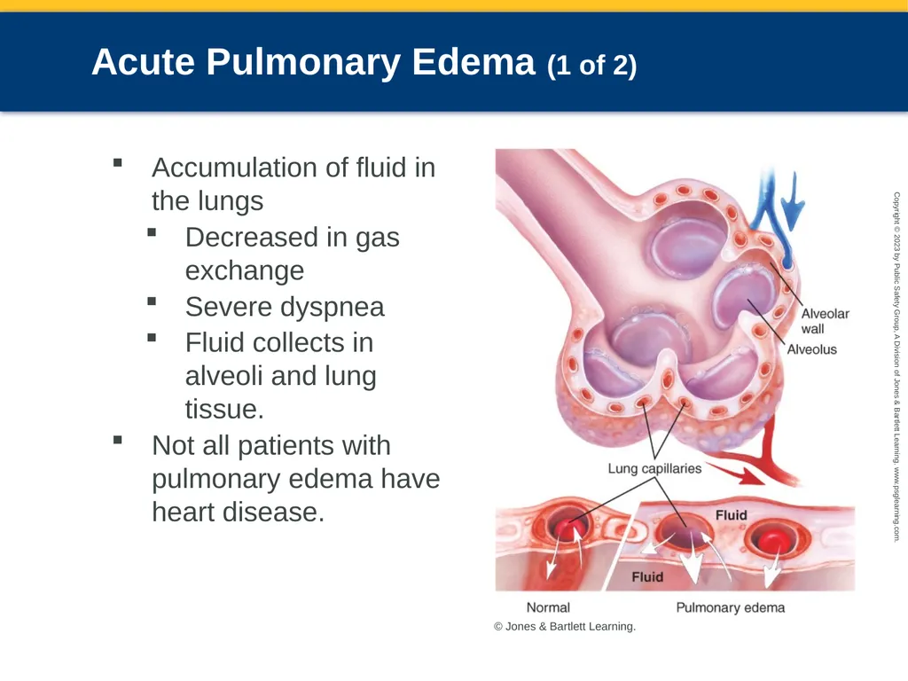
Acute Pulmonary Edema (1 of 2) Accumulation of fluid in the lungs
Decreased in gas exchange
Severe dyspnea
Fluid collects in alveoli and lung tissue.
Not all patients with pulmonary edema have heart disease. © Jones & Bartlett Learning.<br>
Decreased in gas exchange
Severe dyspnea
Fluid collects in alveoli and lung tissue.
Not all patients with pulmonary edema have heart disease. © Jones & Bartlett Learning.<br>
25
Acute Pulmonary Edema (2 of 2) Patients with pulmonary edema may also present with:
Dyspnea
Orthopnea
Fatigue
Reduced exercise capacity
Pulmonary crackles (formerly called rales)<br>
Dyspnea
Orthopnea
Fatigue
Reduced exercise capacity
Pulmonary crackles (formerly called rales)<br>
26
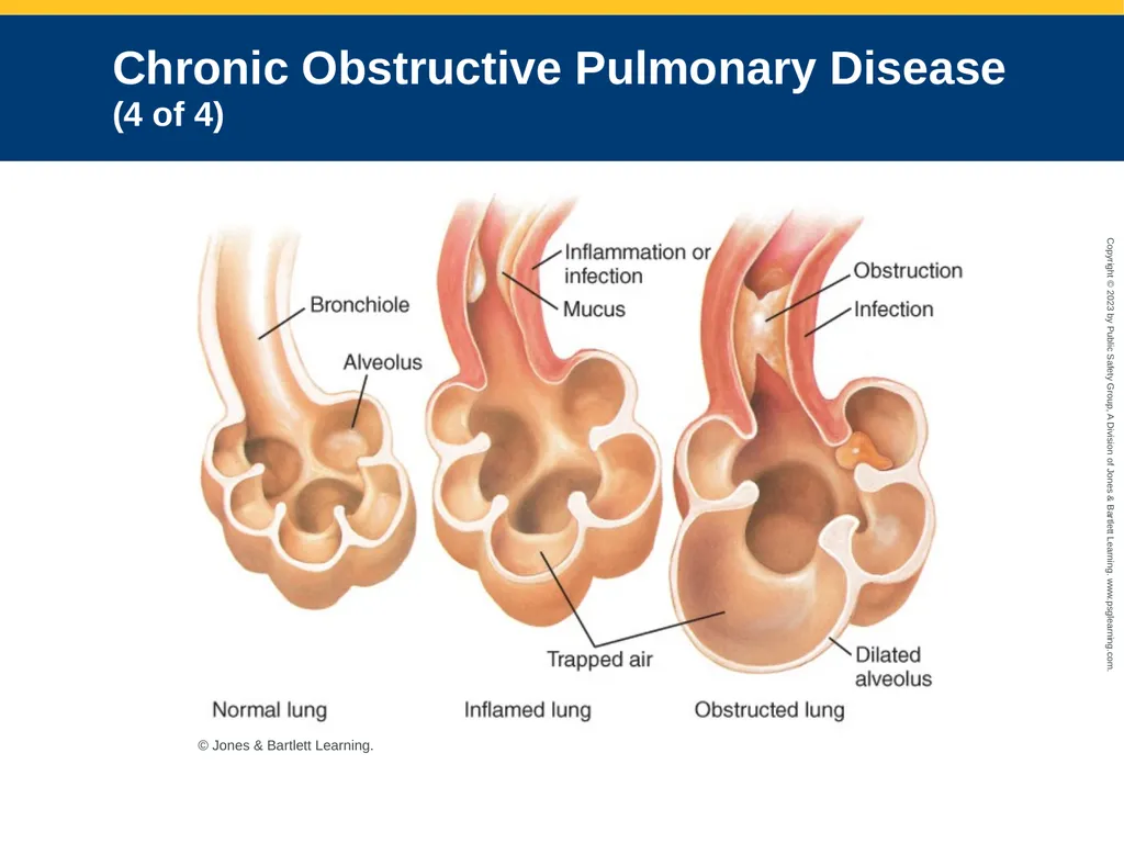
Chronic Obstructive Pulmonary Disease (1 of 4) Slow process of dilation and disruption of the airways and alveoli
Cigarette smoking is most common cause.
Obstruction occurs in the bronchioles.
Chronic bronchitis results from overgrowth of airway mucus glands and excess secretion of mucus.<br>
Cigarette smoking is most common cause.
Obstruction occurs in the bronchioles.
Chronic bronchitis results from overgrowth of airway mucus glands and excess secretion of mucus.<br>
27
Chronic Obstructive Pulmonary Disease (2 of 4) Pneumonia develops when passages are persistently obstructed.
Repeated episodes can lead to COPD
Emphysema
Most common form of COPD
Characterized by destruction of alveolar walls related to destruction of pulmonary surfactant<br>
Repeated episodes can lead to COPD
Emphysema
Most common form of COPD
Characterized by destruction of alveolar walls related to destruction of pulmonary surfactant<br>
28
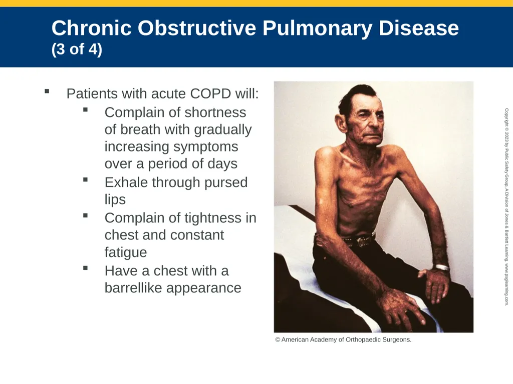
Chronic Obstructive Pulmonary Disease (3 of 4) Patients with acute COPD will:
Complain of shortness of breath with gradually increasing symptoms over a period of days
Exhale through pursed lips
Complain of tightness in chest and constant fatigue
Have a chest with a barrellike appearance © American Academy of Orthopaedic Surgeons.<br>
Complain of shortness of breath with gradually increasing symptoms over a period of days
Exhale through pursed lips
Complain of tightness in chest and constant fatigue
Have a chest with a barrellike appearance © American Academy of Orthopaedic Surgeons.<br>
29
Chronic Obstructive Pulmonary Disease (4 of 4) © Jones & Bartlett Learning.<br>
30
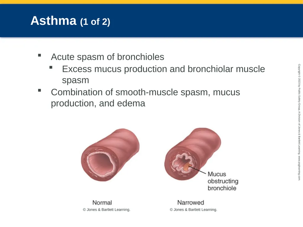
Asthma (1 of 2) Acute spasm of bronchioles
Excess mucus production and bronchiolar muscle spasm
Combination of smooth-muscle spasm, mucus production, and edema © Jones & Bartlett Learning. © Jones & Bartlett Learning.<br>
Excess mucus production and bronchiolar muscle spasm
Combination of smooth-muscle spasm, mucus production, and edema © Jones & Bartlett Learning. © Jones & Bartlett Learning.<br>
31
Asthma (2 of 2) Results from:
Allergic reaction
Exercise
Severe emotional stress
Respiratory infection<br>
Allergic reaction
Exercise
Severe emotional stress
Respiratory infection<br>
32
Anaphylactic Reactions Severe allergic reaction
Characterized by airway swelling
Dilation of blood vessels all over the body
Can cause respiratory distress severe enough to lead to coma and death
Most reactions occur within 30 minutes of exposure to an allergen.<br>
Characterized by airway swelling
Dilation of blood vessels all over the body
Can cause respiratory distress severe enough to lead to coma and death
Most reactions occur within 30 minutes of exposure to an allergen.<br>
33
Spontaneous Pneumothorax (1 of 2) Accumulation of air in pleural space
May be caused by trauma or medical conditions
Diminished breath sounds on affected side © Jones & Bartlett Learning.<br>
May be caused by trauma or medical conditions
Diminished breath sounds on affected side © Jones & Bartlett Learning.<br>
34
Spontaneous Pneumothorax (2 of 2) Patients become acutely dyspneic and typically complain of pleuritic chest pain.
Severe findings:
Altered mental status
Cyanosis, tachycardia
Unilaterally decreased breath sounds
Local hyperresonance to percussion
Subcutaneous emphysema
Tracheal deviation (late sign)<br>
Severe findings:
Altered mental status
Cyanosis, tachycardia
Unilaterally decreased breath sounds
Local hyperresonance to percussion
Subcutaneous emphysema
Tracheal deviation (late sign)<br>
35
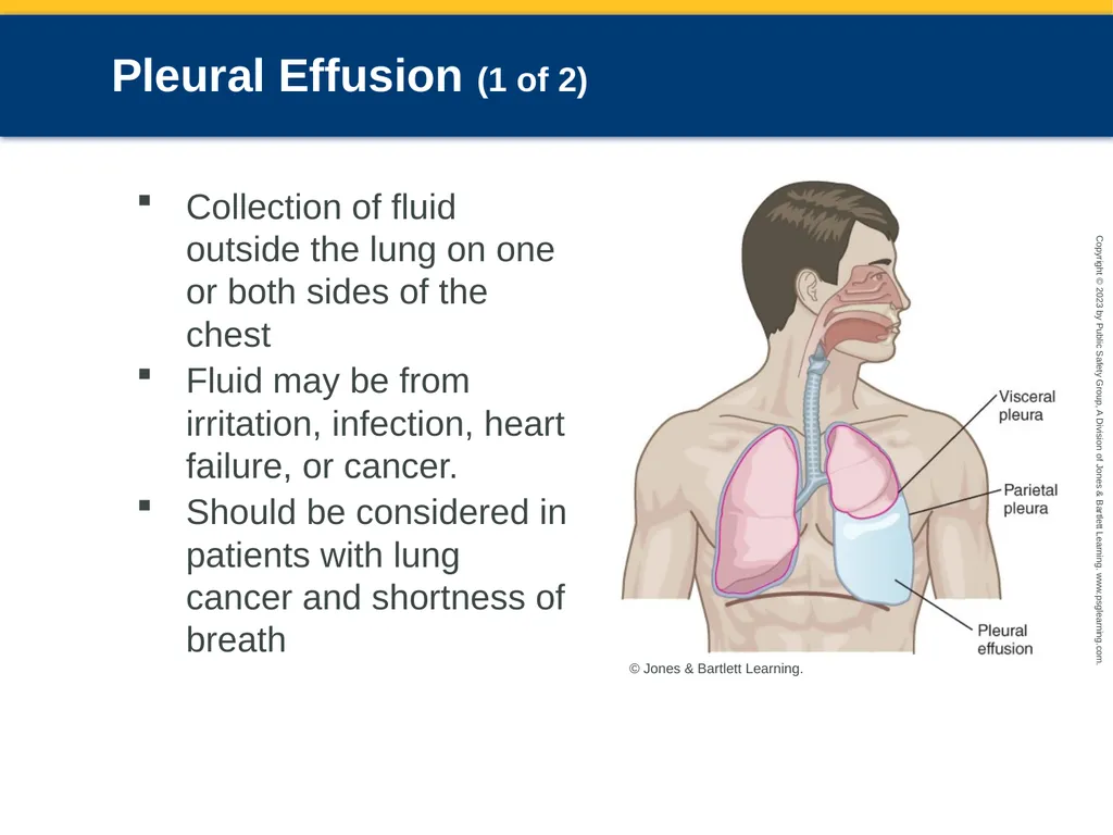
Pleural Effusion (1 of 2) Collection of fluid outside the lung on one or both sides of the chest
Fluid may be from irritation, infection, heart failure, or cancer.
Should be considered in patients with lung cancer and shortness of breath © Jones & Bartlett Learning.<br>
Fluid may be from irritation, infection, heart failure, or cancer.
Should be considered in patients with lung cancer and shortness of breath © Jones & Bartlett Learning.<br>
36
Pleural Effusion (2 of 2) You will hear decreased breath sounds over the region of the chest where fluid has moved the lung away from the chest wall.
Patients usually feel better when they sit upright.
Definitive treatment is fluid removal.<br>
Patients usually feel better when they sit upright.
Definitive treatment is fluid removal.<br>
37
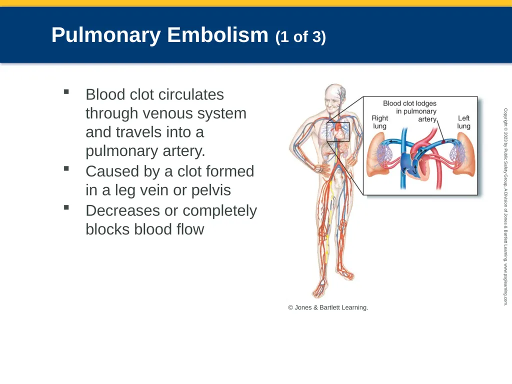
Pulmonary Embolism (1 of 3) Blood clot circulates through venous system and travels into a pulmonary artery.
Caused by a clot formed in a leg vein or pelvis
Decreases or completely blocks blood flow © Jones & Bartlett Learning.<br>
Caused by a clot formed in a leg vein or pelvis
Decreases or completely blocks blood flow © Jones & Bartlett Learning.<br>
38
Pulmonary Embolism (2 of 3) May also occur as the result of:
Damage to the lining of vessels
Hypercoagulability
Slow blood flow in a lower extremity
Risk factors include
Immobilized legs, recent surgery, pregnancy, previous pulmonary embolism, relative with pulmonary embolism, obesity, oral contraceptives, smoking, infection, cancer, sickle cell anemia, prolonged inactivity, being bedridden<br>
Damage to the lining of vessels
Hypercoagulability
Slow blood flow in a lower extremity
Risk factors include
Immobilized legs, recent surgery, pregnancy, previous pulmonary embolism, relative with pulmonary embolism, obesity, oral contraceptives, smoking, infection, cancer, sickle cell anemia, prolonged inactivity, being bedridden<br>
39
Pulmonary Embolism (3 of 3) Signs and symptoms
Dyspnea
Tachycardia
Tachypnea
Varying degrees of hypoxia
Cyanosis
Acute chest pain
Hemoptysis<br>
Dyspnea
Tachycardia
Tachypnea
Varying degrees of hypoxia
Cyanosis
Acute chest pain
Hemoptysis<br>
40
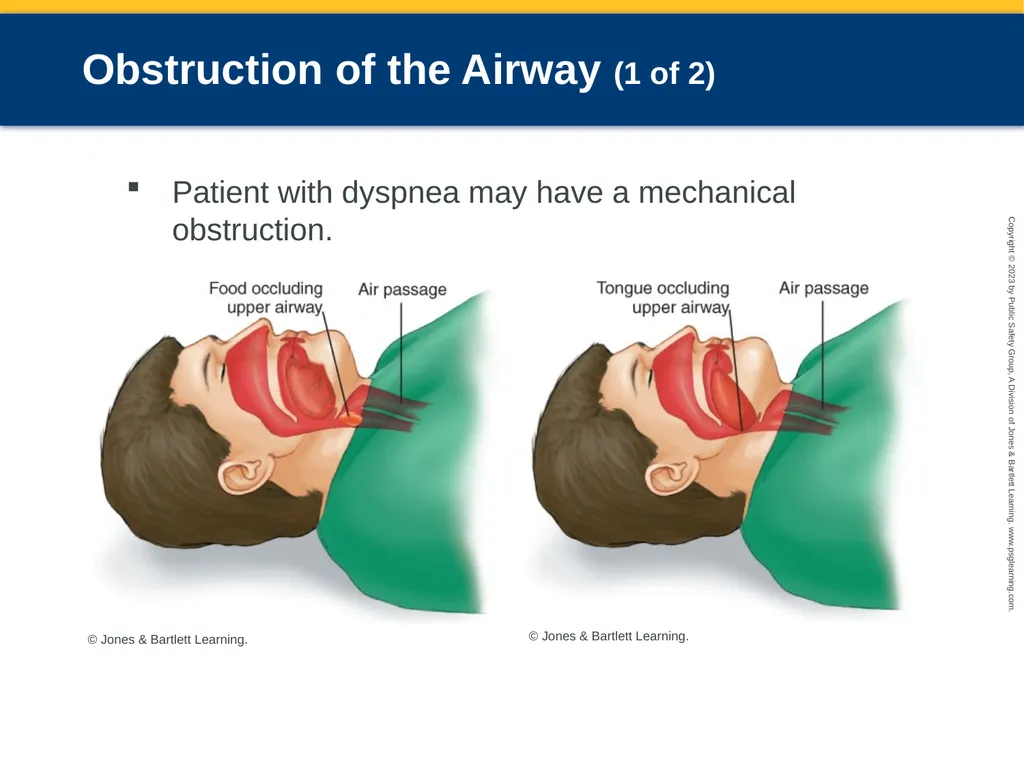
Obstruction of the Airway (1 of 2) Patient with dyspnea may have a mechanical obstruction. © Jones & Bartlett Learning. © Jones & Bartlett Learning.<br>
41
Obstruction of the Airway (2 of 2) In an unresponsive patient, this may be the result of:
Vomitus
Foreign object
Tongue blocking the airway<br>
Vomitus
Foreign object
Tongue blocking the airway<br>
42
Environmental/Industrial Exposure Pesticides, cleaning solutions, chemicals, chlorine, carbon monoxide, and other gases
Some chemicals can combine to create hazardous by-products.
Type of damage largely depends on the water solubility of the toxic gas.
Highly water-soluble gases cause swelling and irritation.
Less water-soluble gases may cause pulmonary edema up to 24 hours later.<br>
Some chemicals can combine to create hazardous by-products.
Type of damage largely depends on the water solubility of the toxic gas.
Highly water-soluble gases cause swelling and irritation.
Less water-soluble gases may cause pulmonary edema up to 24 hours later.<br>
43
Cystic Fibrosis Genetic disorder that affects the lungs and digestive systems
Symptoms range from sinus congestion to wheezing and asthmalike complaints.
Look for respiratory insufficiency, signs of a respiratory infection, and intestinal blockage.
Often causes death in childhood<br>
Symptoms range from sinus congestion to wheezing and asthmalike complaints.
Look for respiratory insufficiency, signs of a respiratory infection, and intestinal blockage.
Often causes death in childhood<br>
44
Age-Related Conditions (1 of 7) Bronchiolitis
Inflammation of bronchioles due to viral infection
Caused by Metapneumovirus and respiratory syncytial virus (RSV)
Respiratory syncytial virus (RSV)
Causes an infection in the lungs and breathing passages
Highly contagious<br>
Inflammation of bronchioles due to viral infection
Caused by Metapneumovirus and respiratory syncytial virus (RSV)
Respiratory syncytial virus (RSV)
Causes an infection in the lungs and breathing passages
Highly contagious<br>
45
Age-Related Conditions (2 of 7) Croup
Due to inflammation and swelling of the pharynx, larynx, and trachea
Often secondary to acute viral infection of the upper respiratory tract
Hallmark signs are stridor and a seal-bark cough.<br>
Due to inflammation and swelling of the pharynx, larynx, and trachea
Often secondary to acute viral infection of the upper respiratory tract
Hallmark signs are stridor and a seal-bark cough.<br>
46
Age-Related Conditions (3 of 7) Epiglottitis
Inflammation of epiglottis
Usually caused by a bacterial infection that produces severe swelling of the flap over the larynx
Patient will appear very sick.
Symptoms include:
Sore throat, high fever, stridor, tripod positioning, drooling<br>
Inflammation of epiglottis
Usually caused by a bacterial infection that produces severe swelling of the flap over the larynx
Patient will appear very sick.
Symptoms include:
Sore throat, high fever, stridor, tripod positioning, drooling<br>
47
Age-Related Conditions (4 of 7) Pneumonia
Caused by an infection of the lung parenchyma
Young children and older adults at greatest risk
Presents as a localized infection in the lungs
Can become systematic
Signs and symptoms
Fever, chills, productive cough with purulent sputum, pleuritic chest pain, excessive mucus<br>
Caused by an infection of the lung parenchyma
Young children and older adults at greatest risk
Presents as a localized infection in the lungs
Can become systematic
Signs and symptoms
Fever, chills, productive cough with purulent sputum, pleuritic chest pain, excessive mucus<br>
48
Age-Related Conditions (5 of 7) Pertussis
Highly contagious airborne bacterial infection
Mostly affects children younger than 6 years
Cold-like symptoms, but coughing spells can last for more than a minute
Feverish, with a “whoop” sound on inspiration after a coughing attack<br>
Highly contagious airborne bacterial infection
Mostly affects children younger than 6 years
Cold-like symptoms, but coughing spells can last for more than a minute
Feverish, with a “whoop” sound on inspiration after a coughing attack<br>
49
Age-Related Conditions (6 of 7) Airway obstruction
Always consider the possibility of a foreign body when a young child becomes short of breath.
Tonsil inflammation may partially occlude the airway.
Dysfunction of a tracheostomy may create an upper airway obstruction.
Obstruction may be in the lower airway: Tracheal trauma, obstructive lung disease, mucus accumulation, smooth muscle spasm<br>
Always consider the possibility of a foreign body when a young child becomes short of breath.
Tonsil inflammation may partially occlude the airway.
Dysfunction of a tracheostomy may create an upper airway obstruction.
Obstruction may be in the lower airway: Tracheal trauma, obstructive lung disease, mucus accumulation, smooth muscle spasm<br>
50
Age-Related Conditions (7 of 7) Heart failure
Heart muscles injured by heart attack or illness
Results in pulmonary edema
Risk factors
Hypertension
History of coronary artery disease
Signs and symptoms include difficulty breathing with exertion, sudden attack of respiratory distress, feeling suffocated, cold sweats, tachycardia.<br>
Heart muscles injured by heart attack or illness
Results in pulmonary edema
Risk factors
Hypertension
History of coronary artery disease
Signs and symptoms include difficulty breathing with exertion, sudden attack of respiratory distress, feeling suffocated, cold sweats, tachycardia.<br>
51
Wet Lungs Versus Dry Lungs Wet lungs: Associated with pulmonary edema
Dry lungs: Associated with COPD
Breath sounds are helpful, but can also be confusing.
Treat the patient, not the breath sounds.<br>
Dry lungs: Associated with COPD
Breath sounds are helpful, but can also be confusing.
Treat the patient, not the breath sounds.<br>
52
Patient Assessment Scene size-up
Use additional PPE if COVID-19 exposure is a possibility
Ensure a safe environment
Determine number of patients, the need for special resources, and the nature of illness.<br>
Use additional PPE if COVID-19 exposure is a possibility
Ensure a safe environment
Determine number of patients, the need for special resources, and the nature of illness.<br>
53
Primary Survey (1 of 4) Recognition of life threats and initiation of resuscitation take priority.
Signs of life-threatening respiratory distress:
Severe cyanosis, absent or abnormal breath sounds, audible stridor, dyspnea, coughing, tachycardia, abdominal breathing, change in respiratory rate or rhythm, pallor and diaphoresis, use of accessory muscles, tripod positioning<br>
Signs of life-threatening respiratory distress:
Severe cyanosis, absent or abnormal breath sounds, audible stridor, dyspnea, coughing, tachycardia, abdominal breathing, change in respiratory rate or rhythm, pallor and diaphoresis, use of accessory muscles, tripod positioning<br>
54
Primary Survey (2 of 4) Note general impression of the patient.
Determine if airway is open and clear.
Determine if patient is breathing.
Provide appropriate oxygen therapy.<br>
Determine if airway is open and clear.
Determine if patient is breathing.
Provide appropriate oxygen therapy.<br>
55
Primary Survey (3 of 4) Assess for shock and bleeding.
Check capillary refill in infants and children.
Check perfusion by evaluating skin color, temperature, and condition.<br>
Check capillary refill in infants and children.
Check perfusion by evaluating skin color, temperature, and condition.<br>
56
Primary Survey (4 of 4) Identify any life threats in your patient.
Problems with airway, breathing, and circulation (ABCs)
Poor initial general impression
Altered mental status
Potential hypoperfusion or shock
Chest pain associated with low blood pressure
Severe pain anywhere
Excessive bleeding<br>
Problems with airway, breathing, and circulation (ABCs)
Poor initial general impression
Altered mental status
Potential hypoperfusion or shock
Chest pain associated with low blood pressure
Severe pain anywhere
Excessive bleeding<br>
57
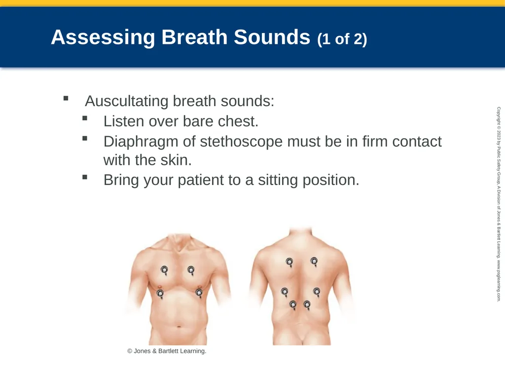
Assessing Breath Sounds (1 of 2) Auscultating breath sounds:
Listen over bare chest.
Diaphragm of stethoscope must be in firm contact with the skin.
Bring your patient to a sitting position. © Jones & Bartlett Learning.<br>
Listen over bare chest.
Diaphragm of stethoscope must be in firm contact with the skin.
Bring your patient to a sitting position. © Jones & Bartlett Learning.<br>
58
Assessing Breath Sounds (2 of 2) Determine whether breath sounds are normal or abnormal.
Abnormal breath sounds include:
Crackles
Rhonchi
Stridor
Wheezing
Pleural friction rub
Snoring sounds<br>
Abnormal breath sounds include:
Crackles
Rhonchi
Stridor
Wheezing
Pleural friction rub
Snoring sounds<br>
59
History Taking (1 of 2) SAMPLE history can be obtained from family or bystanders
Limit questions to pertinent ones.
General state of health
Childhood or adult diseases
Recent surgeries or hospitalizations
Traumatic injuries
Chest pain<br>
Limit questions to pertinent ones.
General state of health
Childhood or adult diseases
Recent surgeries or hospitalizations
Traumatic injuries
Chest pain<br>
60
History Taking (2 of 2) Ask about previous episodes, medication allergies, and current medications.
Pay close attention to the medications the patient is currently taking.
Use OPQRST-I mnemonic to determine the specifics of pain and obtain information about the breathing problem.<br>
Pay close attention to the medications the patient is currently taking.
Use OPQRST-I mnemonic to determine the specifics of pain and obtain information about the breathing problem.<br>
61
Chronic Respiratory Conditions Chronic lower airway obstruction makes it difficult for patients to breathe deeply enough to clear the lungs.
If a new infection occurs in a patient with COPD, arterial oxygen level may fall rapidly.
Patients with asthma may have different triggers.
Try to determine what triggered the attack.<br>
If a new infection occurs in a patient with COPD, arterial oxygen level may fall rapidly.
Patients with asthma may have different triggers.
Try to determine what triggered the attack.<br>
62
Secondary Assessment (1 of 2) Addresses chief complaint
Perform after identifying and treating life threats.
Check for a change in mental status.
Check the patient’s respiratory status.
Look for signs of increased work of breathing.<br>
Perform after identifying and treating life threats.
Check for a change in mental status.
Check the patient’s respiratory status.
Look for signs of increased work of breathing.<br>
63
Secondary Assessment (2 of 2) Assess circulation.
Check for signs of trauma or retractions in the chest.
Obtain vital signs.
Use monitoring devices if available.
Pulse oximeter
End-tidal carbon dioxide (ETCO2) monitor
Peak flowmeter<br>
Check for signs of trauma or retractions in the chest.
Obtain vital signs.
Use monitoring devices if available.
Pulse oximeter
End-tidal carbon dioxide (ETCO2) monitor
Peak flowmeter<br>
64
Reassessment Repeat primary survey, and maintain an open airway.
Monitor patient’s breathing and reassess circulation.
Confirm adequacy of interventions and patient status.
Reassess and record vital signs.<br>
Monitor patient’s breathing and reassess circulation.
Confirm adequacy of interventions and patient status.
Reassess and record vital signs.<br>
65
Emergency Medical Care (1 of 15) Perform standard interventions.
Oxygen to keep the saturation above 94%
Intravenous (IV) line
Psychological support
Decrease the work of breathing
Remove constricting clothing.
Do not make person walk.
Do not lay patient supine.<br>
Oxygen to keep the saturation above 94%
Intravenous (IV) line
Psychological support
Decrease the work of breathing
Remove constricting clothing.
Do not make person walk.
Do not lay patient supine.<br>
66
Emergency Medical Care (2 of 15) Provide supplemental oxygen.
Administer oxygen in the concentrations necessary to be effective.
Pulse oximetry is a useful guide to oxygenation.
Safe to administer oxygen in concentrations less than 50% to almost anyone
Oxygen saturation of 100% should be avoided.
Call for paramedic backup if needed.<br>
Administer oxygen in the concentrations necessary to be effective.
Pulse oximetry is a useful guide to oxygenation.
Safe to administer oxygen in concentrations less than 50% to almost anyone
Oxygen saturation of 100% should be avoided.
Call for paramedic backup if needed.<br>
67
Emergency Medical Care (3 of 15) Administer a bronchodilator.
Patients who do not have bronchospasm usually benefit only slightly from aerosol bronchodilators.
Bronchodilators are of little value in treating conditions such as pneumonia, pulmonary edema, and heart disease.<br>
Patients who do not have bronchospasm usually benefit only slightly from aerosol bronchodilators.
Bronchodilators are of little value in treating conditions such as pneumonia, pulmonary edema, and heart disease.<br>
68
Emergency Medical Care (4 of 15) Respiratory medications
Most common medications used for dyspnea are inhaled beta-agonists.
Secondary bronchoconstriction could be reversed with a bronchodilator.
Common side effects of inhalers: increased pulse rate, nervousness, and muscle tremors<br>
Most common medications used for dyspnea are inhaled beta-agonists.
Secondary bronchoconstriction could be reversed with a bronchodilator.
Common side effects of inhalers: increased pulse rate, nervousness, and muscle tremors<br>
69
Emergency Medical Care (5 of 15) Aerosol therapy
Aerosol nebulizers deliver liquid medications as a fine mist.
To generate optimal particle size, most nebulizers need to have oxygen flow of at least 6 L/min.<br>
Aerosol nebulizers deliver liquid medications as a fine mist.
To generate optimal particle size, most nebulizers need to have oxygen flow of at least 6 L/min.<br>
70
Emergency Medical Care (6 of 15) Aerosol therapy (cont'd)
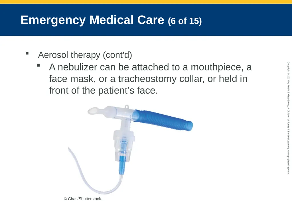
A nebulizer can be attached to a mouthpiece, a face mask, or a tracheostomy collar, or held in front of the patient’s face. © Chas/Shutterstock.<br>
A nebulizer can be attached to a mouthpiece, a face mask, or a tracheostomy collar, or held in front of the patient’s face. © Chas/Shutterstock.<br>
71
Emergency Medical Care (7 of 15) Metered-dose inhalers
Direct medication through the mouth and into the lungs
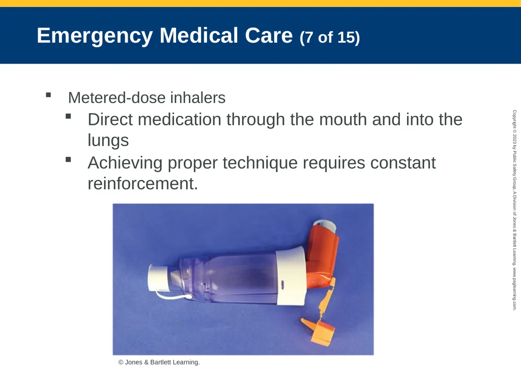
Achieving proper technique requires constant reinforcement. © Jones & Bartlett Learning.<br>
Direct medication through the mouth and into the lungs
Achieving proper technique requires constant reinforcement. © Jones & Bartlett Learning.<br>
72
Emergency Medical Care (8 of 15) Metered-dose inhalers (cont'd)
Tips to avoid common errors when using or administering an MDI:
Ensure the mist enters the lungs.
Have patients inhale deeply and hold their breath.
Keeps the canister and spacer clean.
Have patients rinse mouth after using.<br>
Tips to avoid common errors when using or administering an MDI:
Ensure the mist enters the lungs.
Have patients inhale deeply and hold their breath.
Keeps the canister and spacer clean.
Have patients rinse mouth after using.<br>
73
Emergency Medical Care (9 of 15) Metered-dose inhalers (cont'd)
Contraindications include:
Patient is unable to help coordinate inhalation.
MDI is not prescribed for this patient.
No permission from medical control or protocol
Patient had already met maximum prescribed dose.
Medication is expired.
Other contradictions specific to the medication<br>
Contraindications include:
Patient is unable to help coordinate inhalation.
MDI is not prescribed for this patient.
No permission from medical control or protocol
Patient had already met maximum prescribed dose.
Medication is expired.
Other contradictions specific to the medication<br>
74
Emergency Medical Care (10 of 15) Dry-powder inhalers
Reasonably convenient and easy to use
Rarely used during emergency care
Consider fluid balance.
Give a fluid bolus to younger patients.
In older adults or patients with cardiac dysfunction, administering too much fluid could cause pulmonary edema.
Have an IV in place.<br>
Reasonably convenient and easy to use
Rarely used during emergency care
Consider fluid balance.
Give a fluid bolus to younger patients.
In older adults or patients with cardiac dysfunction, administering too much fluid could cause pulmonary edema.
Have an IV in place.<br>
75
Emergency Medical Care (11 of 15) Support or assist ventilation.
If patient becomes fatigued, breathing might need to be supported more aggressively.
Some patients may simply require bag-mask ventilation for a short period.
Gastric distention and vomiting can occur from overaggressive ventilation.<br>
If patient becomes fatigued, breathing might need to be supported more aggressively.
Some patients may simply require bag-mask ventilation for a short period.
Gastric distention and vomiting can occur from overaggressive ventilation.<br>
76
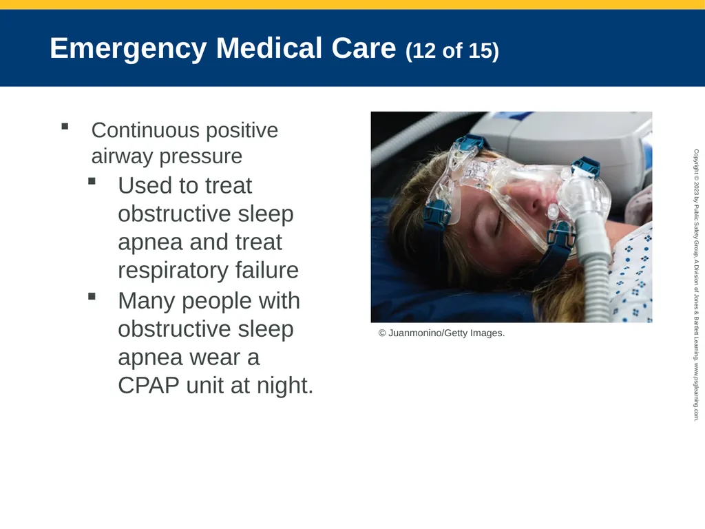
Emergency Medical Care (12 of 15) Continuous positive airway pressure
Used to treat obstructive sleep apnea and treat respiratory failure
Many people with obstructive sleep apnea wear a CPAP unit at night. © Juanmonino/Getty Images.<br>
Used to treat obstructive sleep apnea and treat respiratory failure
Many people with obstructive sleep apnea wear a CPAP unit at night. © Juanmonino/Getty Images.<br>
77
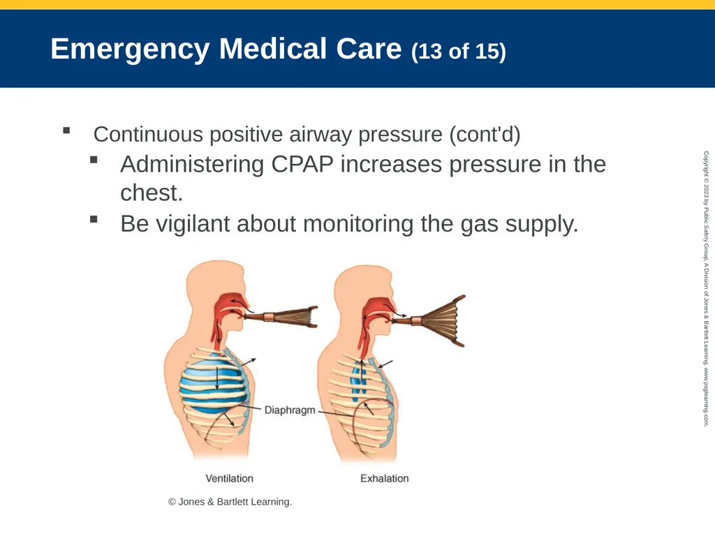
Emergency Medical Care (13 of 15) Continuous positive airway pressure (cont'd)
Administering CPAP increases pressure in the chest.
Be vigilant about monitoring the gas supply. © Jones & Bartlett Learning.<br>
Administering CPAP increases pressure in the chest.
Be vigilant about monitoring the gas supply. © Jones & Bartlett Learning.<br>
78
Emergency Medical Care (14 of 15) Continuous positive airway pressure (cont'd)
The success of CPAP is inversely related to the patient’s respiratory rate.
If this rate increases, the therapy is likely to fail.
If this rate decreases, the therapy is likely to succeed.<br>
The success of CPAP is inversely related to the patient’s respiratory rate.
If this rate increases, the therapy is likely to fail.
If this rate decreases, the therapy is likely to succeed.<br>
79
Emergency Medical Care (15 of 15) Bilevel positive airway pressure
One pressure can be delivered during inspiration and a different pressure can be delivered during exhalation.
Causes a pressure variation in the chest, which allows for more normal blood flow
More complex and expensive<br>
One pressure can be delivered during inspiration and a different pressure can be delivered during exhalation.
Causes a pressure variation in the chest, which allows for more normal blood flow
More complex and expensive<br>
80
Assessment and Management of Specific Conditions (1 of 16) Upper or lower airway diseases
Patients present with signs of severe respiratory impairment
Chief complaint is typically dyspnea, cough, or nocturnal dyspnea.
Wheezing may be present.
Place patient in position of comfort.
Monitor airway, apply high-flow oxygen, and assist ventilation if needed.
Transport and contact medical control.<br>
Patients present with signs of severe respiratory impairment
Chief complaint is typically dyspnea, cough, or nocturnal dyspnea.
Wheezing may be present.
Place patient in position of comfort.
Monitor airway, apply high-flow oxygen, and assist ventilation if needed.
Transport and contact medical control.<br>
81
Assessment and Management of Specific Conditions (2 of 16) Acute pulmonary edema
May be associated with cardiac disease or direct lung damage
Administer 100% oxygen.
If necessary, carefully suction any secretions from airway.
Provide assisted ventilation.
Establish IV access and monitor flow rates carefully.<br>
May be associated with cardiac disease or direct lung damage
Administer 100% oxygen.
If necessary, carefully suction any secretions from airway.
Provide assisted ventilation.
Establish IV access and monitor flow rates carefully.<br>
82
Assessment and Management of Specific Conditions (3 of 16) Aspiration
Inhalation of anything other than gases
Associated with a high mortality rate
Avoid gastric distention when ventilating.
Aggressively monitor the patient’s ability to protect the airway.
Aggressively treat aspiration with suction and airway control if the previous steps fail.<br>
Inhalation of anything other than gases
Associated with a high mortality rate
Avoid gastric distention when ventilating.
Aggressively monitor the patient’s ability to protect the airway.
Aggressively treat aspiration with suction and airway control if the previous steps fail.<br>
83
Assessment and Management of Specific Conditions (4 of 16) Chronic obstructive pulmonary disease (COPD)
Patients often find breathing difficult while lying down.
Assist with prescribed inhaler if there is one.
Auto-PEEP<br>
Patients often find breathing difficult while lying down.
Assist with prescribed inhaler if there is one.
Auto-PEEP<br>
84
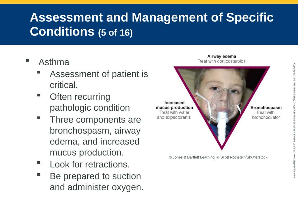
Assessment and Management of Specific Conditions (5 of 16) Asthma
Assessment of patient is critical.
Often recurring pathologic condition
Three components are bronchospasm, airway edema, and increased mucus production.
Look for retractions.
Be prepared to suction and administer oxygen. © Jones & Bartlett Learning; © Scott Rothstein/Shutterstock.<br>
Assessment of patient is critical.
Often recurring pathologic condition
Three components are bronchospasm, airway edema, and increased mucus production.
Look for retractions.
Be prepared to suction and administer oxygen. © Jones & Bartlett Learning; © Scott Rothstein/Shutterstock.<br>
85
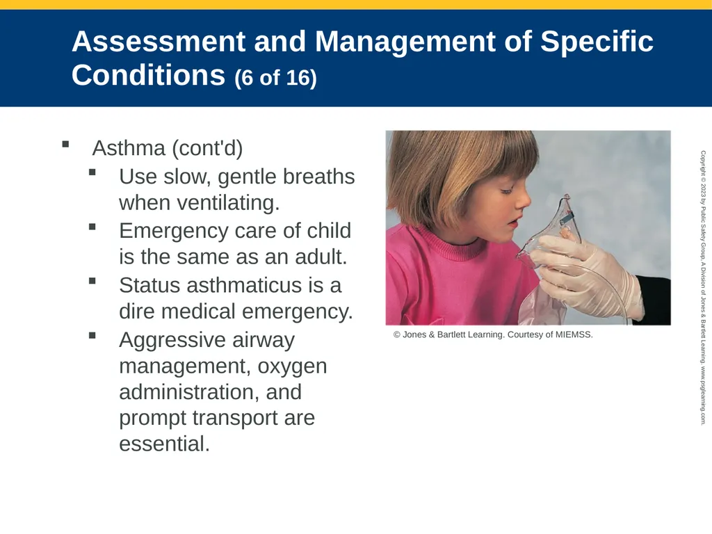
Assessment and Management of Specific Conditions (6 of 16) Asthma (cont'd)
Use slow, gentle breaths when ventilating.
Emergency care of child is the same as an adult.
Status asthmaticus is a dire medical emergency.
Aggressive airway management, oxygen administration, and prompt transport are essential. © Jones & Bartlett Learning. Courtesy of MIEMSS.<br>
Use slow, gentle breaths when ventilating.
Emergency care of child is the same as an adult.
Status asthmaticus is a dire medical emergency.
Aggressive airway management, oxygen administration, and prompt transport are essential. © Jones & Bartlett Learning. Courtesy of MIEMSS.<br>
86
Assessment and Management of Specific Conditions (7 of 16) Anaphylactic reactions
Remove the offending agent.
Maintain the airway.
Administer supplemental oxygen.
Assist breathing as needed.
Spontaneous pneumothorax
Management begins with ABCs.
Provide airway and ventilatory support.
Watch for signs of a tension pneumothorax.<br>
Remove the offending agent.
Maintain the airway.
Administer supplemental oxygen.
Assist breathing as needed.
Spontaneous pneumothorax
Management begins with ABCs.
Provide airway and ventilatory support.
Watch for signs of a tension pneumothorax.<br>
87
Assessment and Management of Specific Conditions (8 of 16) Pleural effusion
Treatment is removal of fluid collected outside the lung.
Provide oxygen and other routine support measures.
Pulmonary embolism
Manage airway, and provide high-flow oxygen.
Initiate CPR for pulseless and apneic patient.
Manage most severe cases as cardiac arrest of unknown origin.<br>
Treatment is removal of fluid collected outside the lung.
Provide oxygen and other routine support measures.
Pulmonary embolism
Manage airway, and provide high-flow oxygen.
Initiate CPR for pulseless and apneic patient.
Manage most severe cases as cardiac arrest of unknown origin.<br>
88
Assessment and Management of Specific Conditions (9 of 16) Hyperventilation
Can be caused by life-threatening illness or panic attack
Give supplemental oxygen and transport.
Interventions for circulatory support and pharmacologic interventions are rarely required.
Provide psychological support for anxiety-related hyperventilation.<br>
Can be caused by life-threatening illness or panic attack
Give supplemental oxygen and transport.
Interventions for circulatory support and pharmacologic interventions are rarely required.
Provide psychological support for anxiety-related hyperventilation.<br>
89
Assessment and Management of Specific Conditions (10 of 16) Obstruction of the airway
Upper airway obstruction may be partial or complete.
If patient can talk and breathe, provide supplemental oxygen and transport.
Remove obstructing body according to basic life support guidelines.
Administer supplemental oxygen.<br>
Upper airway obstruction may be partial or complete.
If patient can talk and breathe, provide supplemental oxygen and transport.
Remove obstructing body according to basic life support guidelines.
Administer supplemental oxygen.<br>
90
Assessment and Management of Specific Conditions (11 of 16) Environmental/industrial exposure
Industrial sites often have their own medical/fire/hazmat teams.
After patient is decontaminated, gather information on substance and cause of dyspnea.
Provide 100% supplemental oxygen or assisted ventilation if necessary.
If upper airway is compromised, aggressive airway management may be required.<br>
Industrial sites often have their own medical/fire/hazmat teams.
After patient is decontaminated, gather information on substance and cause of dyspnea.
Provide 100% supplemental oxygen or assisted ventilation if necessary.
If upper airway is compromised, aggressive airway management may be required.<br>
91
Assessment and Management of Specific Conditions (12 of 16) Bronchiolitis
Treatment is entirely supportive.
Provide supplemental oxygen; be prepared to assist with bag-mask device.
Respiratory syncytial virus.
Look for signs of dehydration.
Treat airway and breathing problems.
Humidified oxygen is helpful, if available.<br>
Treatment is entirely supportive.
Provide supplemental oxygen; be prepared to assist with bag-mask device.
Respiratory syncytial virus.
Look for signs of dehydration.
Treat airway and breathing problems.
Humidified oxygen is helpful, if available.<br>
92
Assessment and Management of Specific Conditions (13 of 16) Croup
Often responds well to humidified oxygen
Allow patient to assume position of comfort.
Avoid agitating patient.
Administer nebulized epinephrine if dictated by local protocol.<br>
Often responds well to humidified oxygen
Allow patient to assume position of comfort.
Avoid agitating patient.
Administer nebulized epinephrine if dictated by local protocol.<br>
93
Assessment and Management of Specific Conditions (14 of 16) Epiglottitis
Keep patient in a position of comfort.
Do not put anything in patient’s mouth.
Give high-flow oxygen.
Pneumonia
Monitor ABCs, provide high-flow oxygen and ventilatory support as needed.
Administration of IV fluids may help.
If high fever is present, cool patient.<br>
Keep patient in a position of comfort.
Do not put anything in patient’s mouth.
Give high-flow oxygen.
Pneumonia
Monitor ABCs, provide high-flow oxygen and ventilatory support as needed.
Administration of IV fluids may help.
If high fever is present, cool patient.<br>
94
Assessment and Management of Specific Conditions (15 of 16) Pertussis
Some infants and younger children should be treated in a hospital.
Watch for signs of dehydration.
Administer oxygen as needed.<br>
Some infants and younger children should be treated in a hospital.
Watch for signs of dehydration.
Administer oxygen as needed.<br>
95
Assessment and Management of Specific Conditions (16 of 16) Airway obstruction in child
Clear the airway.
Provide oxygen and transport.
Heart failure
High blood pressure and low cardiac output often trigger sudden pulmonary edema.
Treatment should consist of airway, ventilatory, and circulatory support.
Consider CPAP.<br>
Clear the airway.
Provide oxygen and transport.
Heart failure
High blood pressure and low cardiac output often trigger sudden pulmonary edema.
Treatment should consist of airway, ventilatory, and circulatory support.
Consider CPAP.<br>
96
Tracheostomy Dysfunction (1 of 2) Children with chronic pulmonary medical conditions may use a home ventilator connected by a tracheostomy tube.
Establish a patent airway, place the patient in position of comfort, and suction to clear the obstruction.<br>
Establish a patent airway, place the patient in position of comfort, and suction to clear the obstruction.<br>
97
Tracheostomy Dysfunction (2 of 2) After the obstruction is clear, oxygenate the patient and treat based on presentation.
Establishing airway patency is the immediate goal for geriatric patients.<br>
Establishing airway patency is the immediate goal for geriatric patients.<br>
98
Epidemic and Pandemic Considerations (1 of 2) Epidemic
Occurs when new cases of a disease occur in a human population and substantially exceed what is expected based on recent experience
Pandemic
Outbreak that occurs on a global scale
Many potentially serious diseases can be passed by the respiratory route.
Influenza<br>
Occurs when new cases of a disease occur in a human population and substantially exceed what is expected based on recent experience
Pandemic
Outbreak that occurs on a global scale
Many potentially serious diseases can be passed by the respiratory route.
Influenza<br>
99
Epidemic and Pandemic Considerations (2 of 2) Wear PPE.
Frequent handwashing is important.
Maintain your vaccinations and stay up to date on the latest CDC recommendations.
Wear a HEPA respirator during aerosol-generating procedures.<br>
Frequent handwashing is important.
Maintain your vaccinations and stay up to date on the latest CDC recommendations.
Wear a HEPA respirator during aerosol-generating procedures.<br>
100
COVID-19 (1 of 7) Contagious respiratory illness
New form of coronavirus (SARS-CoV-2)
Spreads through respiratory droplets
Signs and symptoms can include:
Fever or chills
Cough
Sore throat
Loss of taste or smell<br>
New form of coronavirus (SARS-CoV-2)
Spreads through respiratory droplets
Signs and symptoms can include:
Fever or chills
Cough
Sore throat
Loss of taste or smell<br>
101
COVID-19 (2 of 7) Emergency warning signs include dyspnea, chest pain, confusion, cyanosis, and an inability to wake up or stay awake.
Older adults and those with preexisting medical conditions are at higher risk.<br>
Older adults and those with preexisting medical conditions are at higher risk.<br>
102
COVID-19 (3 of 7) Follow these guidelines to avoid contamination:
Wear proper PPE at all times.
Wash hands often, for at least 20 seconds.
Avoid touching your eyes, nose, and mouth.
Cover your cough or sneeze.
Stay home if you are sick.
Wear a mask that covers your mouth and nose when out in public.<br>
Wear proper PPE at all times.
Wash hands often, for at least 20 seconds.
Avoid touching your eyes, nose, and mouth.
Cover your cough or sneeze.
Stay home if you are sick.
Wear a mask that covers your mouth and nose when out in public.<br>
103
COVID-19 (4 of 7) Follow these guidelines to avoid contamination (cont’d):
Maintain 6 feet between yourself and any person not in your household.
Get a flu shot!
Clean and disinfect frequently touched surfaces/objects daily.
Clean your electronic devices.
Follow manufacturer’s instructions for cleaning and disinfection products.<br>
Maintain 6 feet between yourself and any person not in your household.
Get a flu shot!
Clean and disinfect frequently touched surfaces/objects daily.
Clean your electronic devices.
Follow manufacturer’s instructions for cleaning and disinfection products.<br>
104
COVID-19 (5 of 7) Specific Instructions for the AEMT:
Wear an N95 or higher-level respirator or face mask.
Wear eye protection.
Wear disposable patient examination gloves.
Wear a disposable gown.<br>
Wear an N95 or higher-level respirator or face mask.
Wear eye protection.
Wear disposable patient examination gloves.
Wear a disposable gown.<br>
105
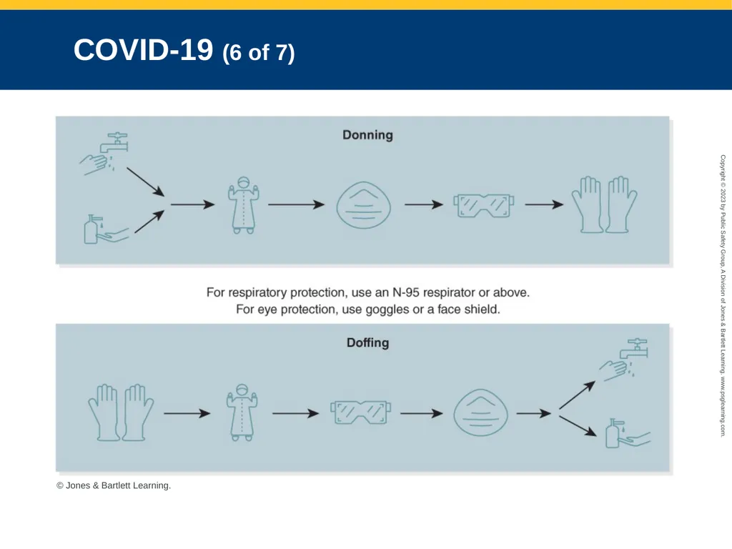
COVID-19 (6 of 7) © Jones & Bartlett Learning.<br>
106
COVID-19 (7 of 7) Specific instructions for protecting the patient:
Have the patient wear a face mask.
Treat the symptoms.
Help the patient find a position of comfort.
Follow local protocols.<br>
Have the patient wear a face mask.
Treat the symptoms.
Help the patient find a position of comfort.
Follow local protocols.<br>