
Retrospective Multicenter Study Of The
KB
Published · 20 slides · 0 views

1 / 1




Description
Retrospective Multicenter Study Of The Neuroimaging Incidence Of CNS Complications In Hospitalized Covid-19 Positive Patients. Scott H. Faro, Arichena Manmatharayan, Benjamin Leiby, Neelu Jain, Feroze B. Mohamed, Kiran S. Talekar, Amish
Related Topics
Share
Embed code
Download this presentation From Below
"Retrospective Multicenter Study Of The" is the property of its rightful owner. Permission is granted to download and print the materials on this website for personal, non-commercial use only, and to display it on your personal computer provided you do not modify the materials and that you retain all copyright notices contained in the materials. By downloading content from our website, you accept the terms of this agreement.
Presentation Transcript
01
Retrospective Multicenter Study Of The Neuroimaging Incidence Of CNS Complications In Hospitalized Covid-19 Positive Patients. Scott H. Faro, Arichena Manmatharayan, Benjamin Leiby, Neelu Jain, Feroze B. Mohamed, Kiran S. Talekar, Amish Doshi, Ivan Jambor, Chang Sanders, Mark Finkelstein, Stephane Kremer, Francois Lersy, Brendan Lindgren, Nathalia M. Figueiredo, Varun Sethi, Simonetta Gerevini, Angela Napolitano, Rajan Jain, Siddhanth Dogra, Jay Pillai, Dan Ryan, Rolf Jager, Francesco Carletti, Asim Mian, Artem Kaliev, Priya Anand, Courtney Takahashi, AK Murat, Rivka Colen, Giancarlo Mansueto, Francesca Pizzini.<br>
02
No Conflicts of Interest<br>
03
The Covid-19 pandemic is a multisystem disorder that extends well beyond the flu-like symptoms and devastating pulmonary involvement.
The pulmonary complications and flu-like symptoms are the most common clinical findings in this patient population.
There have also been reports of relatively less frequent central nervous system (CNS) complications.
We sought to determine the incidence of acute neuroimaging (NI) findings in hospitalized Covid-19 infected patients in 7 US and 4 European University hospitals. Purpose:<br>
The pulmonary complications and flu-like symptoms are the most common clinical findings in this patient population.
There have also been reports of relatively less frequent central nervous system (CNS) complications.
We sought to determine the incidence of acute neuroimaging (NI) findings in hospitalized Covid-19 infected patients in 7 US and 4 European University hospitals. Purpose:<br>
04
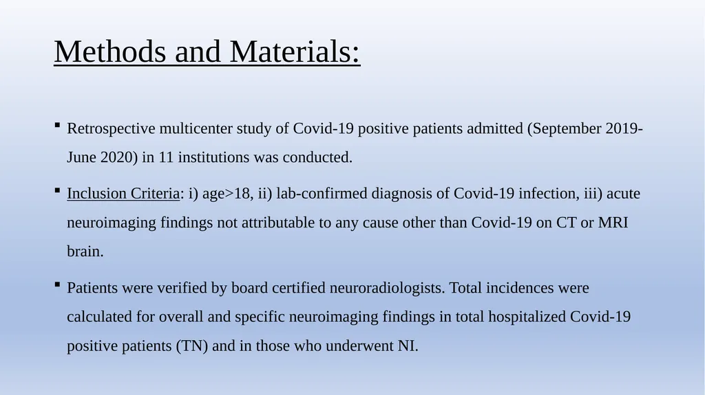
Methods and Materials: Retrospective multicenter study of Covid-19 positive patients admitted (September 2019-June 2020) in 11 institutions was conducted.
Inclusion Criteria: i) age>18, ii) lab-confirmed diagnosis of Covid-19 infection, iii) acute neuroimaging findings not attributable to any cause other than Covid-19 on CT or MRI brain.
Patients were verified by board certified neuroradiologists. Total incidences were calculated for overall and specific neuroimaging findings in total hospitalized Covid-19 positive patients (TN) and in those who underwent NI.<br>
Inclusion Criteria: i) age>18, ii) lab-confirmed diagnosis of Covid-19 infection, iii) acute neuroimaging findings not attributable to any cause other than Covid-19 on CT or MRI brain.
Patients were verified by board certified neuroradiologists. Total incidences were calculated for overall and specific neuroimaging findings in total hospitalized Covid-19 positive patients (TN) and in those who underwent NI.<br>
05
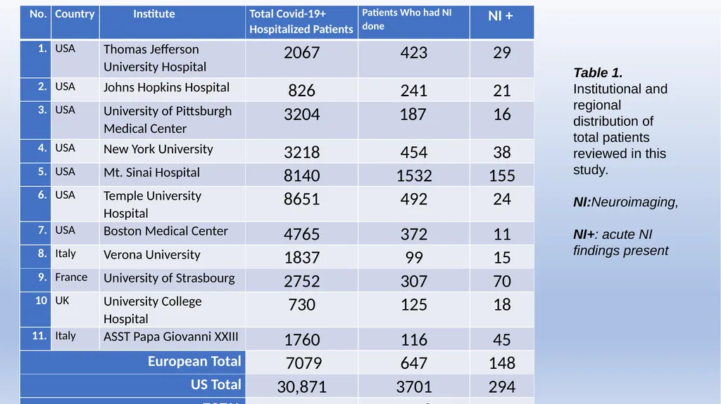
[SF1] Table 1. Institutional and regional distribution of total patients reviewed in this study.
NI:Neuroimaging,
NI+: acute NI findings present<br>
NI:Neuroimaging,
NI+: acute NI findings present<br>
06
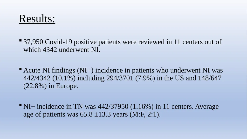
Results: 37,950 Covid-19 positive patients were reviewed in 11 centers out of which 4342 underwent NI.
Acute NI findings (NI+) incidence in patients who underwent NI was 442/4342 (10.1%) including 294/3701 (7.9%) in the US and 148/647 (22.8%) in Europe.
NI+ incidence in TN was 442/37950 (1.16%) in 11 centers. Average age of patients was 65.8 ±13.3 years (M:F, 2:1).<br>
Acute NI findings (NI+) incidence in patients who underwent NI was 442/4342 (10.1%) including 294/3701 (7.9%) in the US and 148/647 (22.8%) in Europe.
NI+ incidence in TN was 442/37950 (1.16%) in 11 centers. Average age of patients was 65.8 ±13.3 years (M:F, 2:1).<br>
07
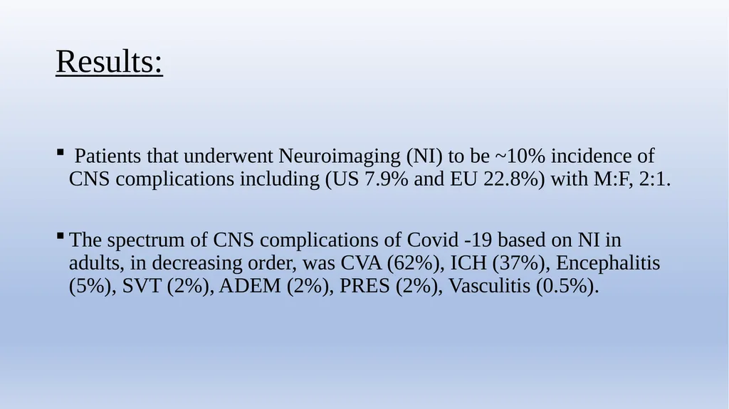
Results: Patients that underwent Neuroimaging (NI) to be ~10% incidence of CNS complications including (US 7.9% and EU 22.8%) with M:F, 2:1.
The spectrum of CNS complications of Covid -19 based on NI in adults, in decreasing order, was CVA (62%), ICH (37%), Encephalitis (5%), SVT (2%), ADEM (2%), PRES (2%), Vasculitis (0.5%).<br>
The spectrum of CNS complications of Covid -19 based on NI in adults, in decreasing order, was CVA (62%), ICH (37%), Encephalitis (5%), SVT (2%), ADEM (2%), PRES (2%), Vasculitis (0.5%).<br>
08
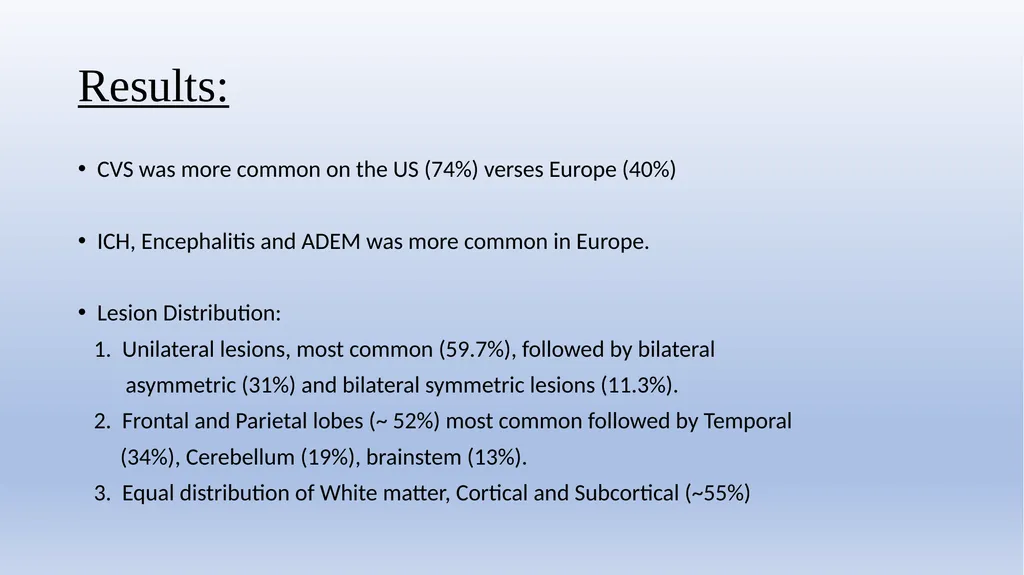
Results: CVS was more common on the US (74%) verses Europe (40%)
ICH, Encephalitis and ADEM was more common in Europe.
Lesion Distribution:
1. Unilateral lesions, most common (59.7%), followed by bilateral
asymmetric (31%) and bilateral symmetric lesions (11.3%).
2. Frontal and Parietal lobes (~ 52%) most common followed by Temporal
(34%), Cerebellum (19%), brainstem (13%).
3. Equal distribution of White matter, Cortical and Subcortical (~55%)<br>
ICH, Encephalitis and ADEM was more common in Europe.
Lesion Distribution:
1. Unilateral lesions, most common (59.7%), followed by bilateral
asymmetric (31%) and bilateral symmetric lesions (11.3%).
2. Frontal and Parietal lobes (~ 52%) most common followed by Temporal
(34%), Cerebellum (19%), brainstem (13%).
3. Equal distribution of White matter, Cortical and Subcortical (~55%)<br>
09
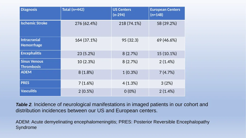
Table 2. Incidence of neurological manifestations in imaged patients in our cohort and distribution incidences between our US and European centers.
ADEM: Acute demyelinating encephalomeningitis; PRES: Posterior Reversible Encephalopathy Syndrome<br>
ADEM: Acute demyelinating encephalomeningitis; PRES: Posterior Reversible Encephalopathy Syndrome<br>
10
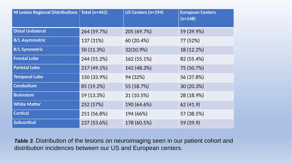
Table 3. Distribution of the lesions on neuroimaging seen in our patient cohort and distribution incidences between our US and European centers.<br>
11
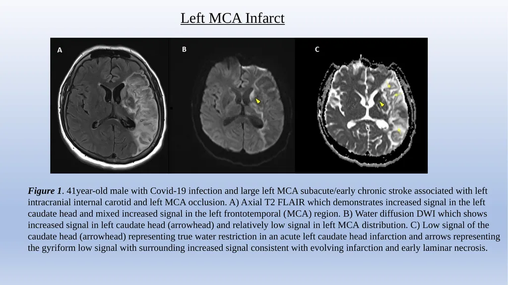
Left MCA Infarct Figure 1. 41year-old male with Covid-19 infection and large left MCA subacute/early chronic stroke associated with left intracranial internal carotid and left MCA occlusion. A) Axial T2 FLAIR which demonstrates increased signal in the left caudate head and mixed increased signal in the left frontotemporal (MCA) region. B) Water diffusion DWI which shows increased signal in left caudate head (arrowhead) and relatively low signal in left MCA distribution. C) Low signal of the caudate head (arrowhead) representing true water restriction in an acute left caudate head infarction and arrows representing the gyriform low signal with surrounding increased signal consistent with evolving infarction and early laminar necrosis.<br>
12
Figure 2. 47-year-old male patient who presented with Covid-19 pneumonia who developed bilateral symmetric diffusion-restricting lesions in the bilateral corona radiata/centrum semiovale, compatible with watershed non-hemorrhagic infarcts.
A) Axial T2 FLAIR shows patchy increased T2 signal within the corona radiata. B) GRE T2* shows no hemorrhage within the infarcts. C) Water restriction DWI sequence shows increased signal within the infarcts and true water restriction (low signal) on the ADC image (D). Acute ACA/MCA Watershed Infarction<br>
A) Axial T2 FLAIR shows patchy increased T2 signal within the corona radiata. B) GRE T2* shows no hemorrhage within the infarcts. C) Water restriction DWI sequence shows increased signal within the infarcts and true water restriction (low signal) on the ADC image (D). Acute ACA/MCA Watershed Infarction<br>
13
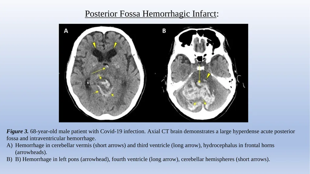
Posterior Fossa Hemorrhagic Infarct: Figure 3. 68-year-old male patient with Covid-19 infection. Axial CT brain demonstrates a large hyperdense acute posterior fossa and intraventricular hemorrhage.
Hemorrhage in cerebellar vermis (short arrows) and third ventricle (long arrow), hydrocephalus in frontal horns (arrowheads).
B) Hemorrhage in left pons (arrowhead), fourth ventricle (long arrow), cerebellar hemispheres (short arrows).<br>
Hemorrhage in cerebellar vermis (short arrows) and third ventricle (long arrow), hydrocephalus in frontal horns (arrowheads).
B) Hemorrhage in left pons (arrowhead), fourth ventricle (long arrow), cerebellar hemispheres (short arrows).<br>
14
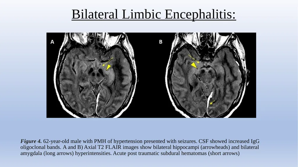
Bilateral Limbic Encephalitis: Figure 4. 62-year-old male with PMH of hypertension presented with seizures. CSF showed increased IgG oligoclonal bands. A and B) Axial T2 FLAIR images show bilateral hippocampi (arrowheads) and bilateral amygdala (long arrows) hyperintensities. Acute post traumatic subdural hematomas (short arrows)<br>
15
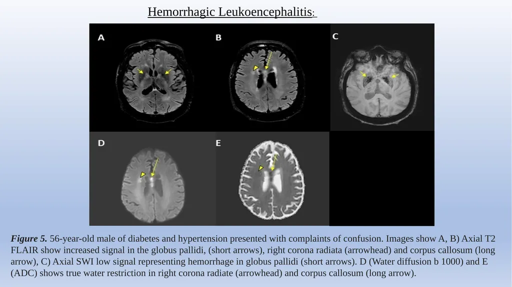
Hemorrhagic Leukoencephalitis: Figure 5. 56-year-old male of diabetes and hypertension presented with complaints of confusion. Images show A, B) Axial T2 FLAIR show increased signal in the globus pallidi, (short arrows), right corona radiata (arrowhead) and corpus callosum (long arrow), C) Axial SWI low signal representing hemorrhage in globus pallidi (short arrows). D (Water diffusion b 1000) and E (ADC) shows true water restriction in right corona radiate (arrowhead) and corpus callosum (long arrow).<br>
16
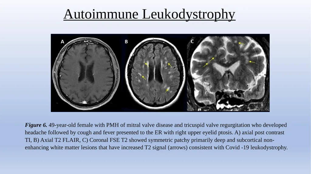
Autoimmune Leukodystrophy Figure 6. 49-year-old female with PMH of mitral valve disease and tricuspid valve regurgitation who developed headache followed by cough and fever presented to the ER with right upper eyelid ptosis. A) axial post contrast TI, B) Axial T2 FLAIR, C) Coronal FSE T2 showed symmetric patchy primarily deep and subcortical non-enhancing white matter lesions that have increased T2 signal (arrows) consistent with Covid -19 leukodystrophy.<br>
17
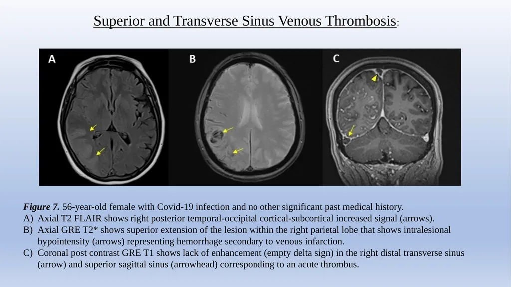
Superior and Transverse Sinus Venous Thrombosis: Figure 7. 56-year-old female with Covid-19 infection and no other significant past medical history.
Axial T2 FLAIR shows right posterior temporal-occipital cortical-subcortical increased signal (arrows).
Axial GRE T2* shows superior extension of the lesion within the right parietal lobe that shows intralesional hypointensity (arrows) representing hemorrhage secondary to venous infarction.
Coronal post contrast GRE T1 shows lack of enhancement (empty delta sign) in the right distal transverse sinus (arrow) and superior sagittal sinus (arrowhead) corresponding to an acute thrombus.<br>
Axial T2 FLAIR shows right posterior temporal-occipital cortical-subcortical increased signal (arrows).
Axial GRE T2* shows superior extension of the lesion within the right parietal lobe that shows intralesional hypointensity (arrows) representing hemorrhage secondary to venous infarction.
Coronal post contrast GRE T1 shows lack of enhancement (empty delta sign) in the right distal transverse sinus (arrow) and superior sagittal sinus (arrowhead) corresponding to an acute thrombus.<br>
18
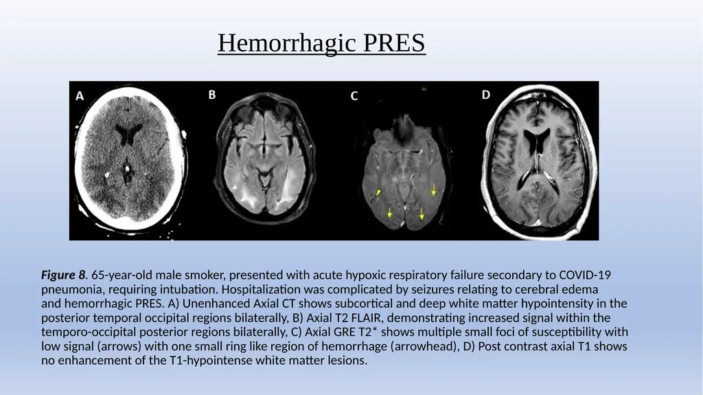
Hemorrhagic PRES Figure 8. 65-year-old male smoker, presented with acute hypoxic respiratory failure secondary to COVID-19 pneumonia, requiring intubation. Hospitalization was complicated by seizures relating to cerebral edema and hemorrhagic PRES. A) Unenhanced Axial CT shows subcortical and deep white matter hypointensity in the posterior temporal occipital regions bilaterally, B) Axial T2 FLAIR, demonstrating increased signal within the temporo-occipital posterior regions bilaterally, C) Axial GRE T2* shows multiple small foci of susceptibility with low signal (arrows) with one small ring like region of hemorrhage (arrowhead), D) Post contrast axial T1 shows no enhancement of the T1-hypointense white matter lesions.<br>
19
Conclusion: In a cohort of ~ 40K we demonstrated an overall incidence of CNS complications in Covid-19 positive patients to be 1.2 %
Patients that underwent Neuroimaging (NI) to be ~10% incidence of CNS complications including (US 7.9% and EU 22.8%) with M:F, 2:1.
The spectrum of CNS complications of Covid -19 based on NI in adults, in decreasing order, was CVA (62%), ICH (37%), Encephalitis (5%), SVT (2%), ADEM (2%), PRES (2%), Vasculitis (0.5%).
In a large international multi-institutional cohort, incidence and spectrum of acute neuroimaging findings helped to characterize the neurological complications of Covid-19 thereby assisting in understanding the full extent of this disease process beyond the devastating pulmonary complications.<br>
Patients that underwent Neuroimaging (NI) to be ~10% incidence of CNS complications including (US 7.9% and EU 22.8%) with M:F, 2:1.
The spectrum of CNS complications of Covid -19 based on NI in adults, in decreasing order, was CVA (62%), ICH (37%), Encephalitis (5%), SVT (2%), ADEM (2%), PRES (2%), Vasculitis (0.5%).
In a large international multi-institutional cohort, incidence and spectrum of acute neuroimaging findings helped to characterize the neurological complications of Covid-19 thereby assisting in understanding the full extent of this disease process beyond the devastating pulmonary complications.<br>
20
Thank You<br>