
Rinderpest Rinderpest is characterized by high
Description: Rinderpest Rinderpest is characterized by high fever, lachrymal discharge, inflammation, hemorrhage, necrosis, Erosions of the epithelium of the mouth and of the digestive tract, profuse diarrhea, and death. The four Ds of Rinderpest:
Related Topics
Download Presentation
"Rinderpest Rinderpest is characterized by high" is the property of its rightful owner. Permission is granted to download and print the materials on this website for personal, non-commercial use only, and to display it on your personal computer provided you do not modify the materials and that you retain all copyright notices contained in the materials. By downloading content from our website, you accept the terms of this agreement.
Presentation Transcript
slide1. Rinderpest Rinderpest is characterized by high fever, lachrymal discharge, inflammation, hemorrhage, necrosis, Erosions of the epithelium of the mouth and of the digestive tract, profuse diarrhea, and death.
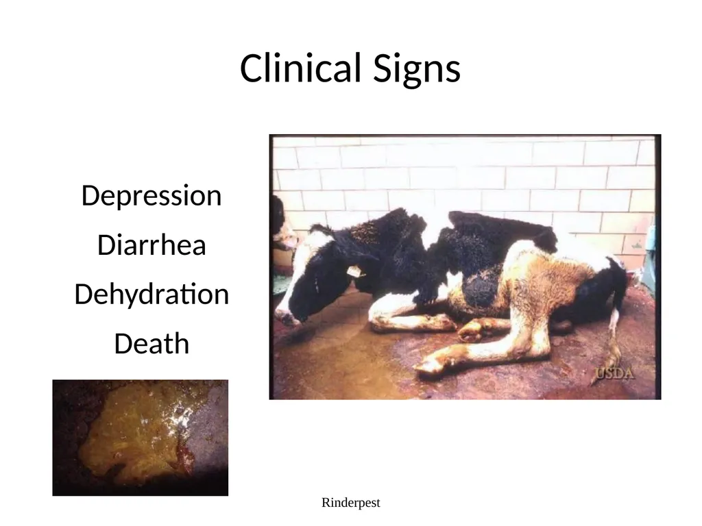
The “four D’s” of Rinderpest:
Depression
Diarrhea
Dehydration
Death Rinderpest<br>
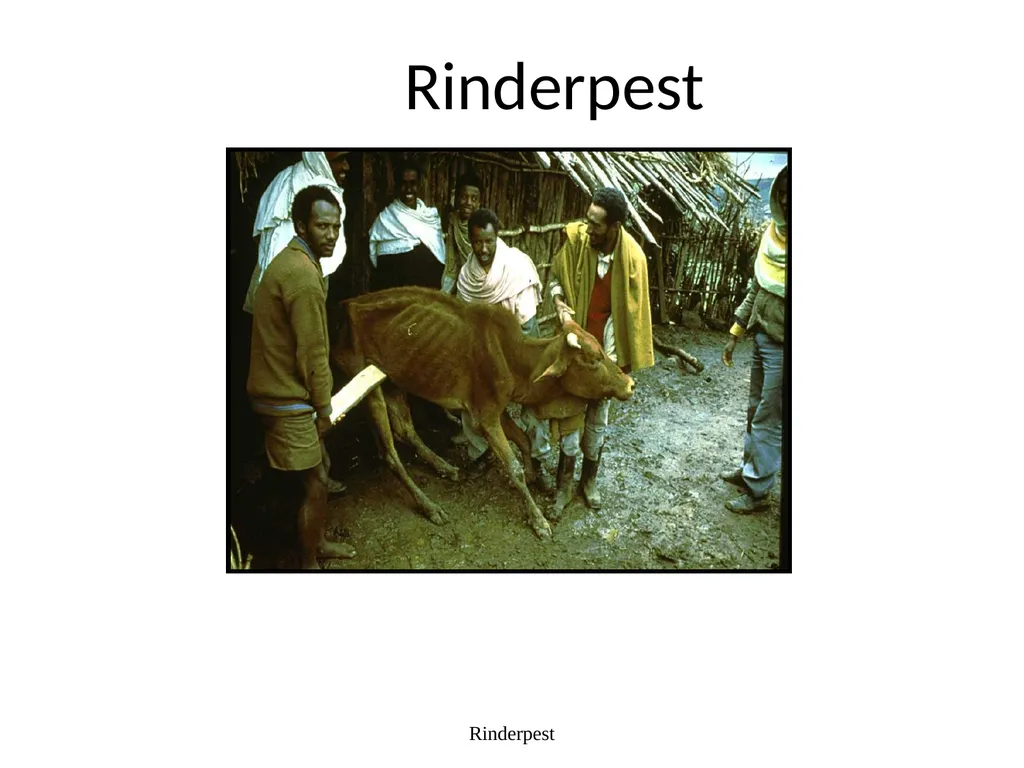
slide2. Rinderpest Rinderpest<br>
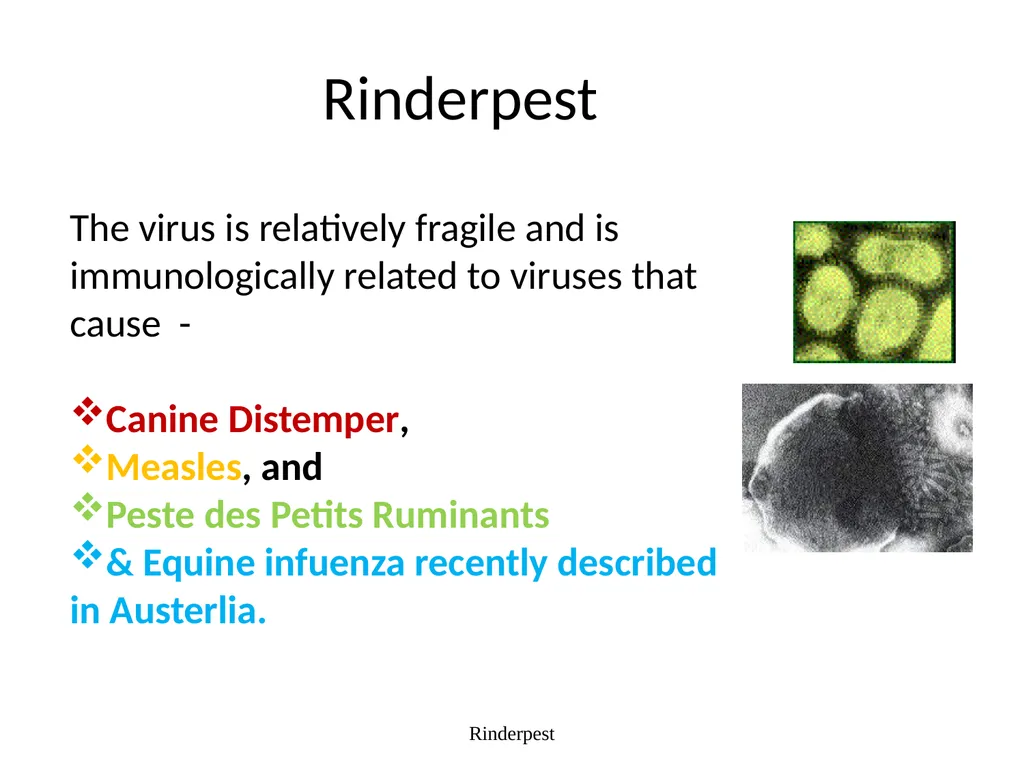
slide3. Rinderpest Rinderpest The virus is relatively fragile and is immunologically related to viruses that cause -
Canine Distemper,
Measles, and
Peste des Petits Ruminants
& Equine infuenza recently described in Austerlia.<br>
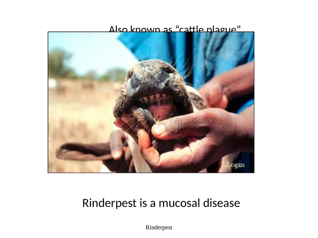
slide4. Rinderpest Also known as “cattle plague” Rinderpest is a mucosal disease<br>
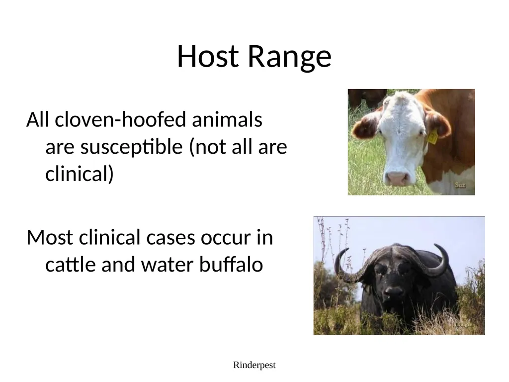
slide5. Host Range All cloven-hoofed animals are susceptible (not all are clinical)
Most clinical cases occur in cattle and water buffalo Rinderpest<br>
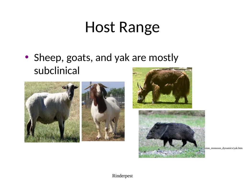
slide6. Host Range Sheep, goats, and yak are mostly subclinical Rinderpest http://www.geo.arizona.edu/dgesl/research/regional/asian_monsoon_dynamics/yak.htm<br>
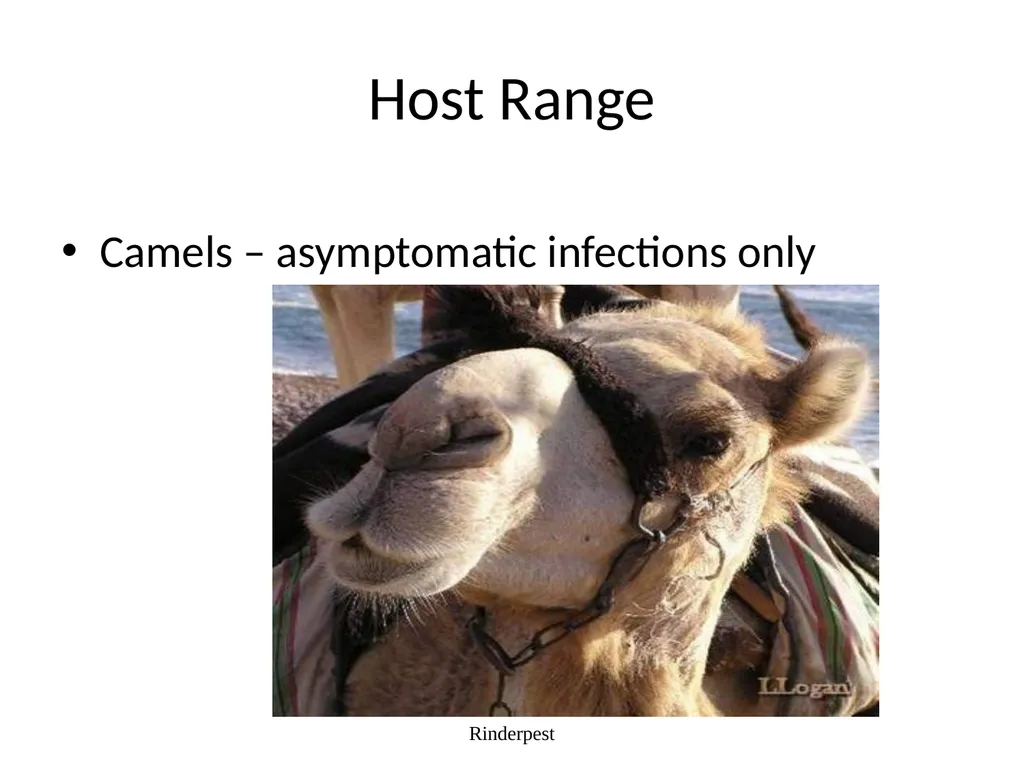
slide7. Host Range Camels – asymptomatic infections only Rinderpest<br>
slide8. Host Range – Wild Animals Most cloven-footed wild animals such as bison and deer
Antelope
Wildebeest
Eland
Giraffe
Hippopotamus
Gazelle
Warthog Rinderpest<br>
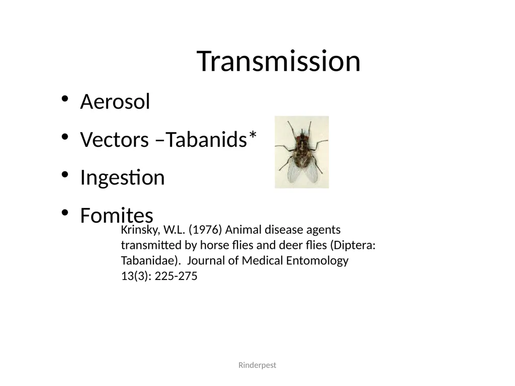
slide9. Transmission Aerosol
Vectors –Tabanids*
Ingestion
Fomites Rinderpest Krinsky, W.L. (1976) Animal disease agents transmitted by horse flies and deer flies (Diptera: Tabanidae). Journal of Medical Entomology 13(3): 225-275<br>
slide10. Transmission There is no vertical transmission, arthropod vector, or carrier state. This makes Rinderpest virus an ideal virus to be targeted for eradication. Rinderpest<br>
slide11. Incubation period Varies with strain of RPV, dosage, and route of exposure (3-15 days)
Normally a range of 3-9 days (can be as short as 3-4 days in experimental infection; also, can be as long as 10-15 days with virus of low virulence)
Duration: 2 or more weeks Rinderpest<br>
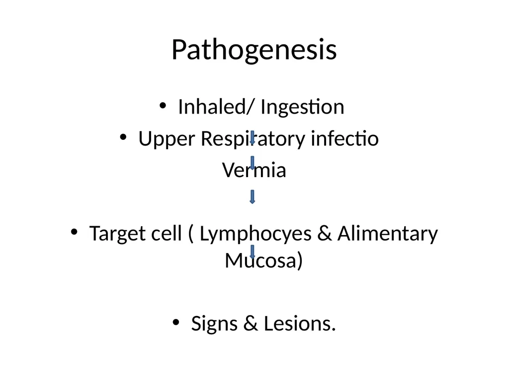
slide12. Pathogenesis Inhaled/ Ingestion
Upper Respiratory infectio
Vermia
Target cell ( Lymphocyes & Alimentary Mucosa)
Signs & Lesions.<br>
slide13. Rinderpest *Virus is present
in blood and secretions
BEFORE
symptoms appear<br>
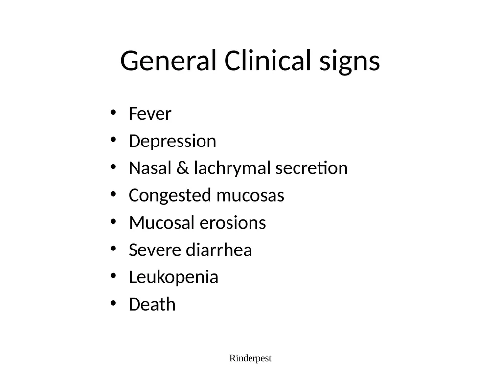
slide14. General Clinical signs Fever
Depression
Nasal & lachrymal secretion
Congested mucosas
Mucosal erosions
Severe diarrhea
Leukopenia
Death Rinderpest<br>
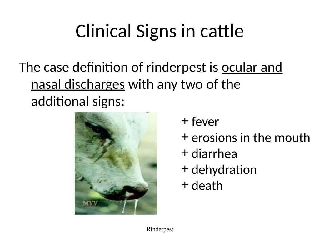
slide15. Clinical Signs in cattle The case definition of rinderpest is ocular and nasal discharges with any two of the additional signs: Rinderpest fever
erosions in the mouth
diarrhea
dehydration
death<br>
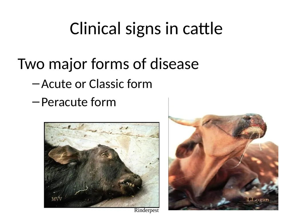
slide16. Clinical signs in cattle Two major forms of disease
Acute or Classic form
Peracute form Rinderpest<br>
slide17. Clinical Signs in cattle (Peracute Form) Most often found in highly susceptible young and newborn animals
No prodromal signs
High fever (104-107 °F)
Congested mucous membranes Rinderpest<br>
slide18. Clinical Signs in cattle (Acute Form) Acute (classic) form characterized by pyrexia, erosive stomatitis, gastroenteritis, dehydration, and death
Four stages
Incubation period
Febrile period
Mucous membrane congestion
Gastrointestinal signs Rinderpest<br>
slide19. Clinical Signs in cattle (Acute Form) Fever - 104 to 107°F (40-42°C)
Serous oculo-nasal discharge
Leukopenia
Depression
Anorexia
Constipation followed by diarrhea
Oral erosions Rinderpest<br>
slide20. Clinical Signs in cattle (Acute Form) Decreases in fever and viral titer
Diarrhea (may be watery or hemorrhagic)
Dehydration, emaciation
Prostration and death 6 to 12 days after onset of illness Rinderpest<br>
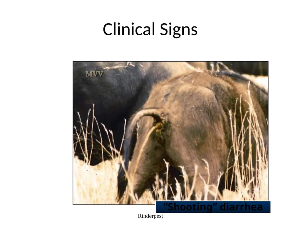
slide21. Clinical Signs Rinderpest “Shooting” diarrhea<br>
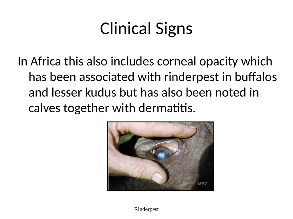
slide22. Clinical Signs In Africa this also includes corneal opacity which has been associated with rinderpest in buffalos and lesser kudus but has also been noted in calves together with dermatitis. Rinderpest<br>
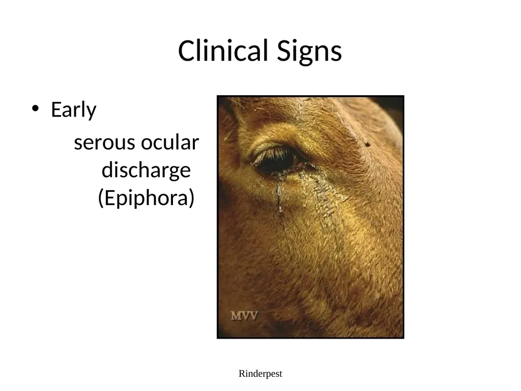
slide23. Clinical Signs Early
serous ocular discharge (Epiphora) Rinderpest<br>
slide24. Clinical Signs Rinderpest Depression
Diarrhea
Dehydration
Death<br>
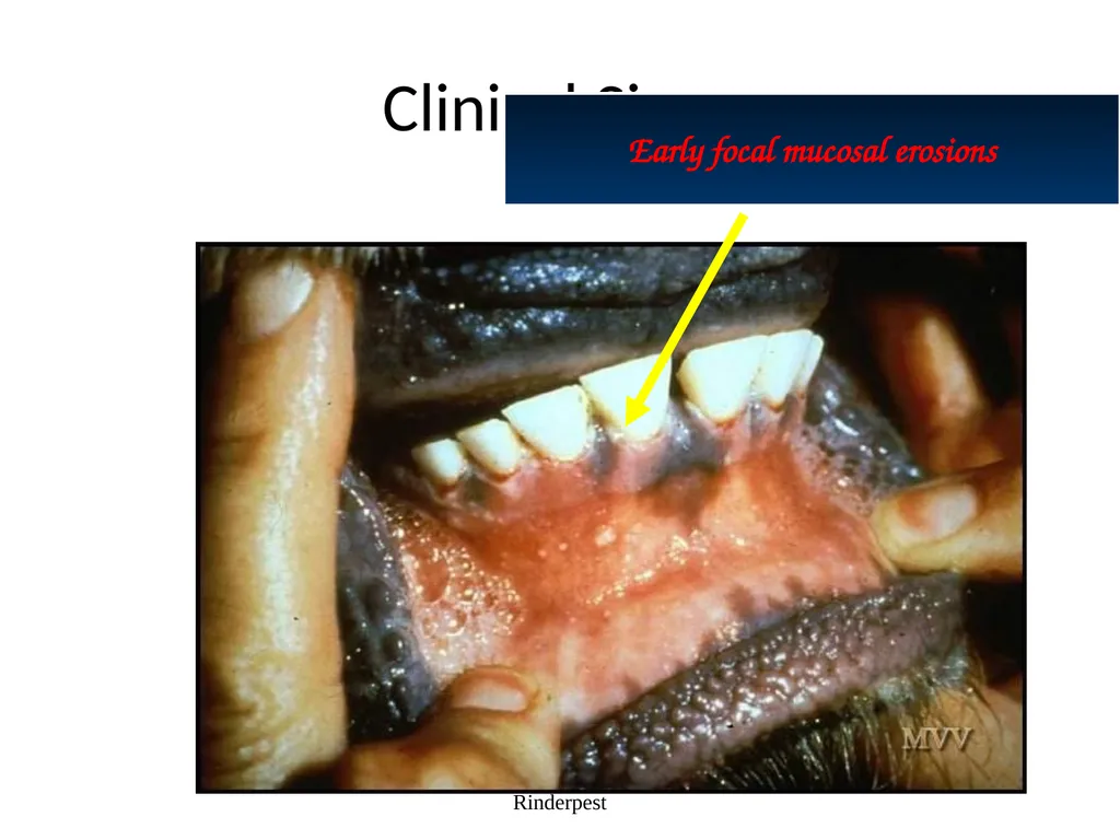
slide25. Clinical Signs Rinderpest Early focal mucosal erosions<br>
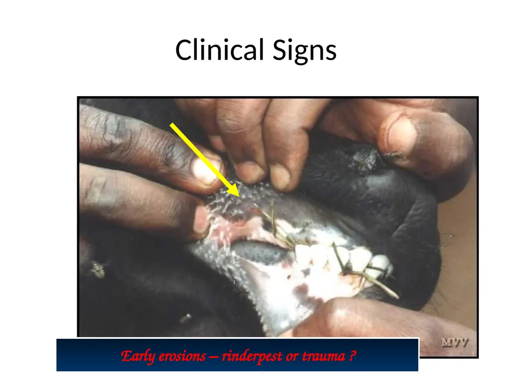
slide26. Clinical Signs Rinderpest Early erosions – rinderpest or trauma ?<br>
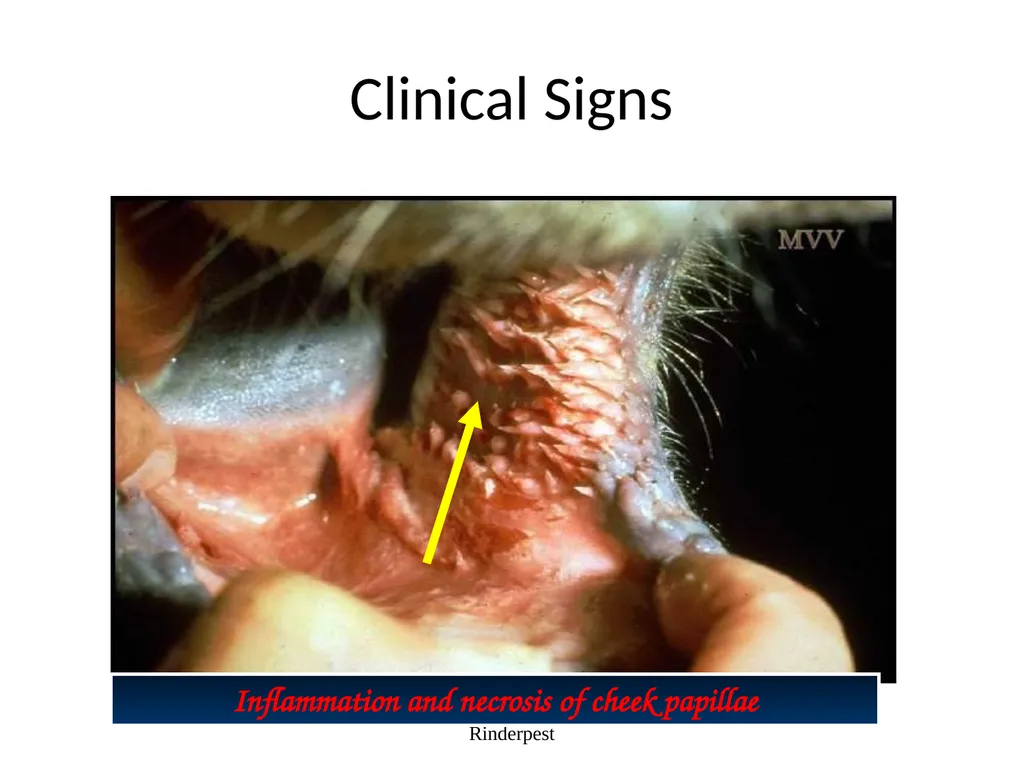
slide27. Clinical Signs Rinderpest Inflammation and necrosis of cheek papillae<br>
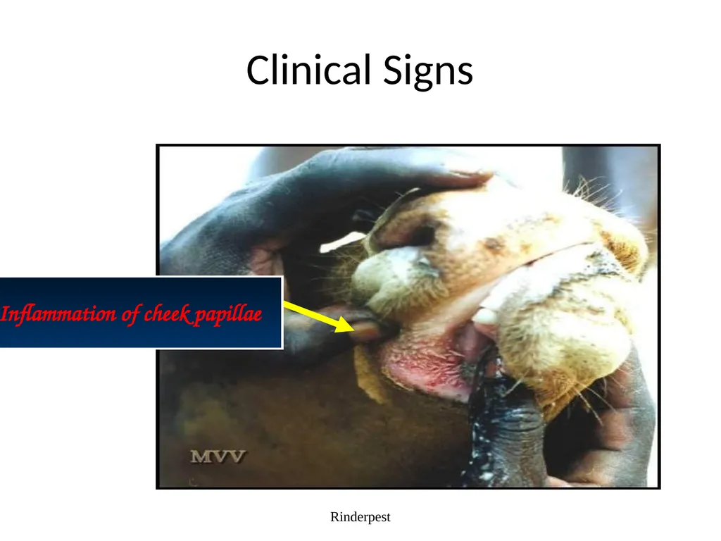
slide28. Clinical Signs Rinderpest Inflammation of cheek papillae<br>
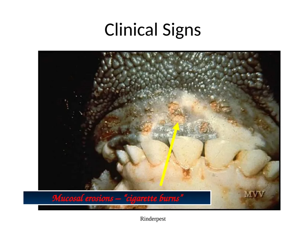
slide29. Clinical Signs Rinderpest Mucosal erosions – “cigarette burns”<br>
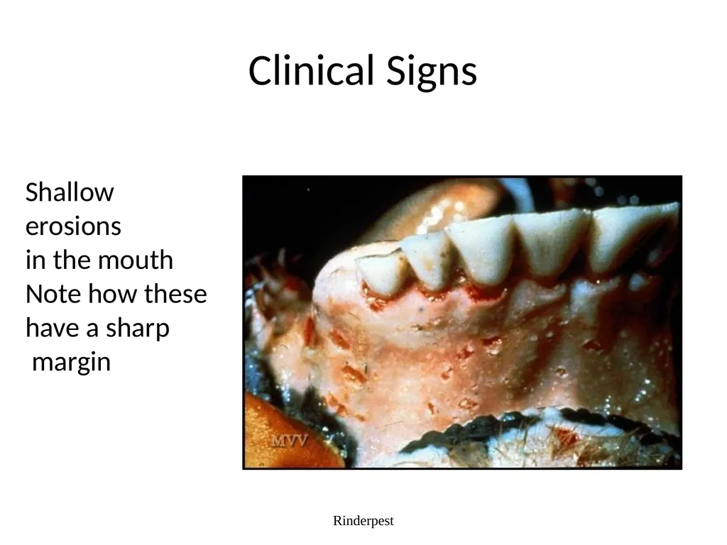
slide30. Clinical Signs Rinderpest Shallow
erosions
in the mouth
Note how these
have a sharp
margin<br>
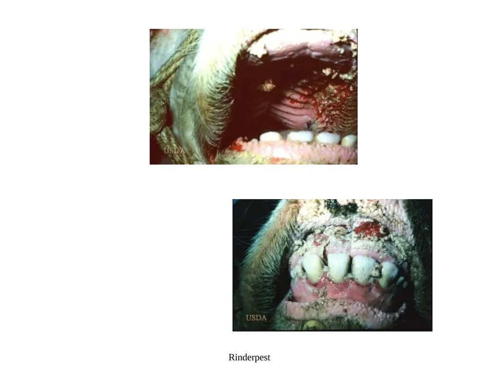
slide31. Rinderpest<br>
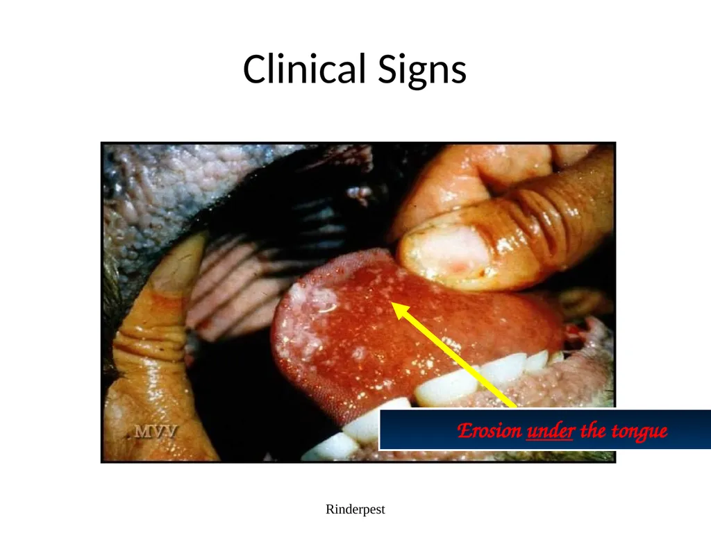
slide32. Clinical Signs Rinderpest Erosion under the tongue<br>
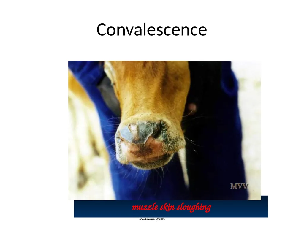
slide33. Convalescence Rinderpest muzzle skin sloughing<br>
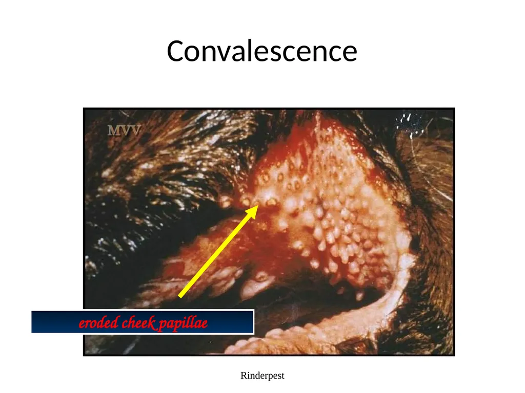
slide34. Convalescence Rinderpest eroded cheek papillae<br>
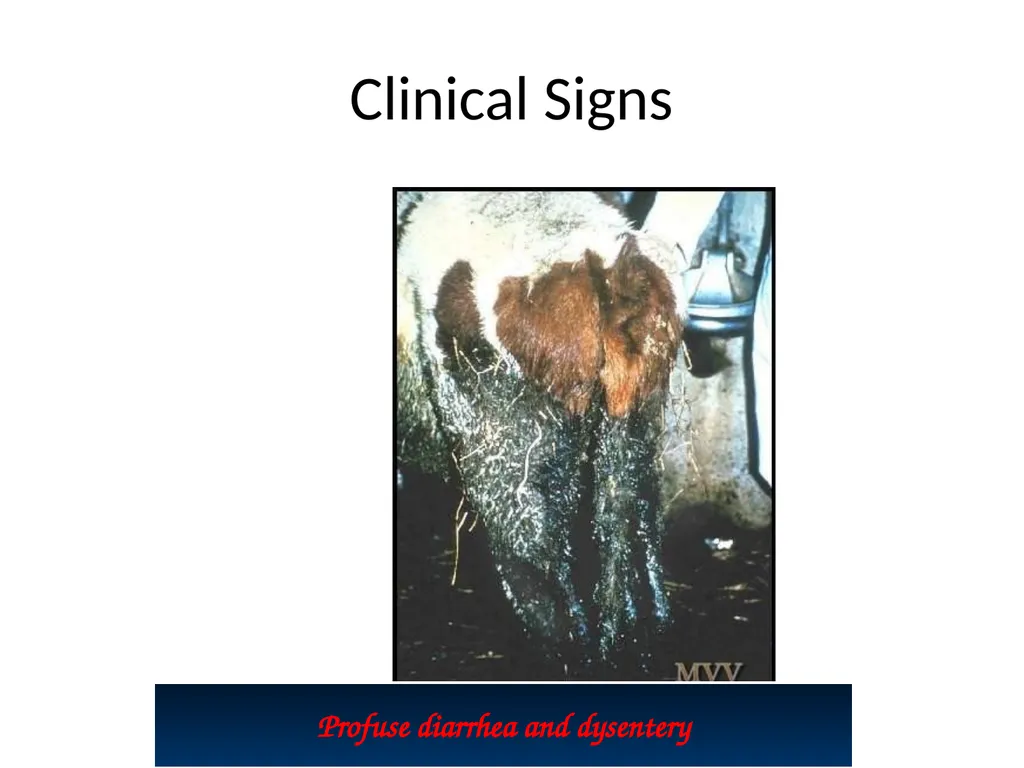
slide35. Clinical Signs Rinderpest Profuse diarrhea and dysentery<br>
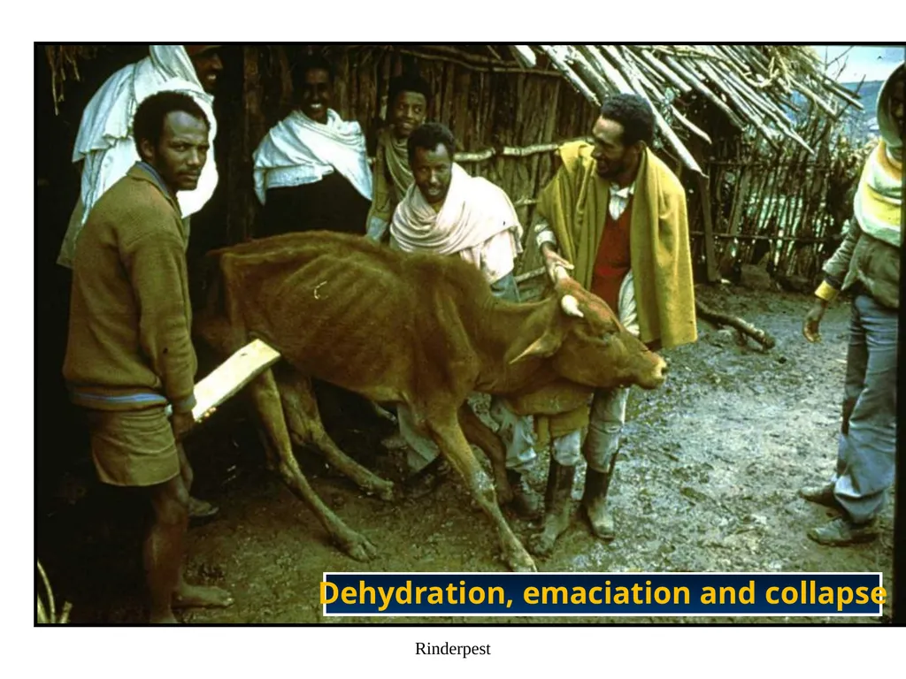
slide36. Rinderpest Dehydration, emaciation and collapse<br>
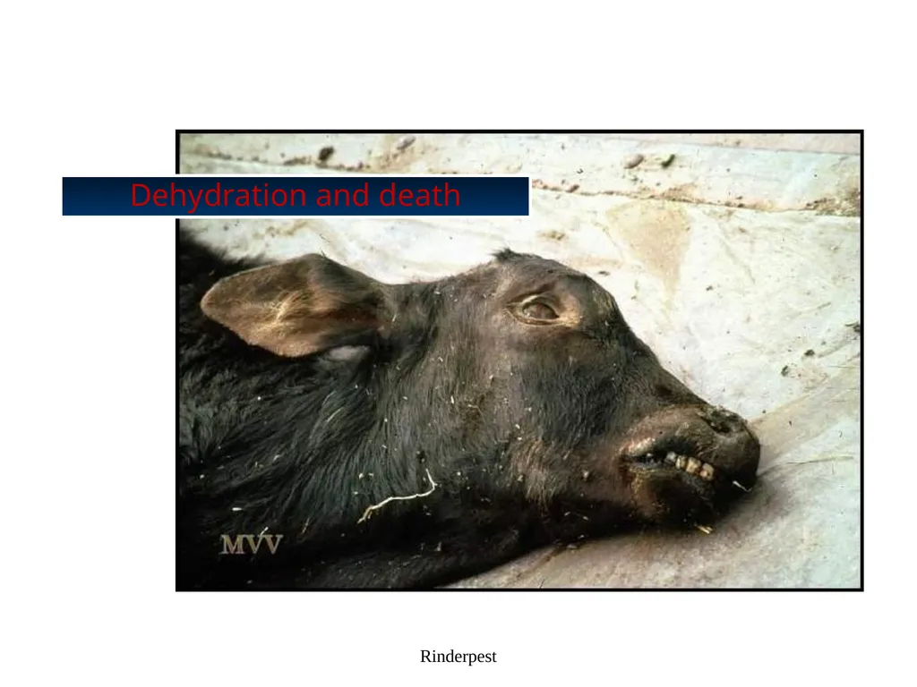
slide37. Rinderpest Dehydration and death<br>
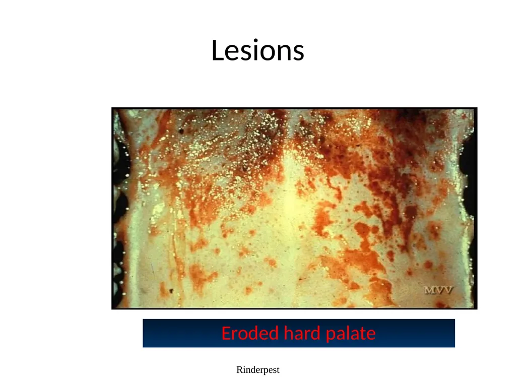
slide38. Lesions Rinderpest Eroded hard palate<br>
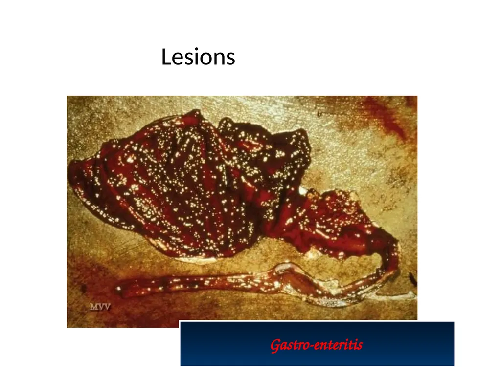
slide39. Rinderpest Gastro-enteritis Lesions<br>
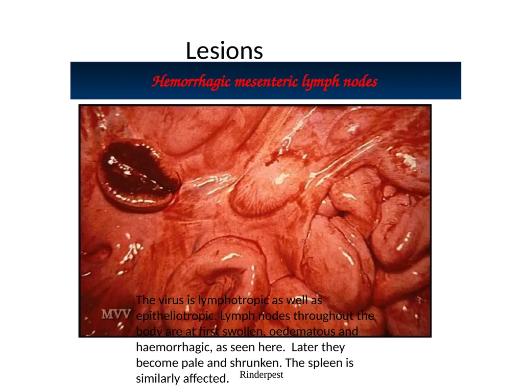
slide40. Rinderpest Hemorrhagic mesenteric lymph nodes Lesions The virus is lymphotropic as well as epitheliotropic. Lymph nodes throughout the body are at first swollen, oedematous and haemorrhagic, as seen here. Later they become pale and shrunken. The spleen is similarly affected.<br>
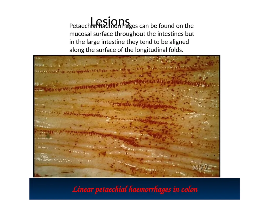
slide41. Rinderpest Linear petaechial haemorrhages in colon Lesions Petaechial haemorrhages can be found on the mucosal surface throughout the intestines but in the large intestine they tend to be aligned along the surface of the longitudinal folds.<br>
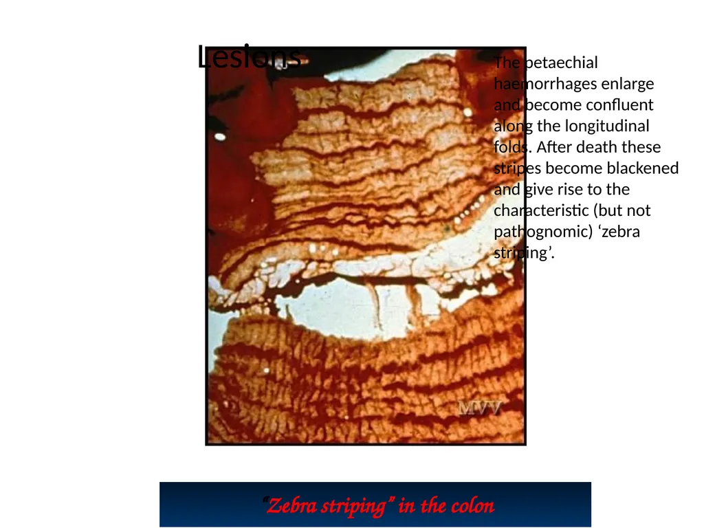
slide42. Rinderpest “Zebra striping” in the colon Lesions The petaechial haemorrhages enlarge and become confluent along the longitudinal folds. After death these stripes become blackened and give rise to the characteristic (but not pathognomic) ‘zebra striping’.<br>
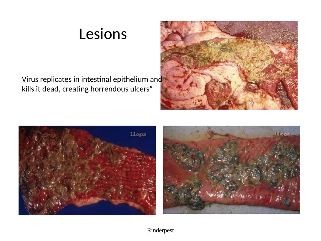
slide43. Rinderpest Lesions Virus replicates in intestinal epithelium and kills it dead, creating horrendous ulcers”<br>
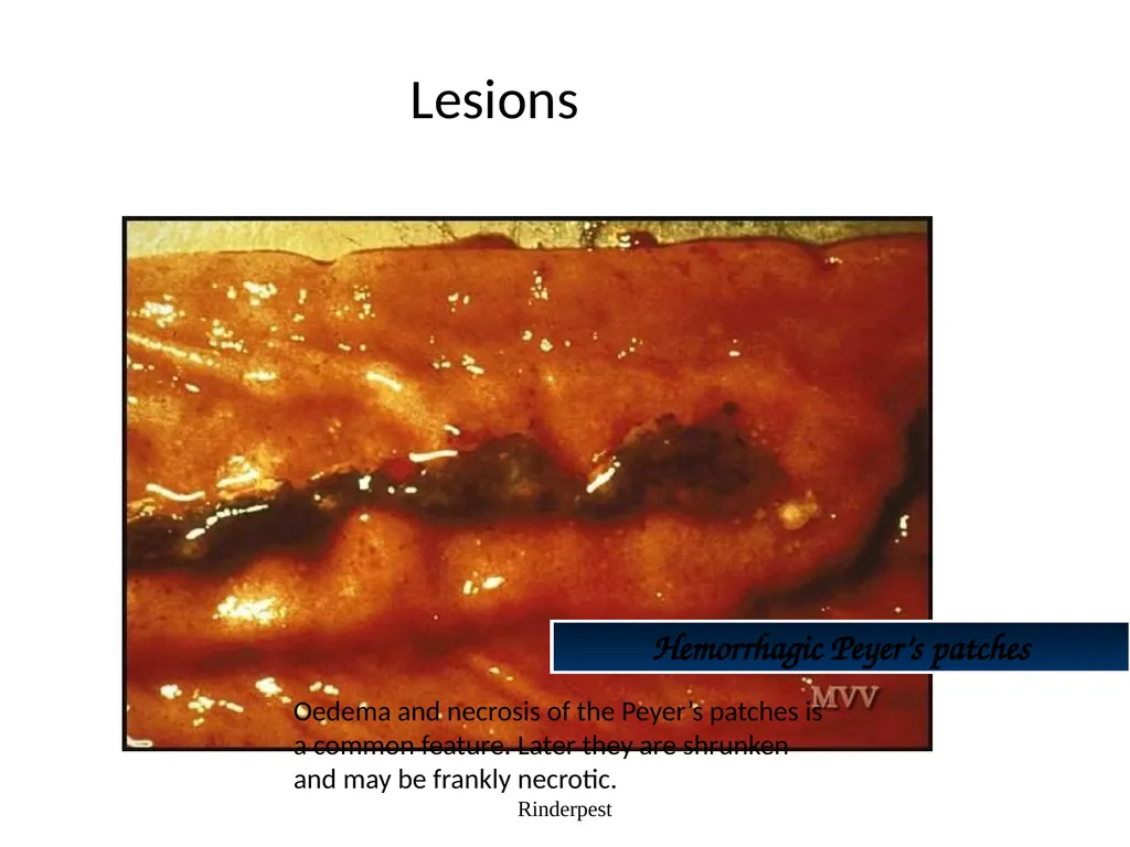
slide44. Rinderpest Hemorrhagic Peyer’s patches Lesions Oedema and necrosis of the Peyer’s patches is a common feature. Later they are shrunken and may be frankly necrotic.<br>
slide45. Diagnosis Samples:
Conjunctival Fluid
Intestinal contents or feces
Whole blood
Lymphoid tissue, lung, intestine
Serum Rinderpest A definitive diagnosis require laboratory confirmation based on detecting viral antigens, the presence of microscopic lesions, and by isolating and identifying the virus. Laboratory confirmation depends on the collection of suitable samples from a group of animals, preferably in the febrile stage with oral lesions rather than from animals already dying, with profuse diarrhea.<br>
slide46. Differential Diagnosis Bovine virus diarrhea
Mucosal disease
Infectious bovine rhinotracheaitis
Malignant catarrhal fever
Vesicular stomatitis
Foot-and-mouth disease Rinderpest<br>
slide47. Differential Diagnosis Salmonellosis
Necrobacillosis
paratuberculosis
Bluetongue / EHD
Mycotic Stomatitis Rinderpest<br>
slide48. Diagnostic Tests Antigen Detection
Antibody Detection
Histopathology Rinderpest Blood should be collected in both serum and EDTA tubes. Swabs of clear tears, necrotic debris from gums and aspiration biopsies from superficial lymph nodes are also recommended.
Virus isolation requires samples of spleen, lymph node and/or tonsils from a freshly euthanized animal<br>
slide49. RINDERPEST Synonym : Cattle plaque
Definition
Acute, highly contagious viral disease of cattle characterized by high fever, necrotic stomatitis, gastroenteritis, diarrhoea and high mortality caused by morbillivirus
In India "Hill Zebu cattle" are more susceptible than "Plain zebu cattle". This disease is characterized by necrosis, and erosions of the mucosa in the respiratory and digestive tracts.
Etiology
Morbillivirus
The virus is antigenically closely related to the viruses of canine distemper, PPR of sheep and goats and measles of humans.
Incidence<br>
slide50. Incidence
The disease has been the foremost cause of death in cattle in most African and Asian countries including India
Rinderpest has not been reported since June 1995 in Indian suncontinent
The seriousness of disease madeto start first Veterinary College in 1762 at Lyons, France.
Susceptibility
Mainly cattle and buffaloes, but also reported in sheep,goat and pigs
Reported in deer, antelope, wild buffaloes, wild boars, bushbuck, warthogs and giraffe
Mortality is 100 % in exotic breeds and 20-50 % mortality in indigenous breeds<br>
slide51. Transmission
Incubation period is 2 to 3 days in experimental inoculation and in contact infection is 6 to 9 days
Virus excreted in body secretions
Ingestion of contaminated feed and water
Inhalation (or) mechanical transmission<br>
slide52. Pathogenesis
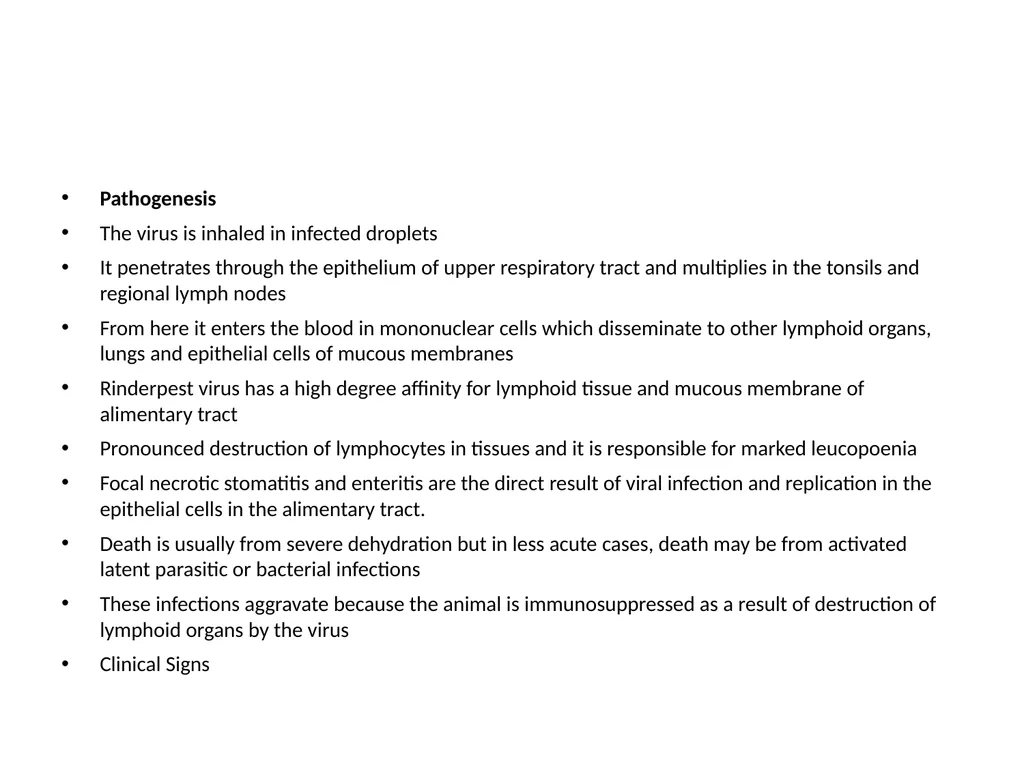
The virus is inhaled in infected droplets
It penetrates through the epithelium of upper respiratory tract and multiplies in the tonsils and regional lymph nodes
From here it enters the blood in mononuclear cells which disseminate to other lymphoid organs, lungs and epithelial cells of mucous membranes
Rinderpest virus has a high degree affinity for lymphoid tissue and mucous membrane of alimentary tract
Pronounced destruction of lymphocytes in tissues and it is responsible for marked leucopoenia
Focal necrotic stomatitis and enteritis are the direct result of viral infection and replication in the epithelial cells in the alimentary tract.
Death is usually from severe dehydration but in less acute cases, death may be from activated latent parasitic or bacterial infections
These infections aggravate because the animal is immunosuppressed as a result of destruction of lymphoid organs by the virus
Clinical Signs<br>
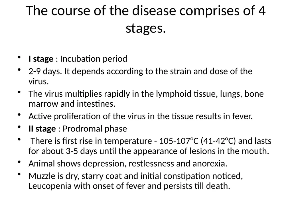
slide53. The course of the disease comprises of 4 stages. I stage : Incubation period
2-9 days. It depends according to the strain and dose of the virus.
The virus multiplies rapidly in the lymphoid tissue, lungs, bone marrow and intestines.
Active proliferation of the virus in the tissue results in fever.
II stage : Prodromal phase
There is first rise in temperature - 105-107°C (41-42°C) and lasts for about 3-5 days until the appearance of lesions in the mouth.
Animal shows depression, restlessness and anorexia.
Muzzle is dry, starry coat and initial constipation noticed, Leucopenia with onset of fever and persists till death.<br>
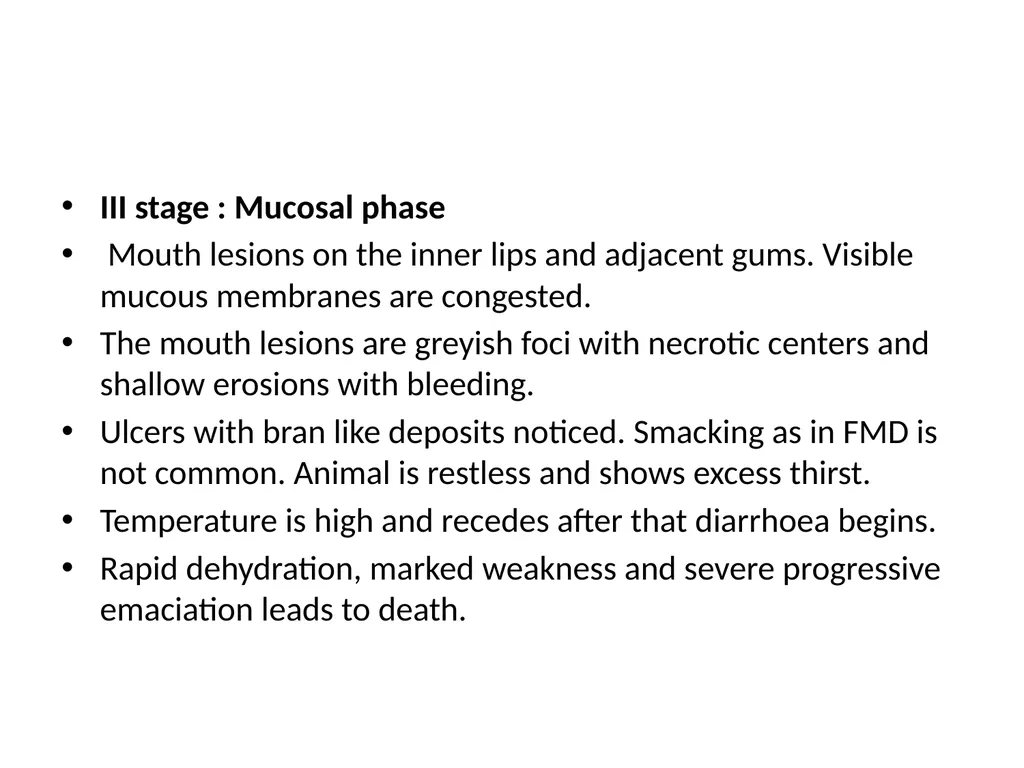
slide54. III stage : Mucosal phase
Mouth lesions on the inner lips and adjacent gums. Visible mucous membranes are congested.
The mouth lesions are greyish foci with necrotic centers and shallow erosions with bleeding.
Ulcers with bran like deposits noticed. Smacking as in FMD is not common. Animal is restless and shows excess thirst.
Temperature is high and recedes after that diarrhoea begins.
Rapid dehydration, marked weakness and severe progressive emaciation leads to death.<br>
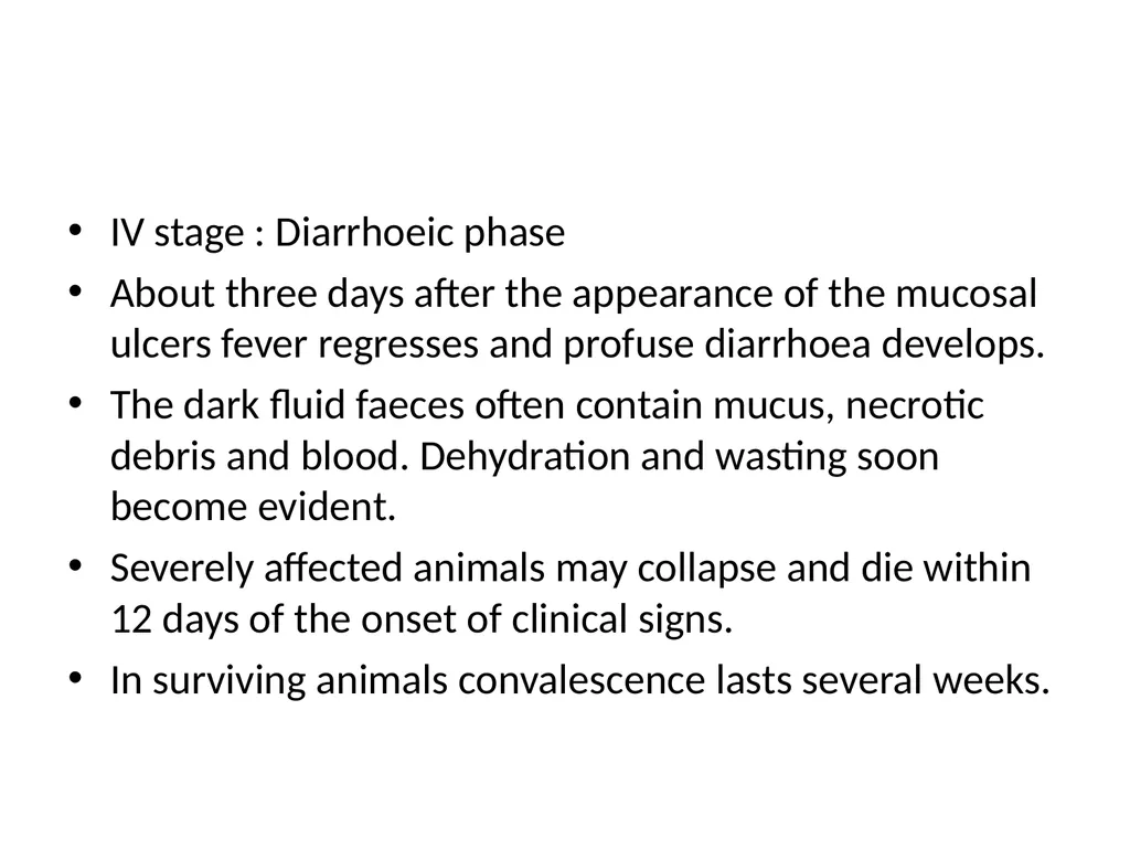
slide55. IV stage : Diarrhoeic phase
About three days after the appearance of the mucosal ulcers fever regresses and profuse diarrhoea develops.
The dark fluid faeces often contain mucus, necrotic debris and blood. Dehydration and wasting soon become evident.
Severely affected animals may collapse and die within 12 days of the onset of clinical signs.
In surviving animals convalescence lasts several weeks.<br>
slide56. V stage : Convalescent phase
Mouth lesions start healing. Rapid regeneration of the affected epithelium noticed.
Slow recuperation of general health. Mortality in cattle, sheep and goats and pigs is 90%.<br>
slide57. Clinical Signs
Fever (104-105 0F), restlessness, dryness of the muzzle and constipation
Other signs include photophobia, excessive thirst, starry coat, retarded rumination, anorexia and excessive salivation
Rashes may develop in those parts of the body where the hair is fine in nature
Mucous membrane of lips, gums and tongue revealed small vesicles resembling bran like deposits
'Shooting diarrhoea' with foetid odour
Dehydration
Marked leucopoenia with drop in lymphocyte count<br>
slide58. Gross lesions
Virus produce lesions in the oral mucosa after settling in the cells following a viremic state
Fore stomach are free
Abomasum reveals necrotic foci and haemorrhagic streaks more seen at the pyloric region.
Folds of abomasum are thick and oedematous
If the disease progress, abomasal mucosa shows irregular ulcers of different size.
The virus has got affinity for lymphoid tissue and in the intestine, peyer's patches are swollen and ulcerated
Duodenum and ileum revealed streaks of haemorrhages and erosions<br>
slide59. In the large intestine, ileo caecal valve may be markedly swollen
Linear haemorrhages on the folds of mucosa of rectum appear like 'Zebra marking' which is pathognomonic in Rinder pest
Lesions are more severe in large intestine with ulceration and diphtheitic patches
Liver: Chronic passive congestion resulting from cardiac and pulmonary complications
Petechiae and erosions in the larynx
Tracheal haemorrhages
Alveolar and interstitial emphysema
Subepithelial and subendocardial haemorrhages
Petechiae and erosions may be seen in the bladder and vagina
Purulent conjunctivitis and ulceration of cornea may be noticed
In sheep and goat, mouth lesions are usually not seen<br>
slide60. Microscopic lesions
Epithelial surface reveals ulcers, haemorrhages, oedema, necrosis and leucocytic infiltration along with multinucleated cells
Eosinophilic cytoplasmic inclusion bodies form in the mucosal epithelial cells and giant cells. Intranuclear inclusion bodies are rare.
Diagnosis
Symptoms and lesions
Complement fixation test (CFT)
Agar Gel Precipitation Test (AGPT)
Virus isolation and diagnosis on tissue cultures
Using specific cDNA probes, isolates of rinderpest and PPR viruses can be differentiated presently
Polymerase chain Reaction (PCR)
Eradicated science<br>
The “four D’s” of Rinderpest:
Depression
Diarrhea
Dehydration
Death Rinderpest<br>
slide2. Rinderpest Rinderpest<br>
slide3. Rinderpest Rinderpest The virus is relatively fragile and is immunologically related to viruses that cause -
Canine Distemper,
Measles, and
Peste des Petits Ruminants
& Equine infuenza recently described in Austerlia.<br>
slide4. Rinderpest Also known as “cattle plague” Rinderpest is a mucosal disease<br>
slide5. Host Range All cloven-hoofed animals are susceptible (not all are clinical)
Most clinical cases occur in cattle and water buffalo Rinderpest<br>
slide6. Host Range Sheep, goats, and yak are mostly subclinical Rinderpest http://www.geo.arizona.edu/dgesl/research/regional/asian_monsoon_dynamics/yak.htm<br>
slide7. Host Range Camels – asymptomatic infections only Rinderpest<br>
slide8. Host Range – Wild Animals Most cloven-footed wild animals such as bison and deer
Antelope
Wildebeest
Eland
Giraffe
Hippopotamus
Gazelle
Warthog Rinderpest<br>
slide9. Transmission Aerosol
Vectors –Tabanids*
Ingestion
Fomites Rinderpest Krinsky, W.L. (1976) Animal disease agents transmitted by horse flies and deer flies (Diptera: Tabanidae). Journal of Medical Entomology 13(3): 225-275<br>
slide10. Transmission There is no vertical transmission, arthropod vector, or carrier state. This makes Rinderpest virus an ideal virus to be targeted for eradication. Rinderpest<br>
slide11. Incubation period Varies with strain of RPV, dosage, and route of exposure (3-15 days)
Normally a range of 3-9 days (can be as short as 3-4 days in experimental infection; also, can be as long as 10-15 days with virus of low virulence)
Duration: 2 or more weeks Rinderpest<br>
slide12. Pathogenesis Inhaled/ Ingestion
Upper Respiratory infectio
Vermia
Target cell ( Lymphocyes & Alimentary Mucosa)
Signs & Lesions.<br>
slide13. Rinderpest *Virus is present
in blood and secretions
BEFORE
symptoms appear<br>
slide14. General Clinical signs Fever
Depression
Nasal & lachrymal secretion
Congested mucosas
Mucosal erosions
Severe diarrhea
Leukopenia
Death Rinderpest<br>
slide15. Clinical Signs in cattle The case definition of rinderpest is ocular and nasal discharges with any two of the additional signs: Rinderpest fever
erosions in the mouth
diarrhea
dehydration
death<br>
slide16. Clinical signs in cattle Two major forms of disease
Acute or Classic form
Peracute form Rinderpest<br>
slide17. Clinical Signs in cattle (Peracute Form) Most often found in highly susceptible young and newborn animals
No prodromal signs
High fever (104-107 °F)
Congested mucous membranes Rinderpest<br>
slide18. Clinical Signs in cattle (Acute Form) Acute (classic) form characterized by pyrexia, erosive stomatitis, gastroenteritis, dehydration, and death
Four stages
Incubation period
Febrile period
Mucous membrane congestion
Gastrointestinal signs Rinderpest<br>
slide19. Clinical Signs in cattle (Acute Form) Fever - 104 to 107°F (40-42°C)
Serous oculo-nasal discharge
Leukopenia
Depression
Anorexia
Constipation followed by diarrhea
Oral erosions Rinderpest<br>
slide20. Clinical Signs in cattle (Acute Form) Decreases in fever and viral titer
Diarrhea (may be watery or hemorrhagic)
Dehydration, emaciation
Prostration and death 6 to 12 days after onset of illness Rinderpest<br>
slide21. Clinical Signs Rinderpest “Shooting” diarrhea<br>
slide22. Clinical Signs In Africa this also includes corneal opacity which has been associated with rinderpest in buffalos and lesser kudus but has also been noted in calves together with dermatitis. Rinderpest<br>
slide23. Clinical Signs Early
serous ocular discharge (Epiphora) Rinderpest<br>
slide24. Clinical Signs Rinderpest Depression
Diarrhea
Dehydration
Death<br>
slide25. Clinical Signs Rinderpest Early focal mucosal erosions<br>
slide26. Clinical Signs Rinderpest Early erosions – rinderpest or trauma ?<br>
slide27. Clinical Signs Rinderpest Inflammation and necrosis of cheek papillae<br>
slide28. Clinical Signs Rinderpest Inflammation of cheek papillae<br>
slide29. Clinical Signs Rinderpest Mucosal erosions – “cigarette burns”<br>
slide30. Clinical Signs Rinderpest Shallow
erosions
in the mouth
Note how these
have a sharp
margin<br>
slide31. Rinderpest<br>
slide32. Clinical Signs Rinderpest Erosion under the tongue<br>
slide33. Convalescence Rinderpest muzzle skin sloughing<br>
slide34. Convalescence Rinderpest eroded cheek papillae<br>
slide35. Clinical Signs Rinderpest Profuse diarrhea and dysentery<br>
slide36. Rinderpest Dehydration, emaciation and collapse<br>
slide37. Rinderpest Dehydration and death<br>
slide38. Lesions Rinderpest Eroded hard palate<br>
slide39. Rinderpest Gastro-enteritis Lesions<br>
slide40. Rinderpest Hemorrhagic mesenteric lymph nodes Lesions The virus is lymphotropic as well as epitheliotropic. Lymph nodes throughout the body are at first swollen, oedematous and haemorrhagic, as seen here. Later they become pale and shrunken. The spleen is similarly affected.<br>
slide41. Rinderpest Linear petaechial haemorrhages in colon Lesions Petaechial haemorrhages can be found on the mucosal surface throughout the intestines but in the large intestine they tend to be aligned along the surface of the longitudinal folds.<br>
slide42. Rinderpest “Zebra striping” in the colon Lesions The petaechial haemorrhages enlarge and become confluent along the longitudinal folds. After death these stripes become blackened and give rise to the characteristic (but not pathognomic) ‘zebra striping’.<br>
slide43. Rinderpest Lesions Virus replicates in intestinal epithelium and kills it dead, creating horrendous ulcers”<br>
slide44. Rinderpest Hemorrhagic Peyer’s patches Lesions Oedema and necrosis of the Peyer’s patches is a common feature. Later they are shrunken and may be frankly necrotic.<br>
slide45. Diagnosis Samples:
Conjunctival Fluid
Intestinal contents or feces
Whole blood
Lymphoid tissue, lung, intestine
Serum Rinderpest A definitive diagnosis require laboratory confirmation based on detecting viral antigens, the presence of microscopic lesions, and by isolating and identifying the virus. Laboratory confirmation depends on the collection of suitable samples from a group of animals, preferably in the febrile stage with oral lesions rather than from animals already dying, with profuse diarrhea.<br>
slide46. Differential Diagnosis Bovine virus diarrhea
Mucosal disease
Infectious bovine rhinotracheaitis
Malignant catarrhal fever
Vesicular stomatitis
Foot-and-mouth disease Rinderpest<br>
slide47. Differential Diagnosis Salmonellosis
Necrobacillosis
paratuberculosis
Bluetongue / EHD
Mycotic Stomatitis Rinderpest<br>
slide48. Diagnostic Tests Antigen Detection
Antibody Detection
Histopathology Rinderpest Blood should be collected in both serum and EDTA tubes. Swabs of clear tears, necrotic debris from gums and aspiration biopsies from superficial lymph nodes are also recommended.
Virus isolation requires samples of spleen, lymph node and/or tonsils from a freshly euthanized animal<br>
slide49. RINDERPEST Synonym : Cattle plaque
Definition
Acute, highly contagious viral disease of cattle characterized by high fever, necrotic stomatitis, gastroenteritis, diarrhoea and high mortality caused by morbillivirus
In India "Hill Zebu cattle" are more susceptible than "Plain zebu cattle". This disease is characterized by necrosis, and erosions of the mucosa in the respiratory and digestive tracts.
Etiology
Morbillivirus
The virus is antigenically closely related to the viruses of canine distemper, PPR of sheep and goats and measles of humans.
Incidence<br>
slide50. Incidence
The disease has been the foremost cause of death in cattle in most African and Asian countries including India
Rinderpest has not been reported since June 1995 in Indian suncontinent
The seriousness of disease madeto start first Veterinary College in 1762 at Lyons, France.
Susceptibility
Mainly cattle and buffaloes, but also reported in sheep,goat and pigs
Reported in deer, antelope, wild buffaloes, wild boars, bushbuck, warthogs and giraffe
Mortality is 100 % in exotic breeds and 20-50 % mortality in indigenous breeds<br>
slide51. Transmission
Incubation period is 2 to 3 days in experimental inoculation and in contact infection is 6 to 9 days
Virus excreted in body secretions
Ingestion of contaminated feed and water
Inhalation (or) mechanical transmission<br>
slide52. Pathogenesis
The virus is inhaled in infected droplets
It penetrates through the epithelium of upper respiratory tract and multiplies in the tonsils and regional lymph nodes
From here it enters the blood in mononuclear cells which disseminate to other lymphoid organs, lungs and epithelial cells of mucous membranes
Rinderpest virus has a high degree affinity for lymphoid tissue and mucous membrane of alimentary tract
Pronounced destruction of lymphocytes in tissues and it is responsible for marked leucopoenia
Focal necrotic stomatitis and enteritis are the direct result of viral infection and replication in the epithelial cells in the alimentary tract.
Death is usually from severe dehydration but in less acute cases, death may be from activated latent parasitic or bacterial infections
These infections aggravate because the animal is immunosuppressed as a result of destruction of lymphoid organs by the virus
Clinical Signs<br>
slide53. The course of the disease comprises of 4 stages. I stage : Incubation period
2-9 days. It depends according to the strain and dose of the virus.
The virus multiplies rapidly in the lymphoid tissue, lungs, bone marrow and intestines.
Active proliferation of the virus in the tissue results in fever.
II stage : Prodromal phase
There is first rise in temperature - 105-107°C (41-42°C) and lasts for about 3-5 days until the appearance of lesions in the mouth.
Animal shows depression, restlessness and anorexia.
Muzzle is dry, starry coat and initial constipation noticed, Leucopenia with onset of fever and persists till death.<br>
slide54. III stage : Mucosal phase
Mouth lesions on the inner lips and adjacent gums. Visible mucous membranes are congested.
The mouth lesions are greyish foci with necrotic centers and shallow erosions with bleeding.
Ulcers with bran like deposits noticed. Smacking as in FMD is not common. Animal is restless and shows excess thirst.
Temperature is high and recedes after that diarrhoea begins.
Rapid dehydration, marked weakness and severe progressive emaciation leads to death.<br>
slide55. IV stage : Diarrhoeic phase
About three days after the appearance of the mucosal ulcers fever regresses and profuse diarrhoea develops.
The dark fluid faeces often contain mucus, necrotic debris and blood. Dehydration and wasting soon become evident.
Severely affected animals may collapse and die within 12 days of the onset of clinical signs.
In surviving animals convalescence lasts several weeks.<br>
slide56. V stage : Convalescent phase
Mouth lesions start healing. Rapid regeneration of the affected epithelium noticed.
Slow recuperation of general health. Mortality in cattle, sheep and goats and pigs is 90%.<br>
slide57. Clinical Signs
Fever (104-105 0F), restlessness, dryness of the muzzle and constipation
Other signs include photophobia, excessive thirst, starry coat, retarded rumination, anorexia and excessive salivation
Rashes may develop in those parts of the body where the hair is fine in nature
Mucous membrane of lips, gums and tongue revealed small vesicles resembling bran like deposits
'Shooting diarrhoea' with foetid odour
Dehydration
Marked leucopoenia with drop in lymphocyte count<br>
slide58. Gross lesions
Virus produce lesions in the oral mucosa after settling in the cells following a viremic state
Fore stomach are free
Abomasum reveals necrotic foci and haemorrhagic streaks more seen at the pyloric region.
Folds of abomasum are thick and oedematous
If the disease progress, abomasal mucosa shows irregular ulcers of different size.
The virus has got affinity for lymphoid tissue and in the intestine, peyer's patches are swollen and ulcerated
Duodenum and ileum revealed streaks of haemorrhages and erosions<br>
slide59. In the large intestine, ileo caecal valve may be markedly swollen
Linear haemorrhages on the folds of mucosa of rectum appear like 'Zebra marking' which is pathognomonic in Rinder pest
Lesions are more severe in large intestine with ulceration and diphtheitic patches
Liver: Chronic passive congestion resulting from cardiac and pulmonary complications
Petechiae and erosions in the larynx
Tracheal haemorrhages
Alveolar and interstitial emphysema
Subepithelial and subendocardial haemorrhages
Petechiae and erosions may be seen in the bladder and vagina
Purulent conjunctivitis and ulceration of cornea may be noticed
In sheep and goat, mouth lesions are usually not seen<br>
slide60. Microscopic lesions
Epithelial surface reveals ulcers, haemorrhages, oedema, necrosis and leucocytic infiltration along with multinucleated cells
Eosinophilic cytoplasmic inclusion bodies form in the mucosal epithelial cells and giant cells. Intranuclear inclusion bodies are rare.
Diagnosis
Symptoms and lesions
Complement fixation test (CFT)
Agar Gel Precipitation Test (AGPT)
Virus isolation and diagnosis on tissue cultures
Using specific cDNA probes, isolates of rinderpest and PPR viruses can be differentiated presently
Polymerase chain Reaction (PCR)
Eradicated science<br>