Ruminant Stomach Abomasal displacement Gulshan Kumar Dept. of Veterinary Surgery Radiology College of Veterinary Science and Animal Husbandry U.P. Pt. Deen Dayal Upadhyaya Pashuchikitsa Vigyan Vishwavidyalaya evam Go Anusandhan Sansthan,
"Ruminant Stomach –Abomasal displacement Gulshan" is the property of its rightful owner. Permission is granted to
download and print the materials on this website for personal, non-commercial use only, and to display it
on your personal computer provided you do not modify the materials and that you retain all copyright
notices contained in the materials. By downloading content from our website, you accept the terms of this
agreement.
Share
Embed code
Presentation Transcript
01
Ruminant Stomach –Abomasal displacement Gulshan Kumar
Dept. of Veterinary Surgery & Radiology
College of Veterinary Science and Animal Husbandry
U.P. Pt. Deen Dayal Upadhyaya Pashuchikitsa Vigyan Vishwavidyalaya evam Go Anusandhan Sansthan, Mathura- 281 001<br>
02
abomasal displacement The abomasum is suspended by a loose attachment to the abdominal wall, hence can potentially move/float out of its normal position where it can fill up with gas and prevent normal flow of feed through the digestive track (called an abomasal displacement).
Its occurrence is common in large sized, high producing adult dairy cows, and in countries where feed comprises of high concentrates as compared to roughages.
Although LDA and RDA are often considered separately, there is evidence of a common underlying etiology; they may be different manifestations of the same or a similar disease process.<br>
03
abomasal displacement: Ætiology Abomasal hypomotility associated with hypocalcaemia and possibly hypokalaemia, as well as concurrent diseases (mastitis, metritis) associated with endotoxemia and decreased rumen fill, periparturient changes in the position of intra-abdominal organs, and genetic predisposition, particularly in deep-bodied cows are the contributing factors.
It may occur any time during parturition but highest incidences are observed during 3 weeks ante-partum and 4 weeks post-partum.
Excessive production of volatile FAs due to high concentrate ration leads to their passage into the abomasum causing its distension, hence its contractility is reduced (hypomotile).<br>
04
abomasal displacement: Ætiology The gravid uterus lifts the rumen giving the hypomotile abomasum an opportunity to slide to the left of the rumen (between rumen and the abdominal wall), Or
Being less motile, it may float towards right, dorsally and caudally to float between 8th and the 13th rib.
It may occur any time during parturition but highest incidences are observed during 3 weeks ante-partum and 4 weeks post-partum.
Excessive production of volatile FAs due to high concentrate ration leads to their passage into the abomasum causing its distension, hence its contractility is reduced (hypomotile).<br>
05
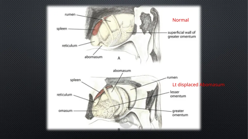
Normal Lt displaced Abomasum<br>
06
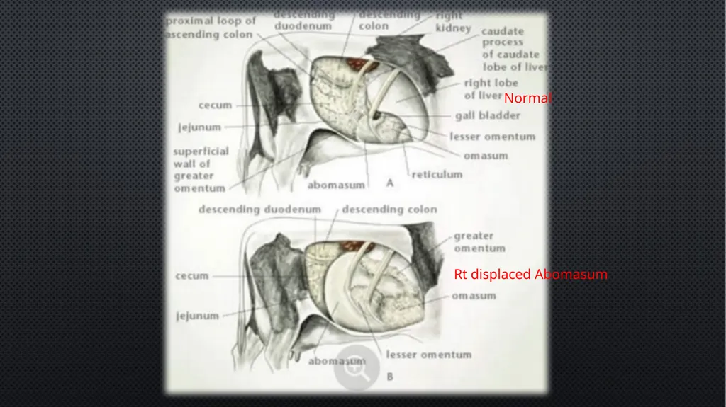
Normal Rt displaced Abomasum<br>
07
Clinical findings Anorexia (intermittent), reduced production, ketosis, body condition loss, dullness and listlessness, mild abdominal pain or discomfort (Shifting weight on limbs), scant and dark faeces.
Sluggish and weak rumen movements.
Chloride-rich fluid (up to 50 L) accumulates in the abomasum,
Hypochloraemic, hypokalaemic metabolic alkalosis develops.
The blood supply to the abomasum, and often the omasum and proximal duodenum, is compromised, eventually resulting in ischemic necrosis of these and dehydration and circulatory failure.
Due to circulatory failure, a metabolic acidosis due to hyper-l-lactatemia and azotaemia may be superimposed on the pre-existing metabolic alkalosis.<br>
08
Clinical findings The most important diagnostic physical finding is a ping on simultaneous auscultation and percussion of the abdomen, (the area marked by a line from the tuber coxae to the point of the elbow), and from the elbow toward the stifle.
The ping characteristic of an LDA is most commonly located in an area between ribs 9 and 13 in the middle to upper third of the left abdomen; however, the ping can be more ventral or more caudal, or both. (Pings associated with a rumen gas cap are usually more dorsal, less resonant, and extend more caudally through the left paralumbar fossa.)
The ping associated with RDA also is most commonly located in the area between ribs 10 and 13 on the right abdomen.<br>
09
Diagnosis Based on history, clinical signs, presence of the characteristic ping on simultaneous auscultation and percussion.
Ultrasonography may be helpful in confirming a diagnosis of LDA and RDA.
Acuteness of the onset of clinical signs, especially rapid heart rate and drop in milk yield, help to differentiate abomasal volvulus from RDA.
“Liptek Test”: an 18G needle is inserted aseptically just below the area of resonant ping in the left abdominal wall in cases of LDA and in the right abdominal region in cases of RDA and the fluid is aspirated. If the pH of the fluid is around 4, abomasal displacement is suspected and a pH of 5 to 7 indicate ruminal contents.<br>
10
treatment The aim of treatment is correction of the displaced abomasum, and fixing the displaced abomasum to prevent recurrence, restoration of gastro intestinal motility, rehydration and correction of metabolic disorders.<br>
11
Treatment: conservative Conservative treatments aim at the release of gases from the abomasum, relief of abomasal impaction and restoration of GI tract motility so that the abomasum return to its normal position.
The major non-surgical technique is to roll the animal from side to side while the animal is in dorsal recumbency.
Calcium boro-gluconate, neostigmine, saline cathartics etc improve the GI tract motility in general. Repeated oral administration of mineral oils and warm saline may help in evacuation of the contents.
Repeated iv isotonic fluid therapy-to treat dehydration.<br>
12
Treatment: surgical Involve decompression, reducing the abomasum to normal anatomical location, and fixation of the omentum/abomasum to the body wall to avoid recurrence.
Left flank omentopexy (Utrecht method):
An 8-9 inch vertical incision is made on the left paralumbar fossa in the standing animal (the displaced abomasum is very likely to be found here).
Insert about 2 m long heavy nonabsorbable suture into the greater omentum near its attachment to the abomasum biting about 7-10 cm of the omental tissue. The other end of the suture should also have a needle.<br>
13
Treatment: surgical Left flank omentopexy (Utrecht method):……contd.
Decompress the abomasum using a needle of 14 G and syringe.
Now, carefully push the abomasum to its normal position.
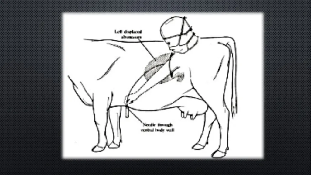
take the needle (cutting) at one end of the suture through the laparotomy incision ventrally and cranially and force it through the ventral wall about 4-5 inches caudally from the xiphisternum and pull it out.
A second needle on the other end of suture is similarly forced through the ventral wall about 3-4 inches caudally from the cranial suture and pulled out. Both the suture ends are then tied and left in place for 4 weeks.<br>
14
Treatment: surgical Right and Left flank abomasopexy:
The procedure is basically similar to omentopexy and the suture is placed in the musculature of the greater curvature of the abomasum.
The left flank approach is used for LDA and right flank approach is used for RDA.
*(A “roll and toggle suture” technique is also used in ventral countries for fixing the abomasum)<br>