Sporogenesis: Microsporogenesis and Megasporogenesis Sporogenesis: Productions of microspores and megaspores is known as sporogenesis. Microspores are produced in anthers (microsporogenesis), while megaspores are produced in ovules
"Sporogenesis: Microsporogenesis and" is the property of its rightful owner. Permission is granted to
download and print the materials on this website for personal, non-commercial use only, and to display it
on your personal computer provided you do not modify the materials and that you retain all copyright
notices contained in the materials. By downloading content from our website, you accept the terms of this
agreement.
Presentation Transcript
01
Sporogenesis: Microsporogenesis and Megasporogenesis Sporogenesis: Productions of microspores and megaspores is known as sporogenesis. Microspores are produced in anthers (microsporogenesis), while megaspores are produced in ovules (megasporogenesis).
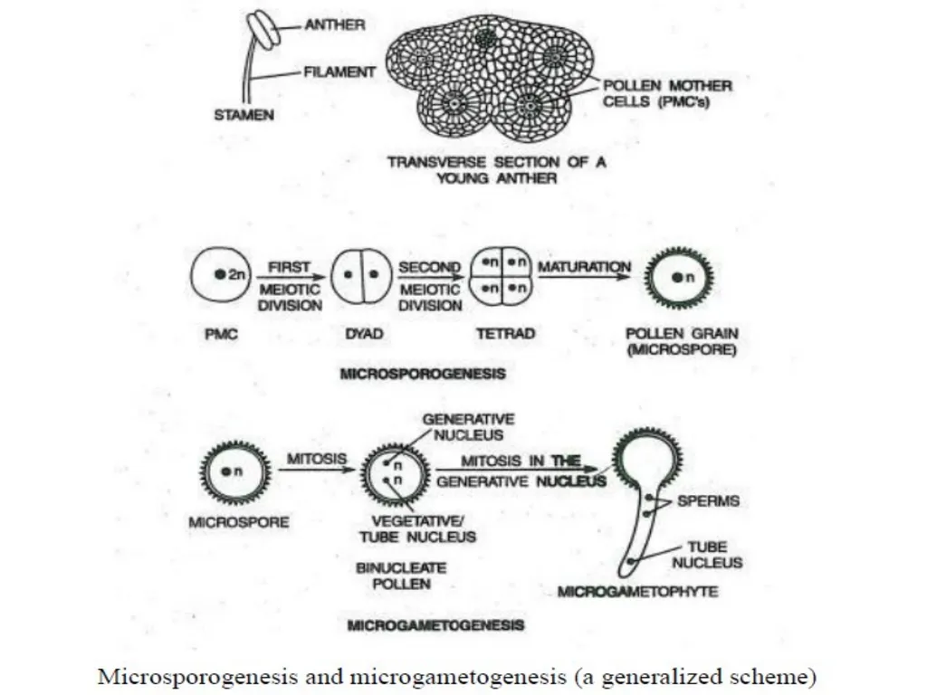
Microsporogenesis: Each anther has four pollen sacs, which contain numerous pollen mother cells (PMCs). Each PMC undergoes meiosis to produce four haploid cells or microspores. This process is known as microsporogenesis (Fig below). The microspores mature into pollen grains mainly by a thickening of their walls.<br>
02
Megasporogenesis: Megasporogenesis occurs in ovules, which are present inside the ovary. A single cell in each ovule differentiates into a megaspore mother cell. The megaspore mother cell undergoes meiosis to produce four haploid megaspores. Three of the megaspores degenerate leaving one functional megaspore per ovule (Fig Below). This completes megasporogenesis.<br>
03
T.S. of anther discloses the following structures:
1. Anther wall:
The fully-fledged anther wall consists of an epidermis accompanied by a layer of endothecium, 2 or 3 middle layers and a single layered tapetum.
2. Epidermis:
It is the outermost layer of anther.
These cells divide frequently by anticlinal division so as to cope up with the rapidly enlarging internal tissue.
Their role is to protect the anther.<br>
04
3. Endothecium:
Normally, the endothecium is single layered and emerges from the parietal cells.
The cells adjacent to endothecium aids in the dehiscence of anther during the course of maturation.
4. Middle layer:
It is 2-3 layered and emerges from parietal cells.
The cells next to endothecium help in the dehiscence of anther and others act as storage centers for starch and other food materials essential for development of pollen.
5. Tapetum:
This is the innermost layer of anther wall and arises from parietal cells. It completely surrounds the sporogenous tissue.
It is made up of single layer of cells, each cell contains dense cytoplasm and obvious nucleus.
It is physiologically vital layer as all the food materials to the sporogenous tissue must pass across it.<br>
05
Sporogenous tissue:
The sporogenous cells may either directly play role as microspore mother cells (or pollen mother cells) or they may go through a few mitoses before entering meiosis.
In a meiotic division, each microspore mother cell yields four haploid microspores. Microspore tetrads represent the group of four microspore.
Cytokinesis takes place either after first meiosis or after second meiosis yielding tetrad.
Mostly all the four spores within a tetrad are totally segregated from one another and form the spores.
Within the callose wall the microspores start to produce their individual wall.
Then the breakdown of the common wall allows the microspores or pollen grains to set free in the anther locule.
Each pollen grain bears haploid nucleus in the pollen cytoplasm surrounded externally by the plasmalemma and pollen wall.
Pollen wall consists of outer uneven exine and inner even intine wall.<br>
06
Micro-gametogenesis:
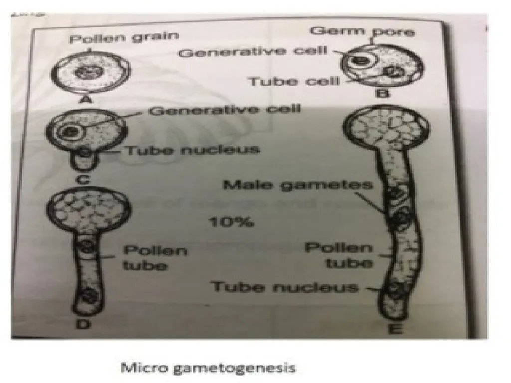
The process of development of male gametes in the microspores or pollen grains is termed as Micro gametogenesis.
Microspores symbolizes the initiation of the male gametophytic generation.
During gametogenesis, the pollen nucleus divides mitotically to yield the unequal cells: The larger one is vegetative cell or tube cell which forms the pollen tube.
The smaller one, situated towards the wall is generative cell which again divides by mitosis to form two sperms (male gametes).<br>
07
After germination of pollen grain, the generative cell may divide inside the pollen grain or in the pollen tube.
At the same time when these alterations are going on, the microspore or pollen grain is also synthesizing its wall. – At one or more loci, the pollen wall is very thin. – These regions are called germpores.
The generative cell divides by a mitosis division.
If this division takes place when they are in anther, the pollens are shed at 3 celled stage and if it takes place after pollen germination, they are shed at 2 celled stage.<br>
08
The exine ruptures and the intine forms a pollen tube.
The vegetative nucleus enters the pollen tube. Now it is known as tube nucleus.
The generative tube nucleus enters the tube and divides to form two generative nuclei which finally form the two male gametes.
The tube nucleus disorganizes.
One of the gamete fuses with the female gamete during fertilization.<br>