SUBJECT : LIFE SCIENCES LEAD TEACHER : SOTSAKA .M DISTRICT : O.R TAMBO INLAND EYE STRUCTURE AND FUNCTION Examination guidelines LESSON OBJECTIVES Study the structure and functions of different parts of the human eye. Explain the structural
"SUBJECT : LIFE SCIENCES LEAD TEACHER : SOTSAKA .M" is the property of its rightful owner. Permission is granted to
download and print the materials on this website for personal, non-commercial use only, and to display it
on your personal computer provided you do not modify the materials and that you retain all copyright
notices contained in the materials. By downloading content from our website, you accept the terms of this
agreement.
Presentation Transcript
01
SUBJECT : LIFE SCIENCES
LEAD TEACHER : SOTSAKA .M
DISTRICT : O.R TAMBO INLAND EYE STRUCTURE AND FUNCTION<br>
02
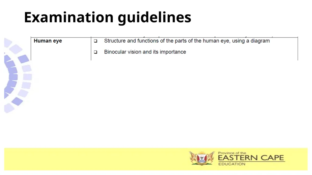
Examination guidelines<br>
03
LESSON OBJECTIVES Study the structure and functions of different parts of the human eye.
Explain the structural adaptations for the various parts to perform their functions.
Explain Binocular vision and stereoscopic vision.<br>
04
Terminology and definitions Refraction: bending of light when light passes through a lens.
Photoreceptors: receptors in the eye that convert light stimuli into nerve impulses.
- Photoreceptors are located on the retina of the eye and are called rod and cone cells.
Binocular vision: the process of looking at an object with both eyes, creating a single visual image.
- Stereoscopic vision: the ability to see the depth and solidity of objects and create a three dimensional presentation of the object.<br>
05
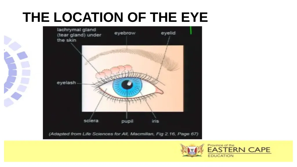
THE LOCATION OF THE EYE<br>
06
THE HUMAN EYE<br>
07
THE STRUCTURE OF THE HUMAN EYE Sclera
Outer covering (the white) of the eye.
Protects the inner parts of the eye
Attachment of the eye muscles. Cornea
Transparent, curved front of the eye.
Refracts the light rays which enter the eye. Aqueous humour
Contains clear fluid that gives shape to the cornea.
Also supply nutrients and oxygen. Iris
Pigmented muscular structure
Consists of inner rings called circular muscles and outer layer of radial muscles
Regulates the amount of light entering the eye (pupil). Lens
Elastic, transparent, biconvex structure.
refracts light and focus it to the retina. Pupil
Opening in Iris allowing light to enter inner eye. Suspensory ligaments
Attaches lens to the ciliary body, helps to alter the shape of the lens Ciliary body and muscle
Enables the lens to change its shape Retina
Inner layer of the eye, contains eye photoreceptors (rods and cones).
Site for which the image is formed.
Helps to convert light stimuli to impulses. Fovea/Yellow spot
Has only cones
Site for clear vision Blind spot
No photoreceptors.
No vision occurs here. Optic nerve
carries impulse from retina to the cerebrum Vitreous Humour
Jelly-like structure, helps to maintain shape of the eyeball.
Plays minor role in refracting light rays.<br>
08
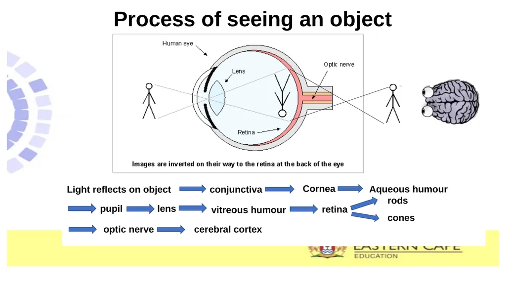
Process of seeing an object Light reflects on object conjunctiva Cornea Aqueous humour pupil lens vitreous humour retina rods cones optic nerve cerebral cortex<br>
09
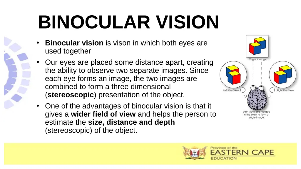
BINOCULAR VISION Binocular vision is vison in which both eyes are used together
Our eyes are placed some distance apart, creating the ability to observe two separate images. Since each eye forms an image, the two images are combined to form a three dimensional (stereoscopic) presentation of the object.
One of the advantages of binocular vision is that it gives a wider field of view and helps the person to estimate the size, distance and depth (stereoscopic) of the object.<br>
10
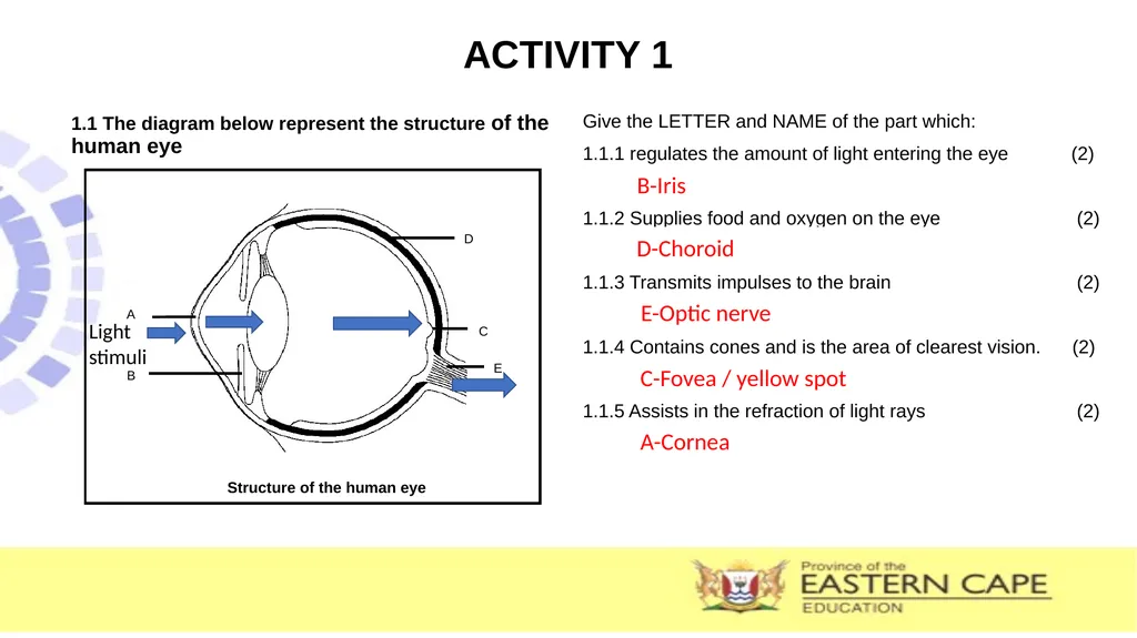
ACTIVITY 1 1.1 The diagram below represent the structure of the human eye Give the LETTER and NAME of the part which:
1.1.1 regulates the amount of light entering the eye (2)
1.1.2 Supplies food and oxygen on the eye (2)
1.1.3 Transmits impulses to the brain (2)
1.1.4 Contains cones and is the area of clearest vision. (2)
1.1.5 Assists in the refraction of light rays (2) Light stimuli B-Iris D-Choroid E-Optic nerve C-Fovea / yellow spot A-Cornea<br>
11
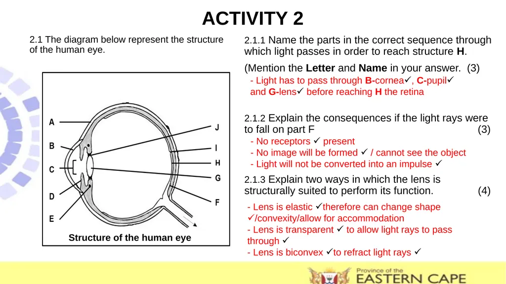
ACTIVITY 2 2.1 The diagram below represent the structure of the human eye. 2.1.1 Name the parts in the correct sequence through which light passes in order to reach structure H.
(Mention the Letter and Name in your answer. (3)
2.1.2 Explain the consequences if the light rays were to fall on part F (3)
2.1.3 Explain two ways in which the lens is structurally suited to perform its function. (4) Structure of the human eye - No receptors present
- No image will be formed / cannot see the object
- Light will not be converted into an impulse - Light has to pass through B-cornea, C-pupil
and G-lens before reaching H the retina - Lens is elastic therefore can change shape /convexity/allow for accommodation
- Lens is transparent to allow light rays to pass through
- Lens is biconvex to refract light rays <br>