Supplement 188: Multi-energy CT Imaging DICOM Working Group 21 Computed Tomography Rationale Multi-energy CT (MECT) uses multiple energies from the X-Ray beam spectrum (conventional CT uses a single (accumulated) X-Ray spectrum). This
"Supplement 188: Multi-energy CT Imaging DICOM" is the property of its rightful owner. Permission is granted to
download and print the materials on this website for personal, non-commercial use only, and to display it
on your personal computer provided you do not modify the materials and that you retain all copyright
notices contained in the materials. By downloading content from our website, you accept the terms of this
agreement.
Presentation Transcript
01
Supplement 188:
Multi-energy CT Imaging DICOM Working Group 21
Computed Tomography<br>
02
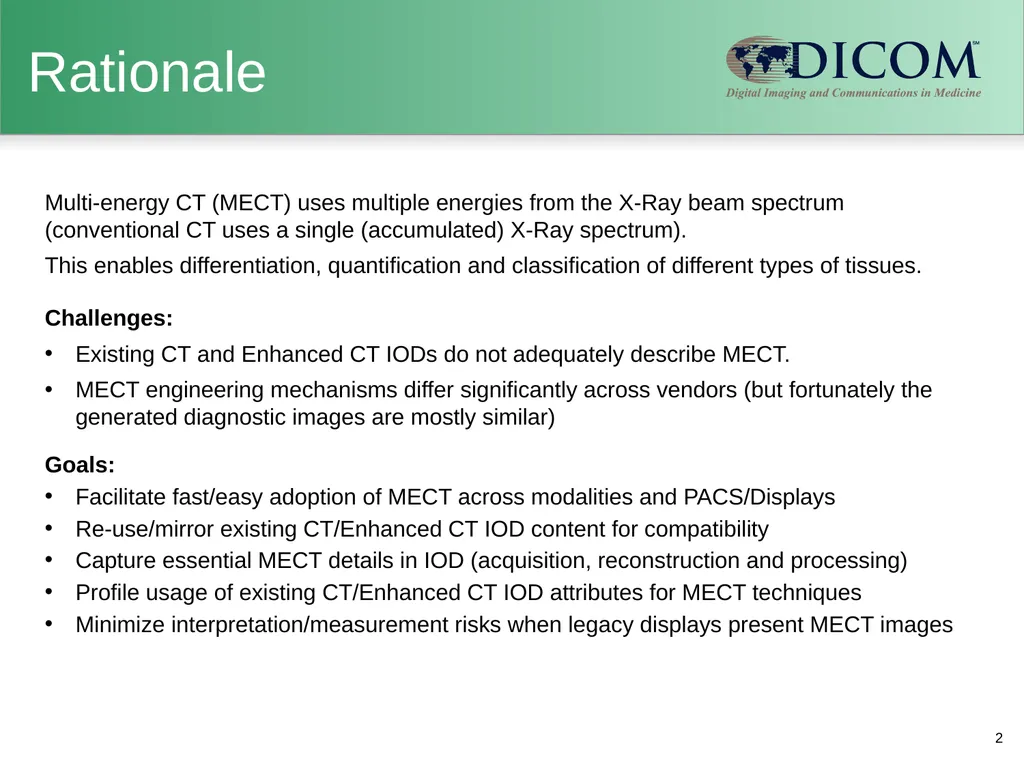
Rationale Multi-energy CT (MECT) uses multiple energies from the X-Ray beam spectrum (conventional CT uses a single (accumulated) X-Ray spectrum).
This enables differentiation, quantification and classification of different types of tissues.
Challenges:
Existing CT and Enhanced CT IODs do not adequately describe MECT.
MECT engineering mechanisms differ significantly across vendors (but fortunately the generated diagnostic images are mostly similar)
Goals:
Facilitate fast/easy adoption of MECT across modalities and PACS/Displays
Re-use/mirror existing CT/Enhanced CT IOD content for compatibility
Capture essential MECT details in IOD (acquisition, reconstruction and processing)
Profile usage of existing CT/Enhanced CT IOD attributes for MECT techniques
Minimize interpretation/measurement risks when legacy displays present MECT images 2<br>
03
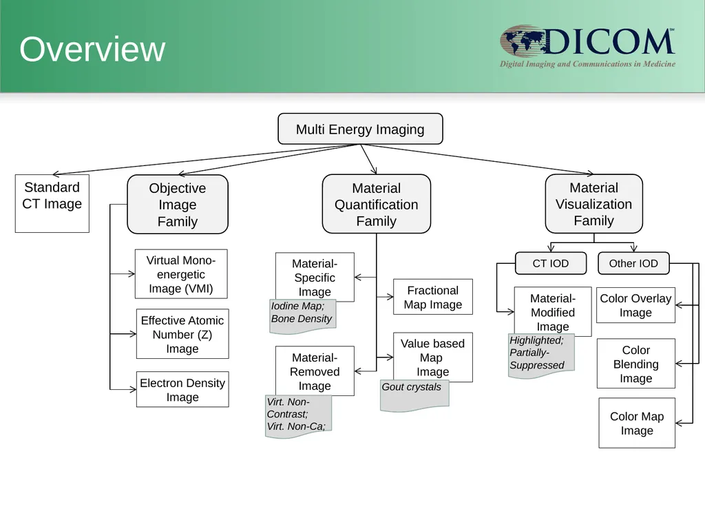
Virtual Mono-energetic Image (VMI) Color Overlay Image Color Blending Image Effective Atomic Number (Z) Image Electron Density Image Color Map Image Multi Energy Imaging Material Quantification
Family Material Visualization
Family Standard CT Image Objective Image
Family Overview Fractional Map Image Value based Map
Image Material-Modified Image Gout crystals Highlighted;
Partially-Suppressed Material-Removed Image Virt. Non-Contrast;
Virt. Non-Ca; CT IOD Other IOD<br>
04
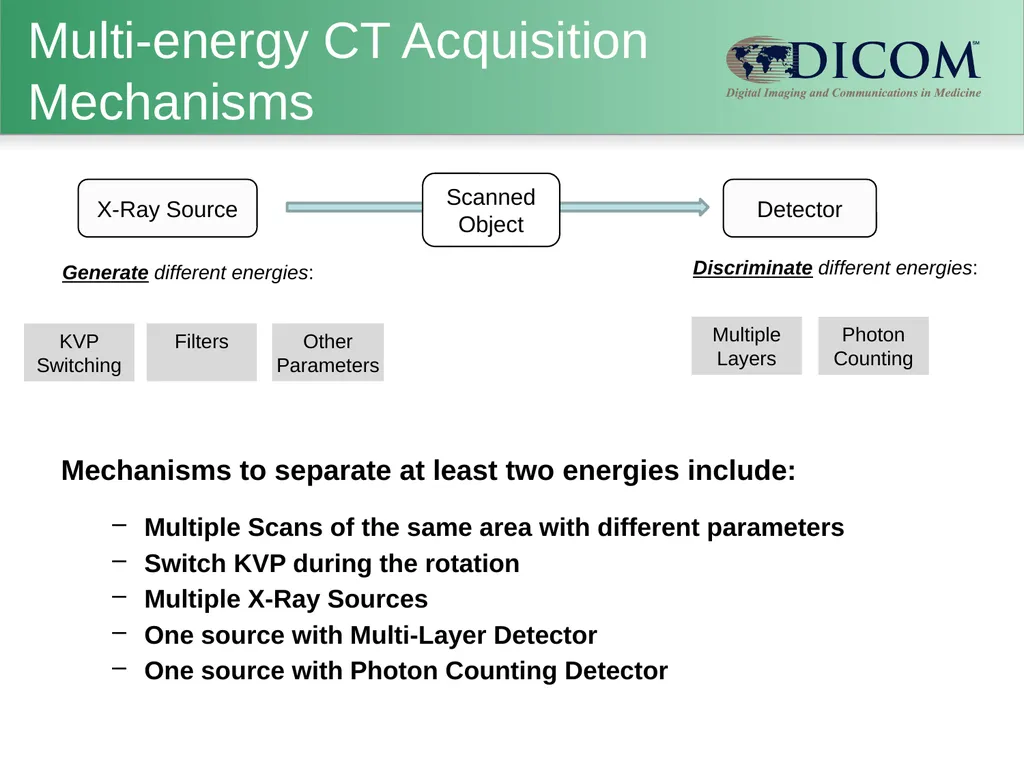
X-Ray Source Generate different energies: KVP Switching Detector Filters Other Parameters Discriminate different energies: Multiple Layers Photon Counting Multi-energy CT Acquisition Mechanisms Mechanisms to separate at least two energies include:
Multiple Scans of the same area with different parameters
Switch KVP during the rotation
Multiple X-Ray Sources
One source with Multi-Layer Detector
One source with Photon Counting Detector Scanned Object<br>
05
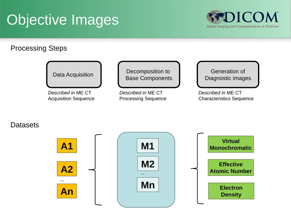
Objective Images Virtual Monochromatic Effective Atomic Number Electron Density Described in ME CT Characteristics Sequence Data Acquisition Decomposition to Base Components Described in ME CT Processing Sequence Described in ME CT Acquisition Sequence A1 A2 M1 M2 Mn … An … Generation of Diagnostic images Datasets Processing Steps<br>
06
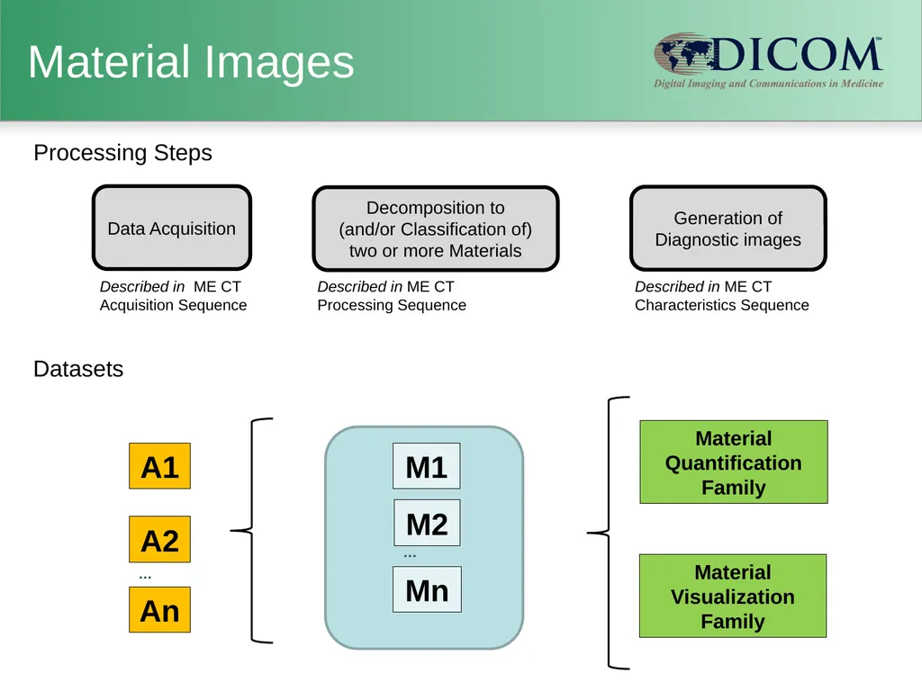
Material Images Data Acquisition A1 A2 Described in ME CT
Processing Sequence Described in ME CT Acquisition Sequence Described in ME CT Characteristics Sequence Material Quantification Family M1 M2 Mn Material Visualization Family Decomposition to
(and/or Classification of)
two or more Materials … An … Generation of Diagnostic images Datasets Processing Steps<br>
07
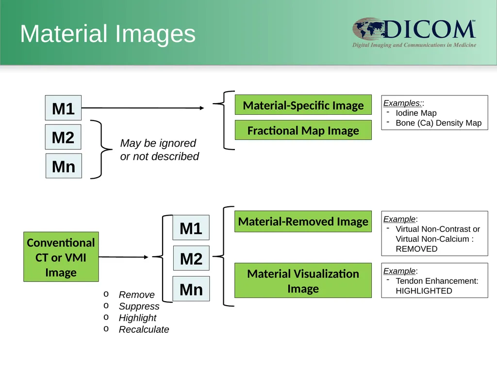
Material Images M1 M2 Material-Specific Image May be ignored or not described M1 M2 Mn Conventional CT or VMI Image Material Visualization Image Remove
Suppress
Highlight
Recalculate Mn Fractional Map Image Material-Removed Image Examples::
Iodine Map
Bone (Ca) Density Map Example:
Virtual Non-Contrast or Virtual Non-Calcium : REMOVED Example:
Tendon Enhancement: HIGHLIGHTED<br>
08
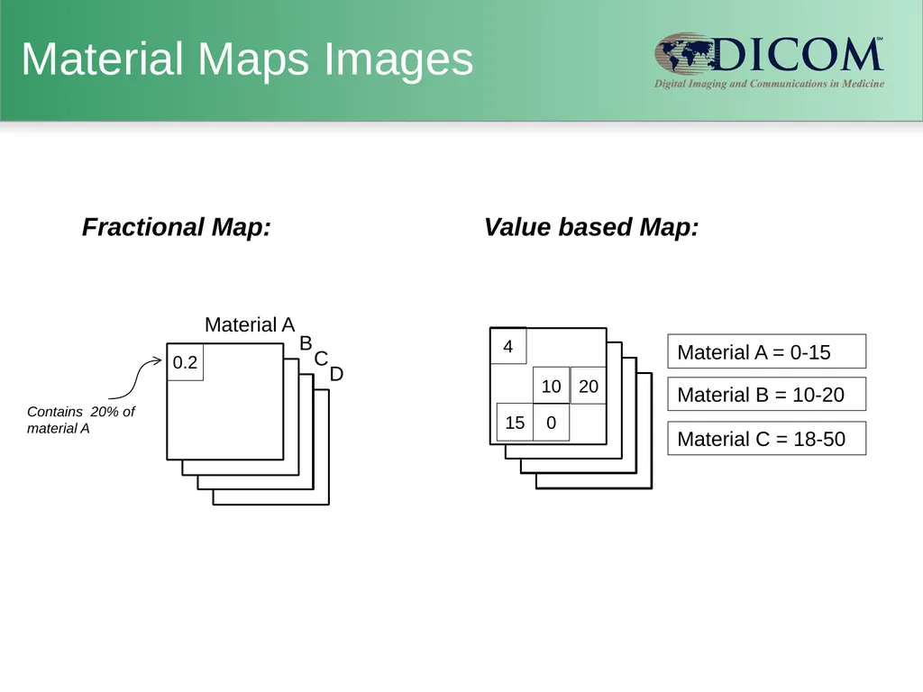
Material Maps Images Material A = 0-15 Material B = 10-20 Material C = 18-50 Contains 20% of material A 0.2 Fractional Map: 4 Value based Map: 20 0 10 15 Material A B C D<br>
09
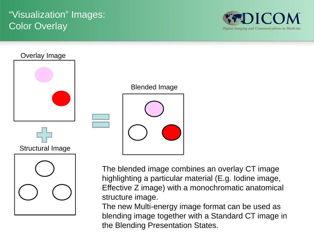
“Visualization” Images: Color Overlay The blended image combines an overlay CT image highlighting a particular material (E.g. Iodine image, Effective Z image) with a monochromatic anatomical structure image.
The new Multi-energy image format can be used as blending image together with a Standard CT image in the Blending Presentation States. Blended Image Structural Image Overlay Image<br>
10
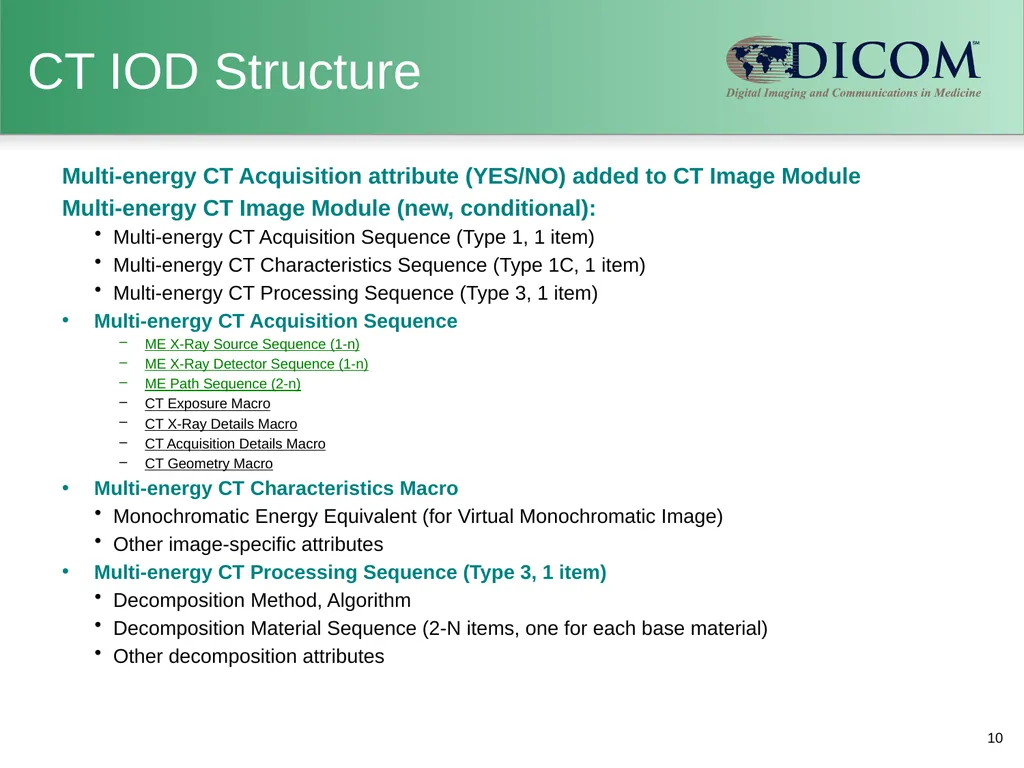
CT IOD Structure Multi-energy CT Acquisition attribute (YES/NO) added to CT Image Module
Multi-energy CT Image Module (new, conditional):
Multi-energy CT Acquisition Sequence (Type 1, 1 item)
Multi-energy CT Characteristics Sequence (Type 1C, 1 item)
Multi-energy CT Processing Sequence (Type 3, 1 item)
Multi-energy CT Acquisition Sequence
ME X-Ray Source Sequence (1-n)
ME X-Ray Detector Sequence (1-n)
ME Path Sequence (2-n)
CT Exposure Macro
CT X-Ray Details Macro
CT Acquisition Details Macro
CT Geometry Macro
Multi-energy CT Characteristics Macro
Monochromatic Energy Equivalent (for Virtual Monochromatic Image)
Other image-specific attributes
Multi-energy CT Processing Sequence (Type 3, 1 item)
Decomposition Method, Algorithm
Decomposition Material Sequence (2-N items, one for each base material)
Other decomposition attributes 10<br>
11
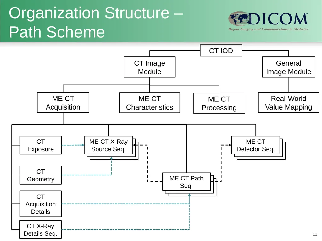
Organization Structure – Path Scheme 11 CT IOD CT Image Module ME CT Acquisition ME CT Characteristics ME CT Processing CT X-Ray Details Seq. CT Exposure CT Acquisition Details CT Geometry General Image Module Real-World Value Mapping<br>