
Surgical Wound Classification (SWC) and Surgical

Description: Surgical Wound Classification (SWC) and Surgical Site Infections (SSI) Emily Dereszkiewicz What is Surgical Wound Classification? Standardized approach to reduce postop infections and help improve patient outcomes Categorizes surgical
Related Topics
Download Presentation
"Surgical Wound Classification (SWC) and Surgical" is the property of its rightful owner. Permission is granted to download and print the materials on this website for personal, non-commercial use only, and to display it on your personal computer provided you do not modify the materials and that you retain all copyright notices contained in the materials. By downloading content from our website, you accept the terms of this agreement.
Presentation Transcript
slide1. Surgical Wound Classification (SWC) and Surgical Site Infections (SSI) Emily Dereszkiewicz<br>
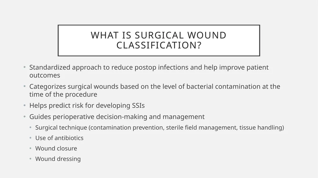
slide2. What is Surgical Wound Classification? Standardized approach to reduce postop infections and help improve patient outcomes
Categorizes surgical wounds based on the level of bacterial contamination at the time of the procedure
Helps predict risk for developing SSIs
Guides perioperative decision-making and management
Surgical technique (contamination prevention, sterile field management, tissue handling)
Use of antibiotics
Wound closure
Wound dressing<br>
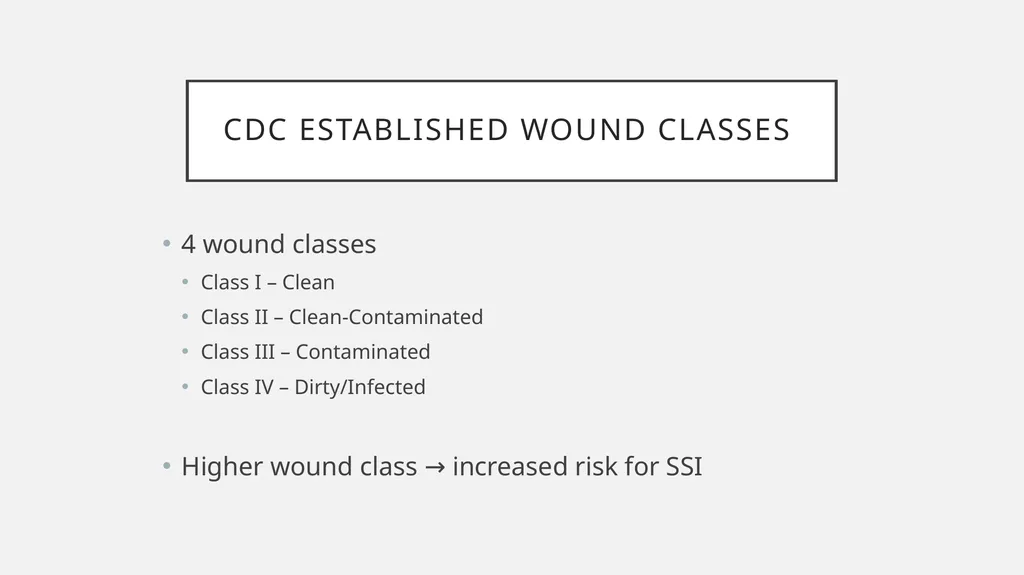
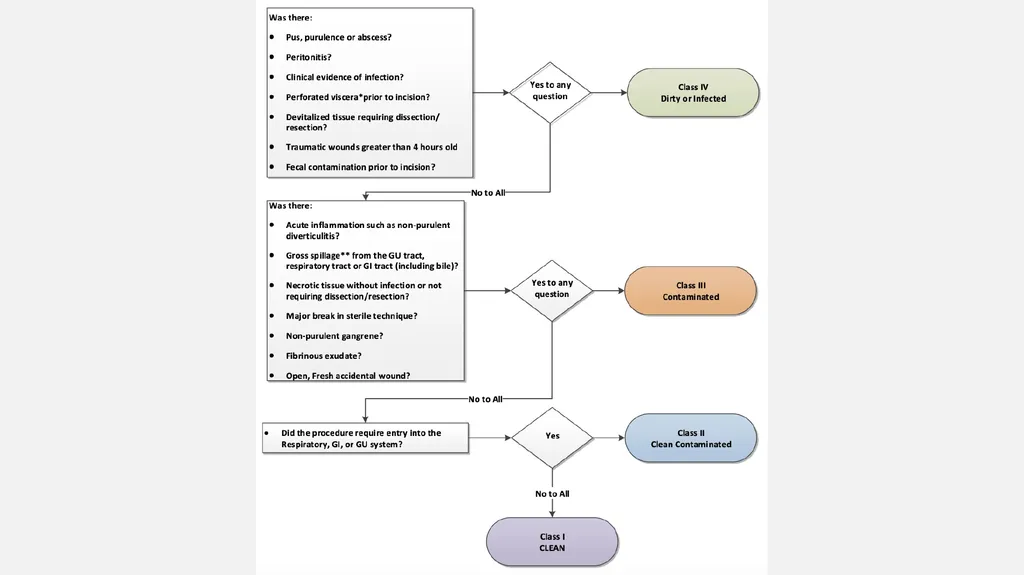
slide3. CDC Established Wound Classes 4 wound classes
Class I – Clean
Class II – Clean-Contaminated
Class III – Contaminated
Class IV – Dirty/Infected
Higher wound class → increased risk for SSI<br>
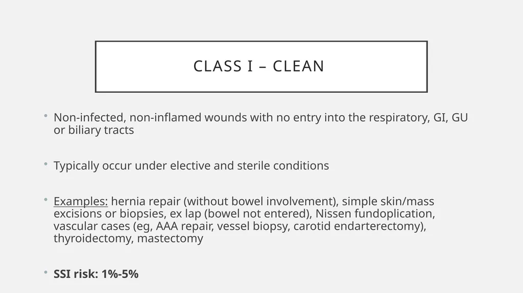
slide4. Class I – Clean Non-infected, non-inflamed wounds with no entry into the respiratory, GI, GU or biliary tracts
Typically occur under elective and sterile conditions
Examples: hernia repair (without bowel involvement), simple skin/mass excisions or biopsies, ex lap (bowel not entered), Nissen fundoplication, vascular cases (eg, AAA repair, vessel biopsy, carotid endarterectomy), thyroidectomy, mastectomy
SSI risk: 1%-5%<br>
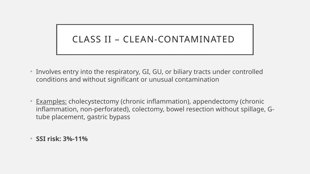
slide5. Class II – Clean-Contaminated Involves entry into the respiratory, GI, GU, or biliary tracts under controlled conditions and without significant or unusual contamination
Examples: cholecystectomy (chronic inflammation), appendectomy (chronic inflammation, non-perforated), colectomy, bowel resection without spillage, G-tube placement, gastric bypass
SSI risk: 3%-11%<br>
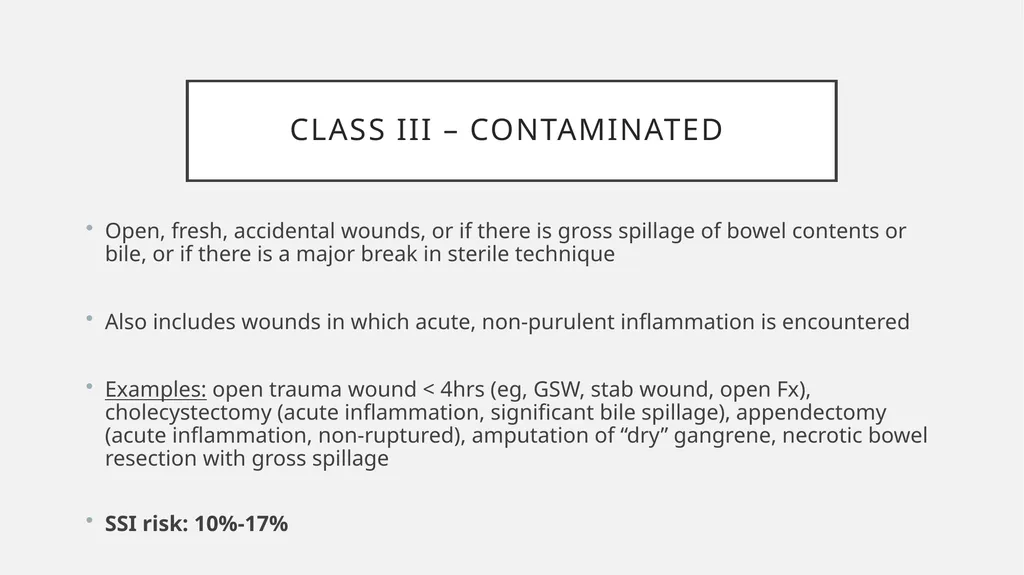
slide6. Class III – Contaminated Open, fresh, accidental wounds, or if there is gross spillage of bowel contents or bile, or if there is a major break in sterile technique
Also includes wounds in which acute, non-purulent inflammation is encountered
Examples: open trauma wound < 4hrs (eg, GSW, stab wound, open Fx), cholecystectomy (acute inflammation, significant bile spillage), appendectomy (acute inflammation, non-ruptured), amputation of “dry” gangrene, necrotic bowel resection with gross spillage
SSI risk: 10%-17%<br>
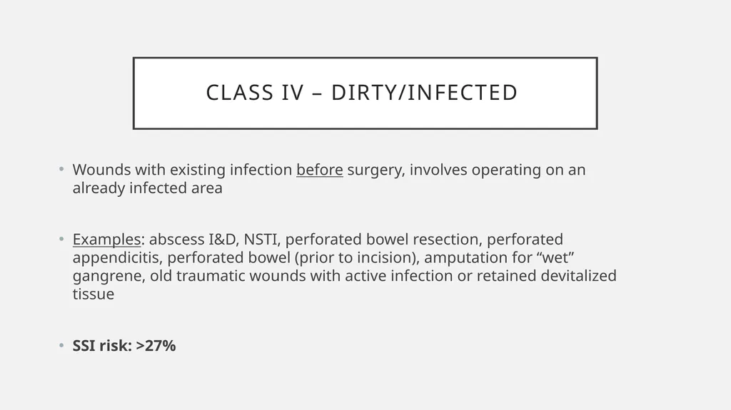
slide7. Class IV – Dirty/Infected Wounds with existing infection before surgery, involves operating on an already infected area
Examples: abscess I&D, NSTI, perforated bowel resection, perforated appendicitis, perforated bowel (prior to incision), amputation for “wet” gangrene, old traumatic wounds with active infection or retained devitalized tissue
SSI risk: >27%<br>
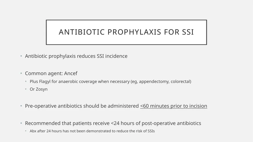
slide9. Antibiotic Prophylaxis for SSI Antibiotic prophylaxis reduces SSI incidence
Common agent: Ancef
Plus Flagyl for anaerobic coverage when necessary (eg, appendectomy, colorectal)
Or Zosyn
Pre-operative antibiotics should be administered <60 minutes prior to incision
Recommended that patients receive <24 hours of post-operative antibiotics
Abx after 24 hours has not been demonstrated to reduce the risk of SSIs<br>
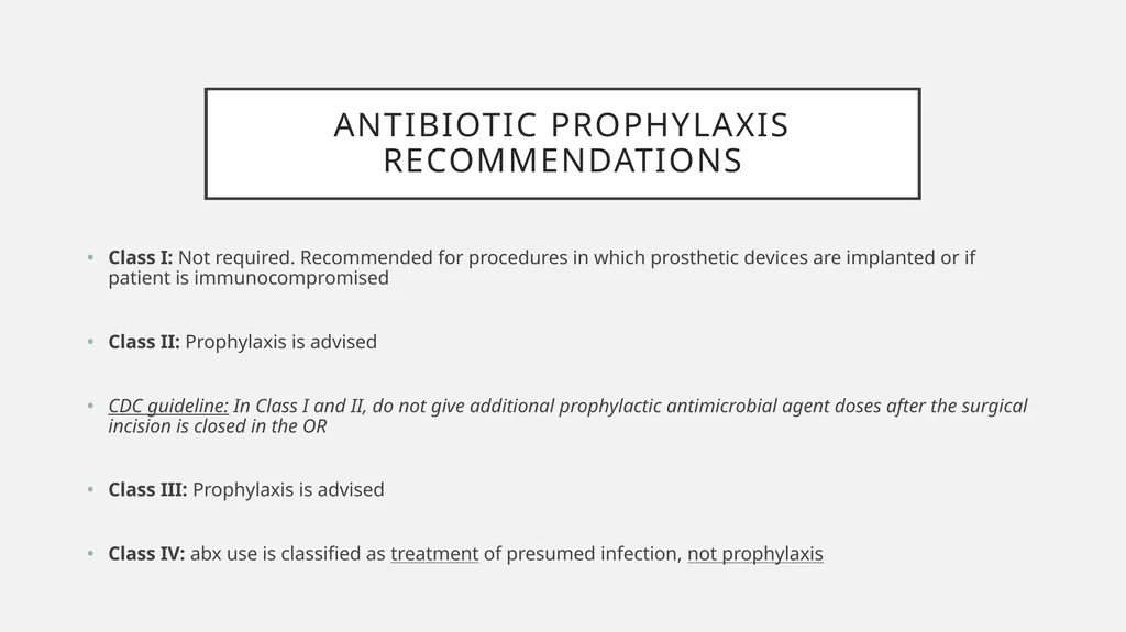
slide10. Antibiotic Prophylaxis Recommendations Class I: Not required. Recommended for procedures in which prosthetic devices are implanted or if patient is immunocompromised
Class II: Prophylaxis is advised
CDC guideline: In Class I and II, do not give additional prophylactic antimicrobial agent doses after the surgical incision is closed in the OR
Class III: Prophylaxis is advised
Class IV: abx use is classified as treatment of presumed infection, not prophylaxis<br>
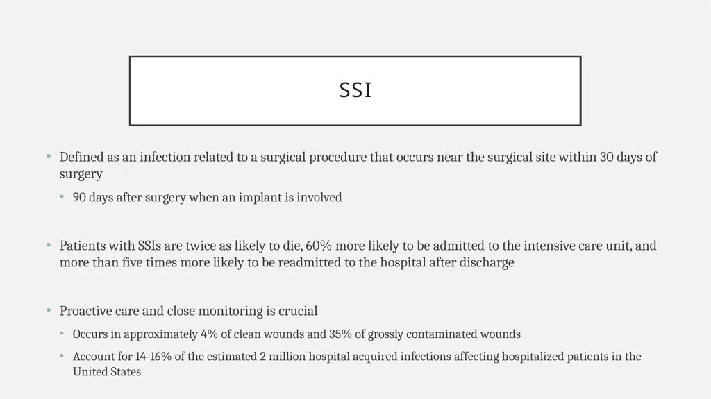
slide11. SSI Defined as an infection related to a surgical procedure that occurs near the surgical site within 30 days of surgery
90 days after surgery when an implant is involved
Patients with SSIs are twice as likely to die, 60% more likely to be admitted to the intensive care unit, and more than five times more likely to be readmitted to the hospital after discharge
Proactive care and close monitoring is crucial
Occurs in approximately 4% of clean wounds and 35% of grossly contaminated wounds
Account for 14-16% of the estimated 2 million hospital acquired infections affecting hospitalized patients in the United States<br>
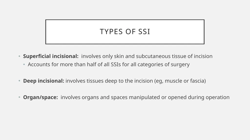
slide12. Types of SSI Superficial incisional: involves only skin and subcutaneous tissue of incision
Accounts for more than half of all SSIs for all categories of surgery
Deep incisional: involves tissues deep to the incision (eg, muscle or fascia)
Organ/space: involves organs and spaces manipulated or opened during operation<br>
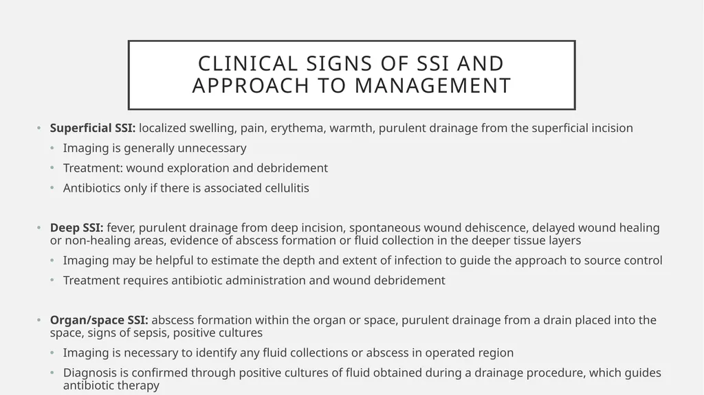
slide13. Clinical Signs of SSI and Approach to Management Superficial SSI: localized swelling, pain, erythema, warmth, purulent drainage from the superficial incision
Imaging is generally unnecessary
Treatment: wound exploration and debridement
Antibiotics only if there is associated cellulitis
Deep SSI: fever, purulent drainage from deep incision, spontaneous wound dehiscence, delayed wound healing or non-healing areas, evidence of abscess formation or fluid collection in the deeper tissue layers
Imaging may be helpful to estimate the depth and extent of infection to guide the approach to source control
Treatment requires antibiotic administration and wound debridement
Organ/space SSI: abscess formation within the organ or space, purulent drainage from a drain placed into the space, signs of sepsis, positive cultures
Imaging is necessary to identify any fluid collections or abscess in operated region
Diagnosis is confirmed through positive cultures of fluid obtained during a drainage procedure, which guides antibiotic therapy<br>
slide2. What is Surgical Wound Classification? Standardized approach to reduce postop infections and help improve patient outcomes
Categorizes surgical wounds based on the level of bacterial contamination at the time of the procedure
Helps predict risk for developing SSIs
Guides perioperative decision-making and management
Surgical technique (contamination prevention, sterile field management, tissue handling)
Use of antibiotics
Wound closure
Wound dressing<br>
slide3. CDC Established Wound Classes 4 wound classes
Class I – Clean
Class II – Clean-Contaminated
Class III – Contaminated
Class IV – Dirty/Infected
Higher wound class → increased risk for SSI<br>
slide4. Class I – Clean Non-infected, non-inflamed wounds with no entry into the respiratory, GI, GU or biliary tracts
Typically occur under elective and sterile conditions
Examples: hernia repair (without bowel involvement), simple skin/mass excisions or biopsies, ex lap (bowel not entered), Nissen fundoplication, vascular cases (eg, AAA repair, vessel biopsy, carotid endarterectomy), thyroidectomy, mastectomy
SSI risk: 1%-5%<br>
slide5. Class II – Clean-Contaminated Involves entry into the respiratory, GI, GU, or biliary tracts under controlled conditions and without significant or unusual contamination
Examples: cholecystectomy (chronic inflammation), appendectomy (chronic inflammation, non-perforated), colectomy, bowel resection without spillage, G-tube placement, gastric bypass
SSI risk: 3%-11%<br>
slide6. Class III – Contaminated Open, fresh, accidental wounds, or if there is gross spillage of bowel contents or bile, or if there is a major break in sterile technique
Also includes wounds in which acute, non-purulent inflammation is encountered
Examples: open trauma wound < 4hrs (eg, GSW, stab wound, open Fx), cholecystectomy (acute inflammation, significant bile spillage), appendectomy (acute inflammation, non-ruptured), amputation of “dry” gangrene, necrotic bowel resection with gross spillage
SSI risk: 10%-17%<br>
slide7. Class IV – Dirty/Infected Wounds with existing infection before surgery, involves operating on an already infected area
Examples: abscess I&D, NSTI, perforated bowel resection, perforated appendicitis, perforated bowel (prior to incision), amputation for “wet” gangrene, old traumatic wounds with active infection or retained devitalized tissue
SSI risk: >27%<br>
slide9. Antibiotic Prophylaxis for SSI Antibiotic prophylaxis reduces SSI incidence
Common agent: Ancef
Plus Flagyl for anaerobic coverage when necessary (eg, appendectomy, colorectal)
Or Zosyn
Pre-operative antibiotics should be administered <60 minutes prior to incision
Recommended that patients receive <24 hours of post-operative antibiotics
Abx after 24 hours has not been demonstrated to reduce the risk of SSIs<br>
slide10. Antibiotic Prophylaxis Recommendations Class I: Not required. Recommended for procedures in which prosthetic devices are implanted or if patient is immunocompromised
Class II: Prophylaxis is advised
CDC guideline: In Class I and II, do not give additional prophylactic antimicrobial agent doses after the surgical incision is closed in the OR
Class III: Prophylaxis is advised
Class IV: abx use is classified as treatment of presumed infection, not prophylaxis<br>
slide11. SSI Defined as an infection related to a surgical procedure that occurs near the surgical site within 30 days of surgery
90 days after surgery when an implant is involved
Patients with SSIs are twice as likely to die, 60% more likely to be admitted to the intensive care unit, and more than five times more likely to be readmitted to the hospital after discharge
Proactive care and close monitoring is crucial
Occurs in approximately 4% of clean wounds and 35% of grossly contaminated wounds
Account for 14-16% of the estimated 2 million hospital acquired infections affecting hospitalized patients in the United States<br>
slide12. Types of SSI Superficial incisional: involves only skin and subcutaneous tissue of incision
Accounts for more than half of all SSIs for all categories of surgery
Deep incisional: involves tissues deep to the incision (eg, muscle or fascia)
Organ/space: involves organs and spaces manipulated or opened during operation<br>
slide13. Clinical Signs of SSI and Approach to Management Superficial SSI: localized swelling, pain, erythema, warmth, purulent drainage from the superficial incision
Imaging is generally unnecessary
Treatment: wound exploration and debridement
Antibiotics only if there is associated cellulitis
Deep SSI: fever, purulent drainage from deep incision, spontaneous wound dehiscence, delayed wound healing or non-healing areas, evidence of abscess formation or fluid collection in the deeper tissue layers
Imaging may be helpful to estimate the depth and extent of infection to guide the approach to source control
Treatment requires antibiotic administration and wound debridement
Organ/space SSI: abscess formation within the organ or space, purulent drainage from a drain placed into the space, signs of sepsis, positive cultures
Imaging is necessary to identify any fluid collections or abscess in operated region
Diagnosis is confirmed through positive cultures of fluid obtained during a drainage procedure, which guides antibiotic therapy<br>