SYSTEMIC CANDIDIASIS Dr. Priyanka Chaturvedi
Description: SYSTEMIC CANDIDIASIS Dr. Priyanka Chaturvedi Morphological classification of fungus Yeast: Cryptococcus Yeast like: Candida Molds: Aspergillus, Mucor, Rhizopus, Rhizomucor Dimorphic: Penicillium, Histoplasma capsulatum SYSTEMIC CANDIDIASIS
Related Topics
Download Presentation
"SYSTEMIC CANDIDIASIS Dr. Priyanka Chaturvedi" is the property of its rightful owner. Permission is granted to download and print the materials on this website for personal, non-commercial use only, and to display it on your personal computer provided you do not modify the materials and that you retain all copyright notices contained in the materials. By downloading content from our website, you accept the terms of this agreement.
Presentation Transcript
slide1. SYSTEMIC CANDIDIASIS Dr. Priyanka Chaturvedi<br>
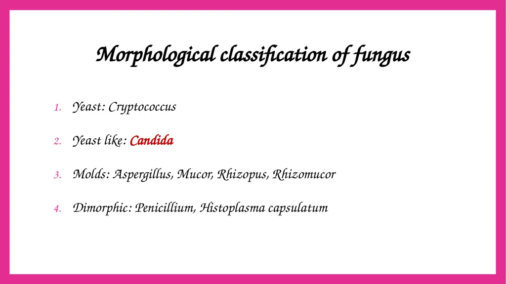
slide2. Morphological classification of fungus Yeast: Cryptococcus
Yeast like: Candida
Molds: Aspergillus, Mucor, Rhizopus, Rhizomucor
Dimorphic: Penicillium, Histoplasma capsulatum<br>
slide3. SYSTEMIC CANDIDIASIS Candidiasis is the most common fungal disease in humans.
Candida affects the skin, mucosa, various internal organs and blood.
Caused by Candida, a yeast like fungus that produces pseudohyphae.<br>
slide4. SYSTEMIC CANDIDIASIS CANDIDA Candida albicans Non-albicans candida
(NAC)<br>
slide5. Non-albicans candida (NAC) Candida tropicalis
Candida auris
Candida parapsilosis
Candida glabrata
Candida krusie
Candida kefyr
Candida dublinensis
Candida gullerimondii
Candida haemulonii<br>
slide6. Pathogenesis It is worldwide in distribution.
Candidiasis accounts for the most common fungal infection in humans, both in HIV and non-HIV infected people.<br>
slide7. Predisposing Factors Physiological state: Extremes of age (infancy, old age), pregnancy
Low immunity: steroid or immunosuppressive drugs, post-transplantation, malignancy, HIV-infected people<br>
slide8. Predisposing Factors Patients on broad spectrum antibiotics
Others: Diabetes mellitus, febrile neutropenia and zinc or iron deficiency<br>
slide9. Virulence Factors Adhesins
Enzymes: Aspartyl proteinases and Serine proteinases ā helps in tissue invasion
Toxins: Glycoprotein extracts of Candida cell wall are pyrogenic<br>
slide10. Virulence Factors Pseudohyphae: Presence of pseudohyphae indicates active infection
Phenotypic switching - Ability to transform between phenotypic forms in the tissue: YEAST AND PSEUDOHYPHAE.<br>
slide11. Clinical Manifestations Invasive candidiasis
Mucosal candidiasis
Cutaneous candidiasis
Allergic candidiasis<br>
slide12. Laboratory diagnosis of Candidiasis Sample collection:
Blood
Sputum
Urine
Endotracheal aspirate
Pus swab<br>
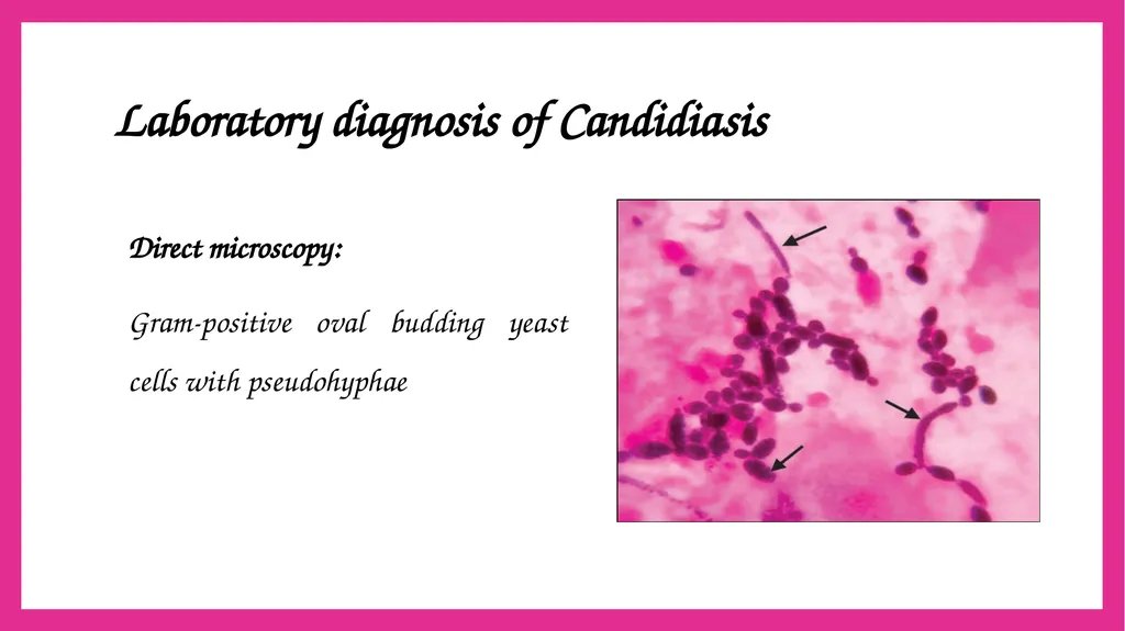
slide13. Laboratory diagnosis of Candidiasis Direct microscopy:
Gram-positive oval budding yeast cells with pseudohyphae<br>
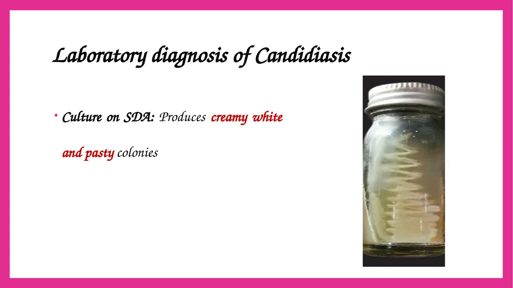
slide14. Laboratory diagnosis of Candidiasis Culture on SDA: Produces creamy white and pasty colonies<br>
slide15. Laboratory diagnosis of Candidiasis Tests for species identification:
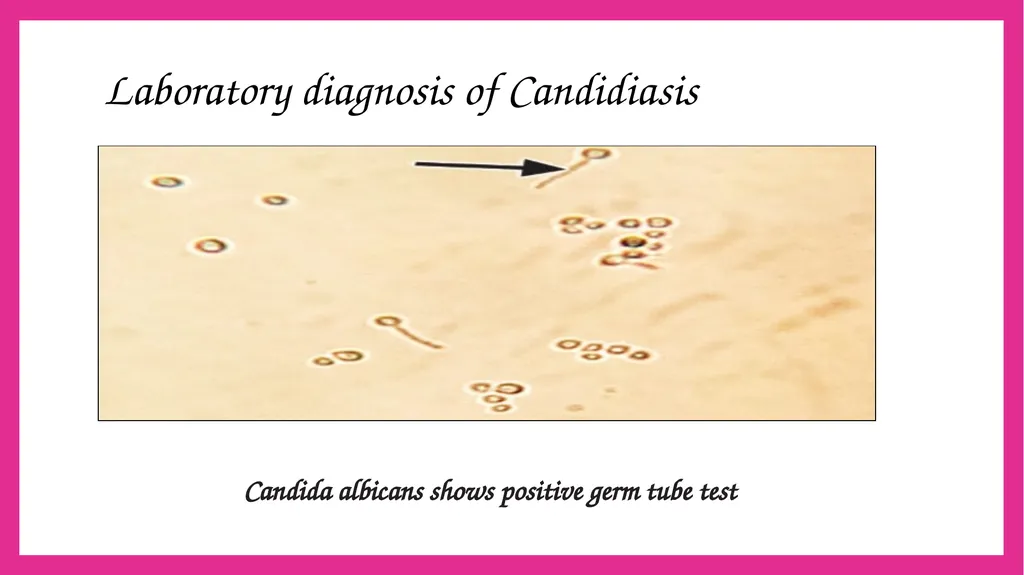
Germ tube test (positive for Candida albicans)
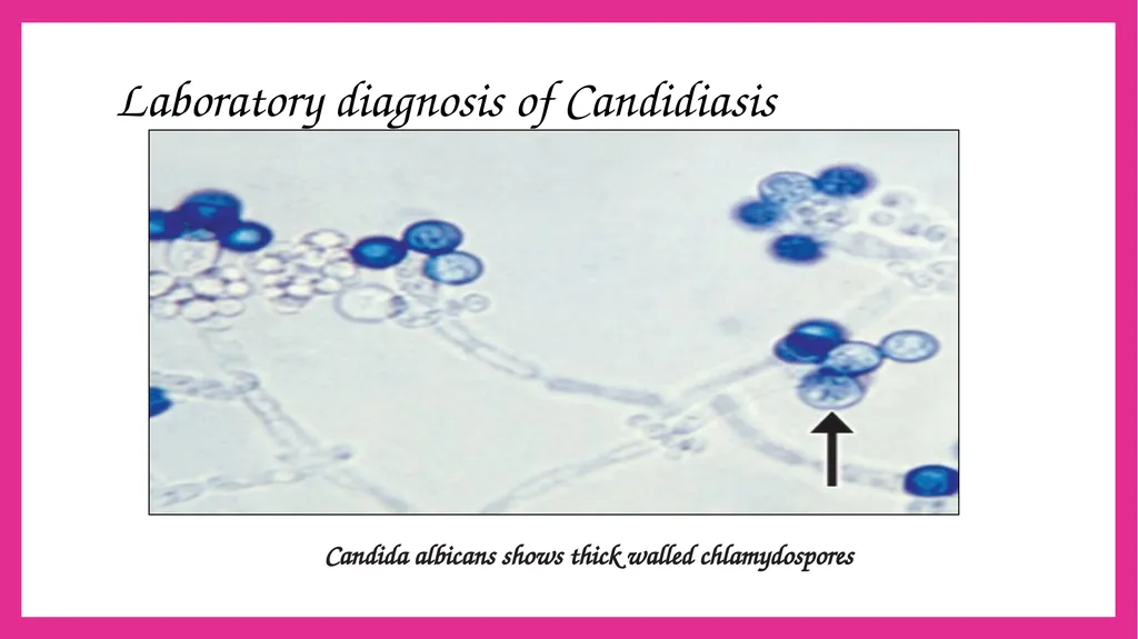
Dalmau plate culture for chlamydospore production
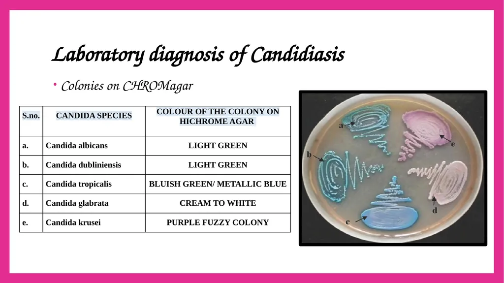
Colonies on CHROMagar<br>
slide16. Laboratory diagnosis of Candidiasis Candida albicans shows positive germ tube test<br>
slide17. Laboratory diagnosis of Candidiasis Candida albicans shows thick walled chlamydospores<br>
slide18. Laboratory diagnosis of Candidiasis Colonies on CHROMagar<br>
slide19. Laboratory diagnosis of Candidiasis Tests for species identification:
Growth at 45°C (positive for C. albicans)
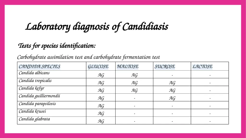
Carbohydrate assimilation test and carbohydrate fermentation test
Molecular methods such as PCR<br>
slide20. Laboratory diagnosis of Candidiasis Tests for species identification:
Carbohydrate assimilation test and carbohydrate fermentation test<br>
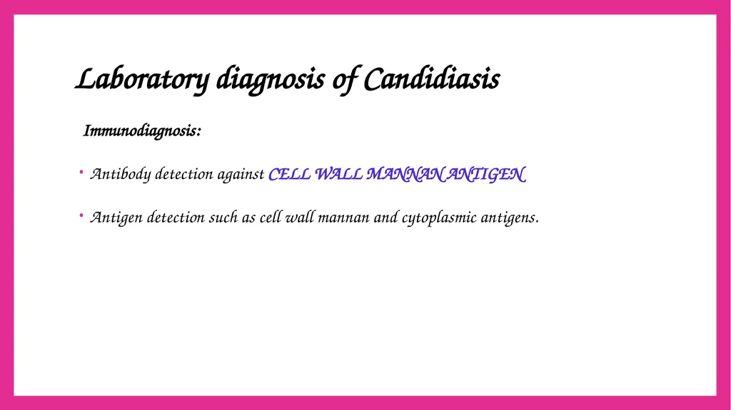
slide21. Laboratory diagnosis of Candidiasis Immunodiagnosis:
Antibody detection against CELL WALL MANNAN ANTIGEN
Antigen detection such as cell wall mannan and cytoplasmic antigens.<br>
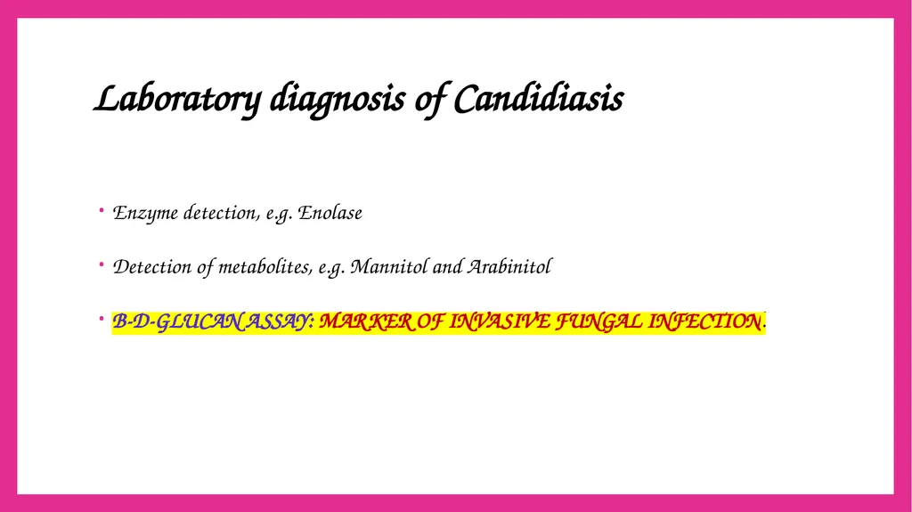
slide22. Laboratory diagnosis of Candidiasis Enzyme detection, e.g. Enolase
Detection of metabolites, e.g. Mannitol and Arabinitol
Ī-D-GLUCAN ASSAY: MARKER OF INVASIVE FUNGAL INFECTION.<br>
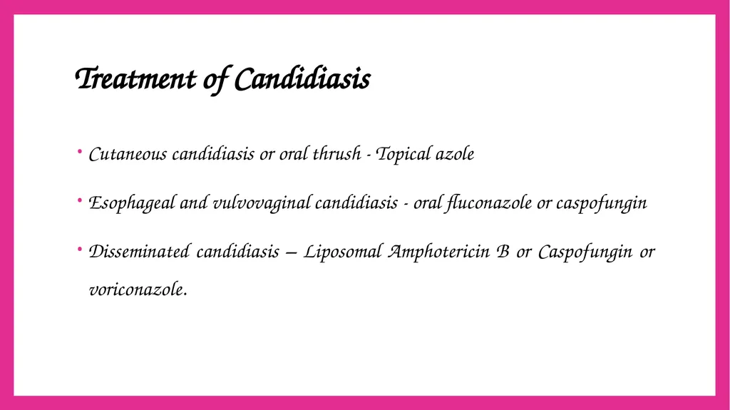
slide23. Treatment of Candidiasis Cutaneous candidiasis or oral thrush - Topical azole
Esophageal and vulvovaginal candidiasis - oral fluconazole or caspofungin
Disseminated candidiasis ā Liposomal Amphotericin B or Caspofungin or voriconazole.<br>
slide24. Treatment of Candidiasis C. glabrata and C. krusei - Intrinsic resistance to azoles and are refractory to treatment with azoles.<br>
slide25. Candida auris Emerging species of Candida, increasingly reported in the recent past.
Manifestations: Cause outbreaks in healthcare settings; produces a wide range of clinical disease from asymptomatic colonization, to candidemia, abscesses, ear infections, etc.<br>
slide26. Candida auris Diagnosis:
Difficult to identify with standard laboratory methods, and it can be misidentified easily as Candida haemulonii
MALDI-TOF are useful for correct identification.<br>
slide27. Candida auris Treatment:
Multidrug resistant candida
Echinocandins are the antifungals given for treatment.<br>

slide28. Thank you<br>
slide2. Morphological classification of fungus Yeast: Cryptococcus
Yeast like: Candida
Molds: Aspergillus, Mucor, Rhizopus, Rhizomucor
Dimorphic: Penicillium, Histoplasma capsulatum<br>
slide3. SYSTEMIC CANDIDIASIS Candidiasis is the most common fungal disease in humans.
Candida affects the skin, mucosa, various internal organs and blood.
Caused by Candida, a yeast like fungus that produces pseudohyphae.<br>
slide4. SYSTEMIC CANDIDIASIS CANDIDA Candida albicans Non-albicans candida
(NAC)<br>
slide5. Non-albicans candida (NAC) Candida tropicalis
Candida auris
Candida parapsilosis
Candida glabrata
Candida krusie
Candida kefyr
Candida dublinensis
Candida gullerimondii
Candida haemulonii<br>
slide6. Pathogenesis It is worldwide in distribution.
Candidiasis accounts for the most common fungal infection in humans, both in HIV and non-HIV infected people.<br>
slide7. Predisposing Factors Physiological state: Extremes of age (infancy, old age), pregnancy
Low immunity: steroid or immunosuppressive drugs, post-transplantation, malignancy, HIV-infected people<br>
slide8. Predisposing Factors Patients on broad spectrum antibiotics
Others: Diabetes mellitus, febrile neutropenia and zinc or iron deficiency<br>
slide9. Virulence Factors Adhesins
Enzymes: Aspartyl proteinases and Serine proteinases ā helps in tissue invasion
Toxins: Glycoprotein extracts of Candida cell wall are pyrogenic<br>
slide10. Virulence Factors Pseudohyphae: Presence of pseudohyphae indicates active infection
Phenotypic switching - Ability to transform between phenotypic forms in the tissue: YEAST AND PSEUDOHYPHAE.<br>
slide11. Clinical Manifestations Invasive candidiasis
Mucosal candidiasis
Cutaneous candidiasis
Allergic candidiasis<br>
slide12. Laboratory diagnosis of Candidiasis Sample collection:
Blood
Sputum
Urine
Endotracheal aspirate
Pus swab<br>
slide13. Laboratory diagnosis of Candidiasis Direct microscopy:
Gram-positive oval budding yeast cells with pseudohyphae<br>
slide14. Laboratory diagnosis of Candidiasis Culture on SDA: Produces creamy white and pasty colonies<br>
slide15. Laboratory diagnosis of Candidiasis Tests for species identification:
Germ tube test (positive for Candida albicans)
Dalmau plate culture for chlamydospore production
Colonies on CHROMagar<br>
slide16. Laboratory diagnosis of Candidiasis Candida albicans shows positive germ tube test<br>
slide17. Laboratory diagnosis of Candidiasis Candida albicans shows thick walled chlamydospores<br>
slide18. Laboratory diagnosis of Candidiasis Colonies on CHROMagar<br>
slide19. Laboratory diagnosis of Candidiasis Tests for species identification:
Growth at 45°C (positive for C. albicans)
Carbohydrate assimilation test and carbohydrate fermentation test
Molecular methods such as PCR<br>
slide20. Laboratory diagnosis of Candidiasis Tests for species identification:
Carbohydrate assimilation test and carbohydrate fermentation test<br>
slide21. Laboratory diagnosis of Candidiasis Immunodiagnosis:
Antibody detection against CELL WALL MANNAN ANTIGEN
Antigen detection such as cell wall mannan and cytoplasmic antigens.<br>
slide22. Laboratory diagnosis of Candidiasis Enzyme detection, e.g. Enolase
Detection of metabolites, e.g. Mannitol and Arabinitol
Ī-D-GLUCAN ASSAY: MARKER OF INVASIVE FUNGAL INFECTION.<br>
slide23. Treatment of Candidiasis Cutaneous candidiasis or oral thrush - Topical azole
Esophageal and vulvovaginal candidiasis - oral fluconazole or caspofungin
Disseminated candidiasis ā Liposomal Amphotericin B or Caspofungin or voriconazole.<br>
slide24. Treatment of Candidiasis C. glabrata and C. krusei - Intrinsic resistance to azoles and are refractory to treatment with azoles.<br>
slide25. Candida auris Emerging species of Candida, increasingly reported in the recent past.
Manifestations: Cause outbreaks in healthcare settings; produces a wide range of clinical disease from asymptomatic colonization, to candidemia, abscesses, ear infections, etc.<br>
slide26. Candida auris Diagnosis:
Difficult to identify with standard laboratory methods, and it can be misidentified easily as Candida haemulonii
MALDI-TOF are useful for correct identification.<br>
slide27. Candida auris Treatment:
Multidrug resistant candida
Echinocandins are the antifungals given for treatment.<br>
slide28. Thank you<br>