04
WALKING CAST Indications
Fractures of the distal radius and tibia, metacarpal/metatarsal bones and phalanges � in large animals.
Comminuted fractures which cannot be treated by other techniques.
In cases where it is necessary to protect the digit from weight bearing.<br>
05
PRINCIPLE Two or three Steinmann pins inserted trans-oseously proximal to the fracture site and connected to a U shaped steel/iron/aluminium frame, allow the patient to rest on the frame and thus relieve the fracture site from direct weight bearing.<br>
06
LOCATION OF PIN PLACEMENT Distal radius and tibial fractures-pins are placed in proximal radius and tibia.
Cannon fractures-pins are placed in the distal radius or tibia.
Phalangeal fractures –pins are placed in the distal metacarpus and metatarsus<br>
07
INSTRUMENTS/MATERIALS REQUIRED Electric /hand drill and key.
Steinmann pins (6-8 mm diameter).
Steel/iron/aluminium frame.
Plaster cast.
Cotton wool & guage.
Traction wire/rope.
Anaesthetic –general/local.<br>
08
POSTOPERATIVE CARE The patient is confined to the stall/rest during the healing period.
The pin sites are watched regularly for osteolytic changes/any purulent secretion. If infection develops, broad spectrum antibiotics are administered.
The cast is removed after the X-ray examination reveals sufficient callus at the fracture site.
After removal of pins the skin and subcutaneous wounds are regularly cleaned and dressed till they are healed.
The healed leg can be temporarily put in a supporting bandage soon after removal of the cast.<br>
09
HANGING PIN CAST Indications :
Fractures of the radius and tibia, specially those at the proximal third of the shaft, which cannot be adequately immobilized by simple cast application as the elbow and stifle joints cannot be included in the cast in large animals.
Principle :
A Steinmann pin inserted transversely through the proximal fracture fragment anchors the full limb cast and thus helps to prevent rotation of the fractured bone and downward slipping of the plaster cast. Unlike with walking cast, with hanging pin cast, the animal bears weight directly on the plastered limb.
The surgical procedure and postoperative care remains the same as with walking cast. Here, generally one or two Steinmann pins are used in the proximal fragment and the metallic frame is not used.<br>
10
Limitations of walking cast/hanging pin cast Generally it is not possible to prevent over ding of fracture fragments incases of oblique/comminuted fractures.
In compound fractures, cast cannot be applied or a window should be provided at the site, which weakens the cast strength.<br>
11
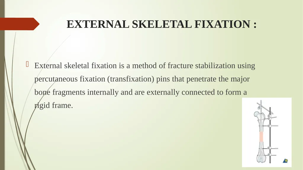
EXTERNAL SKELETAL FIXATION : External skeletal fixation is a method of fracture stabilization using percutaneous fixation (transfixation) pins that penetrate the major bone fragments internally and are externally connected to form a rigid frame.<br>
12
INDICATIONS Open, comminuted or infected fractures of long bones with gross soft tissue damage.
Corrective osteotomy
Limb lengthening procedures
Transarticular application
Mandibular/maxillary fractures
Lumbosacral fractures /luxations<br>
13
ADVANTAGES External skeletal fixation can be used as a closed technique that maximally preserves sources of blood supply to the fracture.
Fracture fixation can be achieved without placing the fixation devices directly through or on the area of injury, which preserves blood supply and increases resistance to infection.
It helps better wound management.
Rigidity of the external fixation device can be adjusted/ dynamized to increase load bearing in late stages.
The technique maintains bone length in the presence of bone defect.
Removal of implant is easy.
The device is reusable.<br>
14
POSTOPERATIVE CARE IN ESF Physical activity is restricted and the animal is confined to reduce the � likelihood of fixator entanglement.
Pin-skin junctions are regularly monitored for evidence of pin tract � sepsis.
The fixator is removed once there is radiographic evidence of � healing.<br>
15
COMPLICATIONS OF ESF Pin tract infection
Loosening of pins
Implant failure
Pressure necrosis of the skin
Soft tissue impalement<br>
16
CONTRAINDICATIONS OF ESF In cases where proper postoperative care of the fixator and animal is not possible.
In cases where more simpler and less- invasive method (external coaptation) would enable successful bone healing.<br>
17
Kirschner-Ehmer (KE) splints : Most commonly used in cats and dogs,
Also used in calves and goats.
Materials required
Steinmann pins/ Krischner wires
Connecting bars.
Clamps<br>
18
TYPES OF K-E SPLINTS The Kirschner splint is available in 3 sizes :
Small for cats and miniature breed dogs,
Medium-for most canines,
Large-for giant breed dogs and
Extra large-for calves and foals.<br>
19
CONT. Available in 2 basic frame configurations :
UNILATERAL- have one or more rods connected to 2 or more clamps that are attached to half pins (a half pin penetrates near cutaneous surface and both the near and far cortices of the bone)
BILATERAL – have a connecting rod on both sides of the limb (medial and lateral), connected to 2 or more full pins that transfix the bone (a full pin penetrates both cortices of the bone and the both near and far cutaneous surfaces of the limb).<br>
20
INDICATIONS Unilateral, uniplanar type is generally used for primary fixation of simple fractures and as a supplement to basic internal fixation methods,
Unilateral, biplanar type is useful for stabilizing fractures of the femur and humerus (more resistant to bending and shear forces than uniplanar type).
Bilateral type (uniplanar/biplanar) provides increased resistance to compressive and rotational forces, and are most commonly applied to fractures of the radius or tibia and sometimes are used to immobilize joints like elbow, carpus, stifle and hock.
The basic principle of fixation apply and the aim is to have the pins engage a minimum of 4 cortices above and below the fracture site.<br>
21
The technique of application of a 4 pin unilateral type –I fixator The surface to which the external fixator is applied varies from bone to bone :
Humerus –craniolateral,
Radius craniomedial,
Femur-lateral,
Tibia-medial.<br>
22
TRANSFIXATION PINNING : Indications :
Compound/comminuted frctures of tibia, radius, metatarsus and metacarpus, which are not generally amenable to cast immobilization in large animals.
Materials required :
Steinmann I M pins/Krischner wires
Side bars (stainless steel, aluminium or acrylic)<br>