/
Central Metatarsal Fractures Central Metatarsal Fractures

Central Metatarsal Fractures - PowerPoint Presentation

stefany-barnette
stefany-barnette . @stefany-barnette
Follow
424 views
Uploaded On 2016-05-30

Central Metatarsal Fractures - PPT Presentation

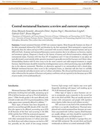
Dan Preece DPM PGY1 Stress Fx Frequency Distribution second metatarsal 52 third metatarsal 35 first metatarsal 8 fourth and fifth metatarsals 5 shorter first metatarsal of Mortons foot ID: 341529

stress fractures fixation metatarsal fractures stress metatarsal fixation foot fracture med bone ankle sports options fxs injuries cases injury

Share:

Link:

Embed:

Download Presentation from below link

Download Presentation The PPT/PDF document "Central Metatarsal Fractures" is the property of its rightful owner. Permission is granted to download and print the materials on this web site for personal, non-commercial use only, and to display it on your personal computer provided you do not modify the materials and that you retain all copyright notices contained in the materials. By downloading content from our website, you accept the terms of this agreement.


Presentation Transcript

Slide1

Central Metatarsal Fractures

Dan Preece DPM, PGY-1Slide2

Stress

Fx

Frequency /Distribution: -second metatarsal 52%-third metatarsal 35%-first metatarsal 8%-fourth and fifth metatarsals 5%**shorter first metatarsal of Morton's foot leads to a higher risk of second metatarsal stress fractures

2: Leabhart J: Stress fractures of the calcaneus. J Bone Joint Surg Am 41:1285-1290, 1959.

1: Drez D Jr, Young J, Johnston R, Parker W: Metatarsal stress fractures. Am J Sports Med 8:123-125, 1980Slide3

Factors that can

increase the risk

of fractures (and stress fractures:-cavus foot conformation-long second metatarsal (e.g. Morton's foot)-metatarsus adductus-amenorrhea (hypoestrogenism)-hyperthyroidism-

osteoporosis -medications -tobacco -alcohol abuse -nutritional problems-anemic disorders

-training errors-poor footwear-improper athletic technique 3: Daffner R, Pavlov H: Stress fractures: Current concepts.

Am J Radiol

159:245-252, 1992.Slide4

Etiology and Description of Stress Fractures:

-

Microfractures  Stress Reactions  Cortical Fracture.- Stress fxs occur by cyclic loading that does not exceed the ultimate breaking limits of plastic deformation of bone.

- One possible cause of stress fracture is muscle fatigue, which decreases shock-absorbing capacity of the extremity.- Repetitive force exerted by a muscle on bone

3: Daffner R, Pavlov H: Stress fractures: Current concepts. Am J Radiol 159:245-252, 1992.Slide5

Biomechanical Causes of Metatarsal Stress

Fxs

: Dorsal strains are significantly reduced by contraction of the plantar flexory musculature.- Fatigue of these muscles during strenuous or prolonged running may result in decreased dissipation of forces by the musculature and increased exposure of the metatarsals to stress.

4: Brukner P, Bradshaw C, Khan KM, et al. Stress fractures: a review of 180 cases. Clin J Sport Med. 1996;6(2):85–9.Slide6

Clinical

Presentation of Stress

Fxs

:

- Metatarsal stress

f

xs

have a much

faster

onset than similar injuries of the tibia and fibula.

- The average time to presentation ranges from 2 to 6 weeks

- C

an occur with a single training event or military exercise.

- Pain to direct palpation and through ROM, also pain, redness, swelling present.

5: Milgrom C, Finestone A, Sharkey N, et al: Metatarsal strains are sufficient to cause fracture during cyclic loading. Foot Ankle Int 23(3):230-235, 2002Slide7

Stress Fractures:

- Imaging: -Plain films may not show changes for > 10 days. Changes may be evident in only 30% to 70% of cases. -Bone Scan: 99Tc bone scan is extremely sensitive, and uptake may

be evident within 24 hours of injury (not specific however) . -MRI is highly sensitive and specific, particularly to

identifying location. -CT is helpful to define a fx line

and

to determine whether the

fx

is

complete

or incomplete.

6: Matheson

G, Clements D, McKenzie D, et al: Stress fractures in athletes: A study of 320 cases.

Am J Sports Med

15:46-58, 1987.Slide8

-Forty-one feet were analyzed with US and dedicated MRI from 37 patients.

-MRI detected 13 fractures in 12 patients.

-US was 83% sensitive, 76% specific. Positive predictive value 59%, and negative predictive value 92%. -In cases of normal radiographs, US is indicated in the diagnosis of metatarsal bone stress fractures, as it is a low cost, noninvasive, rapid, and easy technique with good sensitivity and specificity. US in Dx of Stress Fractures:

7: F Banal, F

Etchepare, B Rouhier, C Rosenberg, V Foltz, S

Rozenberg

. Ultrasound ability in early diagnosis of stress fracture of metatarsal bone. Ann Rheum Dis. 2006 July; 65(7): 977–978. Slide9

Stress

Fx

Treatment: -Activity restriction 4-8+ weeks.-Immobilization: depends on the duration of symptoms before the patient presenting for treatment. Longer duration: more severe injury.Serial radiography used to document bony

union.Correct contributing factors : training techniques and footwear.

Return to activity is allowed when radiographic healing is noted and tenderness has completely resolved. Recurrent stress fractures are uncommon in the absence of metabolic or endocrine

disorders

, and they rarely recur at the same site

8: K

, Hahn S, Chung M, et al: A clinical study of stress fractures in sports activities.

Orthopedics

15:1089-1095, 1991.Slide10

Metatarsal

Fx’s

:-Frequency: 5th  3rd  2nd  1st  4

th (different frequency than stress fx’s)

-MCC: -Direct force: crushing, blunt trauma, penetrating wounds. -Indirect force: twisting injury (forepart of the foot is fixed as the pt turns)Who? Most commonly: athletes, diabetics

(worse

with

longer

duration), men.

Diabetic

neuropathy

:

has been associated

with

osteopenia

in both hands and feet as well as metatarsal fxs.10: DeLee JC, Evans JP, Julian J. Stress fracture of the fifth metatarsal. Am J Sports Med 1983;11:349-353.9: Sammarco GJ: The Jones fracture. Instr Course Lect 42:201-205, 1993.16: Cundy, TF, Edmonds, ME, Watkins, PJ. Osteopenia and metatarsal fractures in diabetic neuropathy. Diabet Med 1985; 2:461.Slide11

A 6-month study showed that metatarsal

fractures accounted for (majority are 1st and 5th met fx): 35% of foot fractures, 6% of foot injuries, 5% of skeletal fractures,

0.2% of emergency department visits.11: SPECTOR FC, KARLIN JM, SCURRAN BL, ET AL: Lesser metatarsal fractures: incidence, management, and review. JAPA

74: 259, 1984.Slide12

Unstable Foot Types Leads to Stress Overload:

hypermobile

1

st

and/or 5th rays causes overload of the lesser mets.

GRF

GRFSlide13

Surgical

Approach:

consider the complex soft tissue anatomy that surrounds, inserts or originates from each of the central metatarsals.Slide14

-Fractures of the proximal shaft and base must be evaluated for ligamentous disruption.

- Manipulation under anesthesia while monitoring with fluoroscopy is recommended.Slide15

Fracture Treatment Options:

Non-dislocated

fractures w/o ligament damage of the second, third, and fourth metatarsal bases rarely need treatment other than solid-soled shoe or, if painful, a cast.Met fxs are often held in good alignment by surrounding ligamentous structures, with exception of met neck and shaft fractures that displace easily.3 to 4 mm of medial or lateral transverse plane deformity and 10° or less of angulation are well tolerated and need not undergo

surgical corrective measures.Healing time may be as much as 3 months from injury. Weight bearing is advanced as tolerated.

12:

Armagan

 OE, Shereff MJ. Injuries to the toes and metatarsal. Foot Ankle Trauma. 2001;32:1–10. Slide16

Fracture Tx Options:

-ORIF is

indicated in metatarsal fractures that are irreducible, involve a joint, or are significantly displaced.Fixation options: -crossed K-wires, -percutaneous pinning,

-circlage wire, -interfrag screw, -plate and screw fixation, -external fixation, -intramedullary

fixation using a Steinmann pin or double- threaded compression screw .

13:Rammelt S,

Heineck

 J,

Zwipp

 H. Metatarsal fractures. Injury. 2004;35:S-B77–S-B86.

14:

Pendarvis

 JA,

Mandracchia

 VJ, Haverstock BD, et al. A new fixation technique for metatarsal fractures. Clin Podiatr Med Surg. 1999;16:643–657Slide17

Evidence for which type of fixation is best in lesser met fractures:

-None

-Didley Squat -Zilch*Best evidence available is “author’s experience”. *Most authors recommend k-wire/steinman pins or other types of intramed fixation.Slide18

Evidence from osteotomies similar to met fractures:

-40 bone models were divided equally into 4 groups: a control group consisting of intact lesser rays; and Weil osteotomies that were fixated with 2

crossed Kirschner wires (0.045-in K-wires), 2.0-mm cortical screws, or cannulated 2.4-mm cortical screws. Result: There was no statistical difference in structural stiffness among the 3 groups of fixation methods.20: Craig T. Jex

, DPM,1 Chanda J. Wan, DPM,2 Steve Rundell, MS. Analysis of Three Types of Fixation of the Weil Osteotomy. The Journal of Foot & Ankle Surgery 45(1):13–19, 2006.Slide19

K-Wire Pinning Technique: (same idea as hammertoe fixation)Slide20

K-Wire Fixation:Slide21

Fixation Options: k-wires Slide22

Comminuted

Fxs

:-Kirschner wire for provisional stability. -Application of mini external fixation devices to the fourth and fifth metatarsal fractures.15: I,

Mosheiff R, Zelgowski A, et al. Crush injuries of the foot with compartment syndrome: immediate one-stage management. Foot Ankle. 1989;9(4):185–189. Slide23

IM Rod FixationSlide24

Fixation Options:Slide25

Fixation Options: plate and screws.Slide26

Fixation Options: k-wires, plate and screws.Slide27

Complications to be aware of:

Significant shortening or angular deviation of mets

-may result in transfer lesions/pressure points/new stress fx Early weight bearing with unstable fixationnon unions

Pin tract infection vs irritation, hematoma, seroma etc.Slide28

References:

1:

Drez D Jr, Young J, Johnston R, Parker W: Metatarsal stress fractures. Am J Sports Med 8:123-125, 1980.2: Leabhart

J: Stress fractures of the calcaneus. J Bone Joint Surg Am 41:1285-1290, 1959.3: Daffner R, Pavlov H: Stress fractures: Current concepts.

Am J Radiol 159:245-252, 1992.4: Brukner P, Bradshaw C, Khan KM, et al. Stress fractures: a review of 180 cases. Clin J Sport Med. 1996;6(2):85–9.

5:

Milgrom

C,

Finestone

A, Sharkey N, et al: Metatarsal strains are sufficient to cause fracture during cyclic loading.

Foot Ankle Int

23(3):230-235, 2002

6: Matheson G, Clements D, McKenzie D, et al: Stress fractures in athletes: A study of 320 cases.

Am J Sports Med

15:46-58, 1987.

7: F Banal, F Etchepare, B Rouhier, C Rosenberg, V Foltz, S Rozenberg. Ultrasound ability in early diagnosis of stress fracture of metatarsal bone. Ann Rheum Dis. 2006 July; 65(7): 977–978. 8: Ha K, Hahn S, Chung M, et al: A clinical study of stress fractures in sports activities. Orthopedics 15:1089-1095, 1991.9: Sammarco GJ: The Jones fracture. Instr Course Lect 42:201-205, 1993.10: DeLee JC, Evans JP, Julian J. Stress fracture of the fifth metatarsal. Am J Sports Med 1983;11:349-353.11: Spector FC, Karlin JM, Scurran BL, et al: Lesser metatarsal fractures: incidence, management, and review. JAPA 74: 259, 1984.12: Armagan OE, Shereff MJ. Injuries to the toes and metatarsal. Foot Ankle Trauma. 2001;32:1–1013: Rammelt S, Heineck J, Zwipp H. Metatarsal fractures. Injury. 2004;35:S-B77–S-B86.14: Pendarvis

 JA, Mandracchia VJ, Haverstock BD, et al. A new fixation technique for metatarsal fractures. Clin Podiatr Med Surg. 1999;16:643–65715: I, Mosheiff R, Zelgowski A, et al. Crush injuries of the foot with compartment syndrome: immediate one-stage management. Foot Ankle. 1989;9(4):185–189.

16:

Cundy

, TF, Edmonds, ME, Watkins, PJ.

Osteopenia

and metatarsal fractures in diabetic neuropathy.

Diabet Med

1985; 2:461.

17: Wolf, SK. Diabetes mellitus and predisposition to athletic pedal fracture.

J Foot Ankle Surg

1998; 37:16.

18: S Papp, R Sanders. Fractures of the Midfoot and Forefoot.

Surgery of the Foot and Ankle

. 8

th

edition. Ch 41 pg 2215.

19: Craig T.

Jex

, DPM,1

Chanda

J. Wan, DPM,2 Steve

Rundell

, MS. Analysis of Three Types of Fixation of the Weil Osteotomy..

JFAS

45(1):13–19, 2006.