
THE MICROSCOPE A Practical Guide Microscopes used
PC
Published · 22 slides · 0 views
1 / 1


Description
THE MICROSCOPE A Practical Guide Microscopes used in clinical practice are light microscopes. They are called light microscopes because they use a beam of light to view specimens. A compound light microscope is the most common microscope
Related Topics
Share
Embed code
Download this presentation From Below
"THE MICROSCOPE A Practical Guide Microscopes used" is the property of its rightful owner. Permission is granted to download and print the materials on this website for personal, non-commercial use only, and to display it on your personal computer provided you do not modify the materials and that you retain all copyright notices contained in the materials. By downloading content from our website, you accept the terms of this agreement.
Presentation Transcript
01
THE MICROSCOPE
A Practical Guide<br>
02
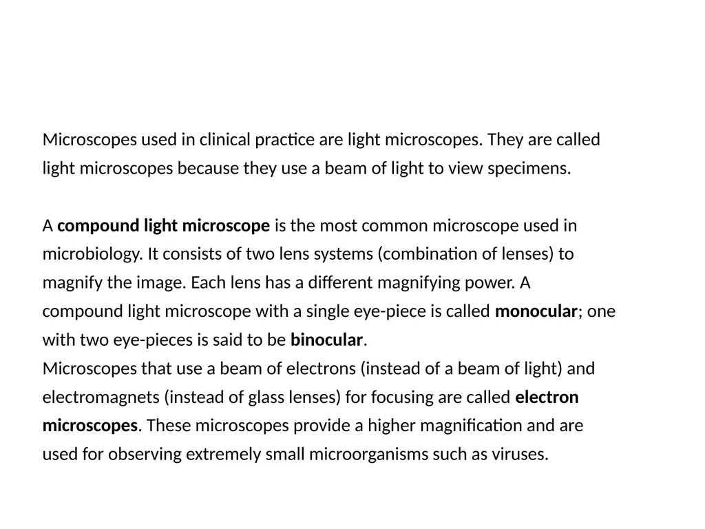
Microscopes used in clinical practice are light microscopes. They are called
light microscopes because they use a beam of light to view specimens.
A compound light microscope is the most common microscope used in
microbiology. It consists of two lens systems (combination of lenses) to
magnify the image. Each lens has a different magnifying power. A
compound light microscope with a single eye-piece is called monocular; one
with two eye-pieces is said to be binocular.
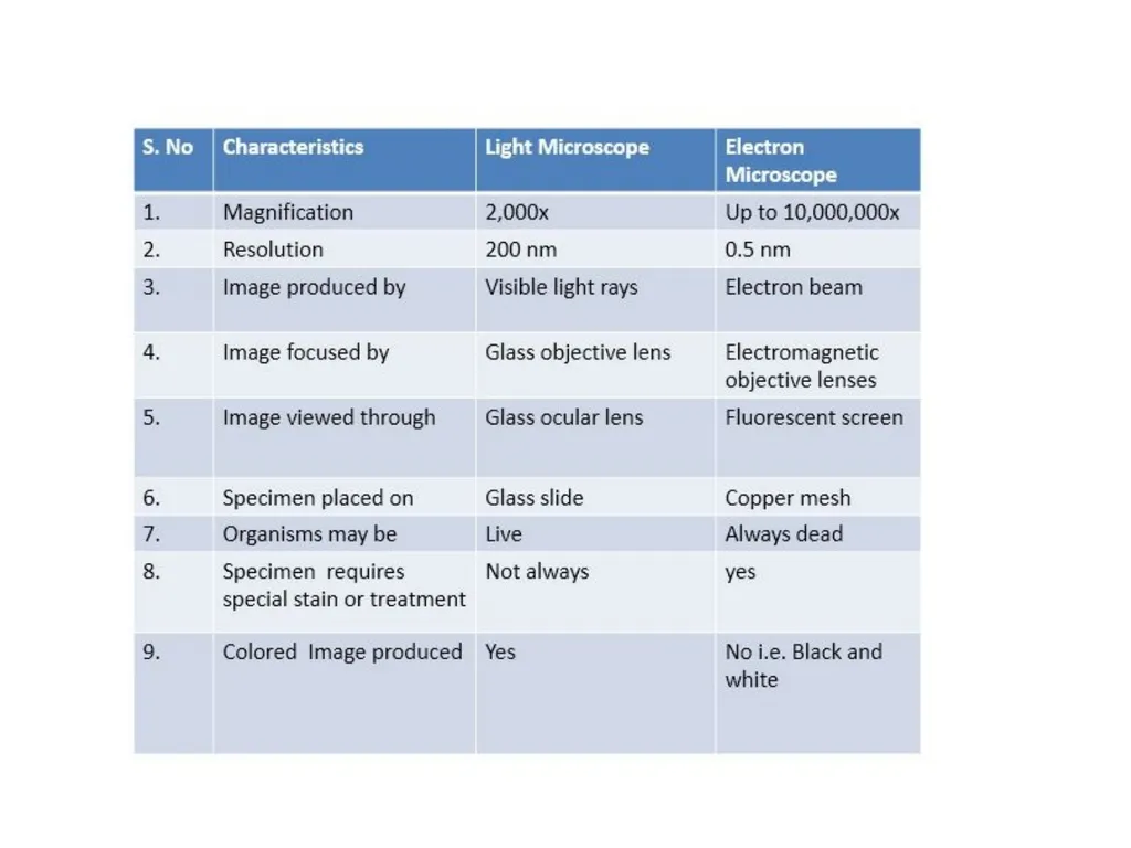
Microscopes that use a beam of electrons (instead of a beam of light) and
electromagnets (instead of glass lenses) for focusing are called electron
microscopes. These microscopes provide a higher magnification and are
used for observing extremely small microorganisms such as viruses.<br>
03
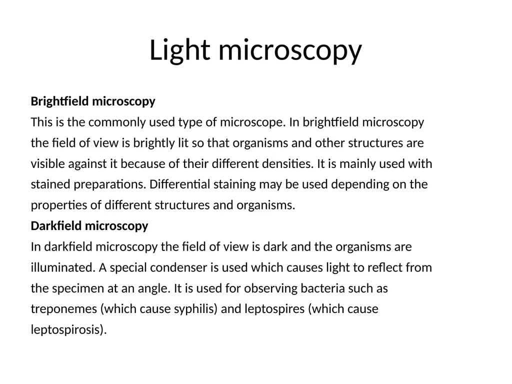
Light microscopy Brightfield microscopy
This is the commonly used type of microscope. In brightfield microscopy
the field of view is brightly lit so that organisms and other structures are
visible against it because of their different densities. It is mainly used with
stained preparations. Differential staining may be used depending on the
properties of different structures and organisms.
Darkfield microscopy
In darkfield microscopy the field of view is dark and the organisms are
illuminated. A special condenser is used which causes light to reflect from
the specimen at an angle. It is used for observing bacteria such as
treponemes (which cause syphilis) and leptospires (which cause
leptospirosis).<br>
04
Phase-contrast microscopy
Phase-contrast microscopy allows the examination of live unstained
organisms. For phase-contrast microscopy, special condensers and objectives
are used. These alter the phase relationships of the light passing through the
object and that passing around it.
Fluorescence microscopy
In fluorescence microscopy specimens are stained with fluorochromes/
fluorochrome complexes. Light of high energy or short wavelengths (from
halogen lamps or mercury vapour lamps) is then used to excite molecules
within the specimen or dye molecules attached to it. These excited
molecules emit light of different wavelengths, often of brilliant colours.
Auramine differential staining for acid-fast bacilli is one application of the
technique; rapid diagnostic kits have been developed using fluorescent
antibodies for identifying many pathogens.<br>
05
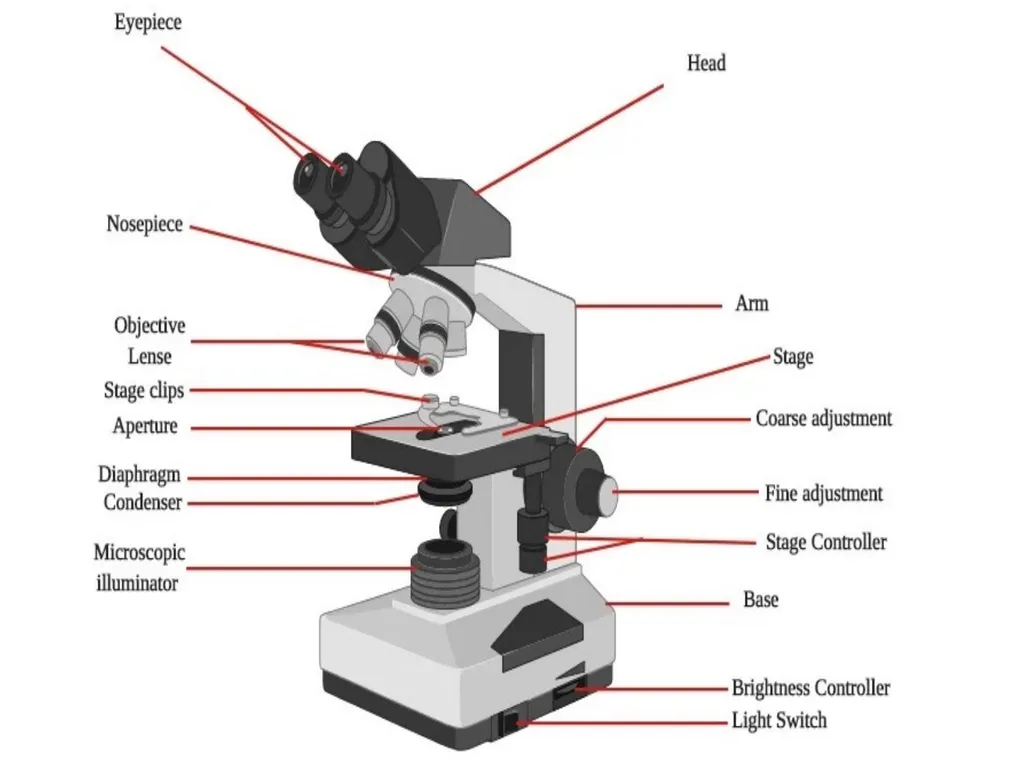
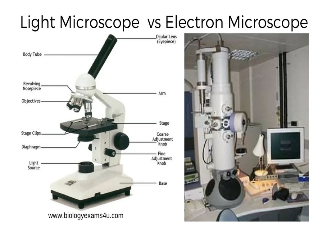
Parts of the Microscope The main parts of the microscope are the eye-pieces, microscope tube, nosepiece, objective, mechanical stage, condenser, coarse and fine focusing knobs, and light source.<br>
06
Eye-pieces
The specimen is viewed through the eye-piece. It has a lens which magnifies the image formed
by the objective. The magnifying power of the eye-piece is in the range 5x20x. A movable pointer may be attached to the inside of the
eye-piece<br>
The specimen is viewed through the eye-piece. It has a lens which magnifies the image formed
by the objective. The magnifying power of the eye-piece is in the range 5x20x. A movable pointer may be attached to the inside of the
eye-piece<br>
07
Mechanical tube length
Mechanical tube length is the distance between the place where the objective is inserted and the top of the draw-tube into which the eyepieces fit.
In modern microscopes it is not tubular; it contains prisms that bend the light coming up, thus providing a comfortable viewing angle. In a binocular tube, the light is also split and sent to both eye-pieces.<br>
08
Nose-piece
The nose-piece is attached under the arm of the microscope tube. The nose-piece houses
the objectives and rotates them.
The objectives are arranged in sequential order of their magnifying power, from lower to higher. This helps to prevent the immersion oil from getting onto the intermediate objectives.<br>
09
Objectives
❍ The image of the specimen first passes through the objective. Objectives with magnifying powers 4x, 10x, 40x and 100x are commonly used. The magnifying power is marked on the lens and is usually colour-coded for easy identification.
The 100x objective is for oil immersion.<br>
❍ The image of the specimen first passes through the objective. Objectives with magnifying powers 4x, 10x, 40x and 100x are commonly used. The magnifying power is marked on the lens and is usually colour-coded for easy identification.
The 100x objective is for oil immersion.<br>
10
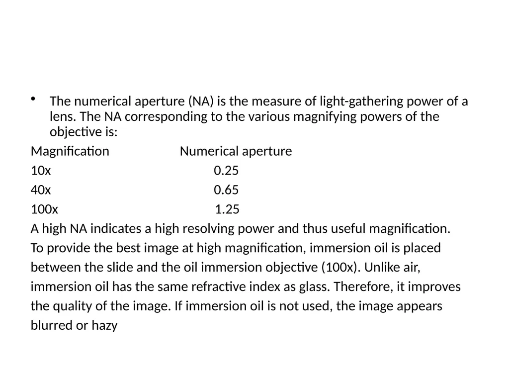
The numerical aperture (NA) is the measure of light-gathering power of a lens. The NA corresponding to the various magnifying powers of the objective is:
Magnification Numerical aperture
10x 0.25
40x 0.65
100x 1.25
A high NA indicates a high resolving power and thus useful magnification.
To provide the best image at high magnification, immersion oil is placed
between the slide and the oil immersion objective (100x). Unlike air,
immersion oil has the same refractive index as glass. Therefore, it improves
the quality of the image. If immersion oil is not used, the image appears
blurred or hazy<br>
11
Mechanical stage
❍ The mechanical stage holds the slide and allows it to be moved to the left, right, forward or backward by rotating the knobs.
❍ It is fitted with fine vernier graduations as on a ruler. This helps in relocating a specific field of examination<br>
❍ The mechanical stage holds the slide and allows it to be moved to the left, right, forward or backward by rotating the knobs.
❍ It is fitted with fine vernier graduations as on a ruler. This helps in relocating a specific field of examination<br>
12
The condenser
Illuminates the specimen and controls the amount of light and contrast. There are different types of condensers.
Some condensers have a rack-and pinion mechanism for up-and-down adjustment
Two-sided mirror
❍ A mirror is the simplest illuminator. The two-sided mirror
provides necessary illumination through reflection of natural or
artificial light. It has two surfaces, one plain for artificial light and
other concave for natural light. It is supported on two sides by a fork
fixed on a mount in a way that permits free rotation.<br>
13
Built-in light sources
An illuminator is built into the base of the microscope. A halogen bulb
provides the best illumination. On top of the illuminator is an in-built filter holder to fit the filter of desired quality.
Filters
❍ Blue filters are used to change the light from ordinary electric bulbs into a more natural white light.
❍ Neutral density filters are used to reduce brightness without changing the colour of the background.
❍ Green filters may be useful in some situations.<br>
14
Immersion oil
❍ Immersion oil must be used with objectives having NA more than 1.0. This increases the resolving power of the objective.
❍ An immersion oil of medium viscosity and refractive index of 1.5 is adequate. Any synthetic non-drying oil with a refractive index of 1.5 and/or as recommended by the manufacturer should be used
Cedar wood oil should not be used as it leaves a sticky residue on the objective. If cedar wood oil is used, particular care then needs to be taken to ensure that the objective is thoroughly and promptly cleaned with xylene after each session of use. Petrol can be used in place of xylene for cleaning if xylene is not available.<br>
15
Coarse and fine focusing knobs
The coarse and fine focusing knobs are used to change the distance between the specimen slide and the objective.
The coarse focusing knob alters this distance rapidly and is used to bring the specimen into the field of view using an objective having low magnification power.
The fine focusing knob changes the distance very slowly and permits better viewing of the object.
One revolution of the fine focusing knob should generally move the mechanical stage by 100 µm. The movement should be smooth and free from jerks.<br>
16
Halogen lamp
Halogen lamps are low wattage, high intensity lamps and are the preferred light source. Though costlier, these have the following advantages over tungsten lamps:
❍ emit white light
❍ have higher luminosity (brighter)
❍ have compact filament
❍ have longer life.<br>
Halogen lamps are low wattage, high intensity lamps and are the preferred light source. Though costlier, these have the following advantages over tungsten lamps:
❍ emit white light
❍ have higher luminosity (brighter)
❍ have compact filament
❍ have longer life.<br>
17
Functioning of the microscope
There are three main optical pieces in the compound light microscope. All three are essential for a sharp and clear image. These are:
❍ Condenser
❍ Objectives
❍ Eye-pieces.<br>
There are three main optical pieces in the compound light microscope. All three are essential for a sharp and clear image. These are:
❍ Condenser
❍ Objectives
❍ Eye-pieces.<br>
18
The condenser illuminates the object by converging a parallel beam of light on it from a built-in or natural source.
The objective forms a magnified inverted (upside down) image of the object. The eye-piece magnifies the image formed by the objective. This image is formed below the plane of the slide.
The total magnification of the microscope is the product of the magnifying powers of the objective and the eye-piece.
For example, if the magnifying power of the eye-piece is 10x and that of the
objective is 100x, then the total magnification of the compound light microscope is: 10x X 100x = 1000-fold magnification.<br>
19
❍ Install the microscope on a sturdy, level table. Equipment and
instruments which generate vibrations, such as centrifuges and
refrigerators, should not be placed on or near this table.
❍ The height of the table should be convenient for the user. As an
alternative or in addition, an adjustable stool should be made available
to make microscopy comfortable.
❍ The table should be away from water, sinks, and racks containing
chemicals, to prevent damage to the microscope from splashes or spills.
❍ If the microscope does not have a built-in light source then the table
should be placed near a window away from direct sunlight and
arrangements made for the provision of a lamp.
❍ In so far as is possible, the microscopy room should be free from dust
and should not be damp.
❍ If the microscope is to be used every day, do not remove it from the site
of installation, provided security is assured.
❍ When the microscope is not in use, keep it covered with a polythene or
plastic cover and take necessary precautions against fungus. MAINTENANCE<br>