
The Skeletal System: Bones and Joints Copyright ©
DJ
Published · 88 slides · 0 views
1 / 1
Description
The Skeletal System: Bones and Joints Copyright 2021 by Jones Bartlett Learning, LLC, an Ascend Learning company 1 Learning Objectives (1 of 1) List the functions of bone Describe the structure of a long bone. Name the three different
Related Topics
Share
Embed code
Download this presentation From Below
"The Skeletal System: Bones and Joints Copyright ©" is the property of its rightful owner. Permission is granted to download and print the materials on this website for personal, non-commercial use only, and to display it on your personal computer provided you do not modify the materials and that you retain all copyright notices contained in the materials. By downloading content from our website, you accept the terms of this agreement.
Presentation Transcript
01
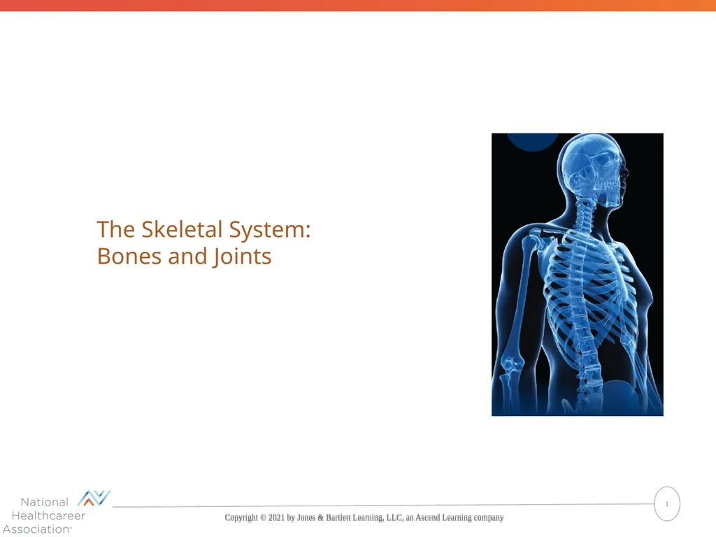
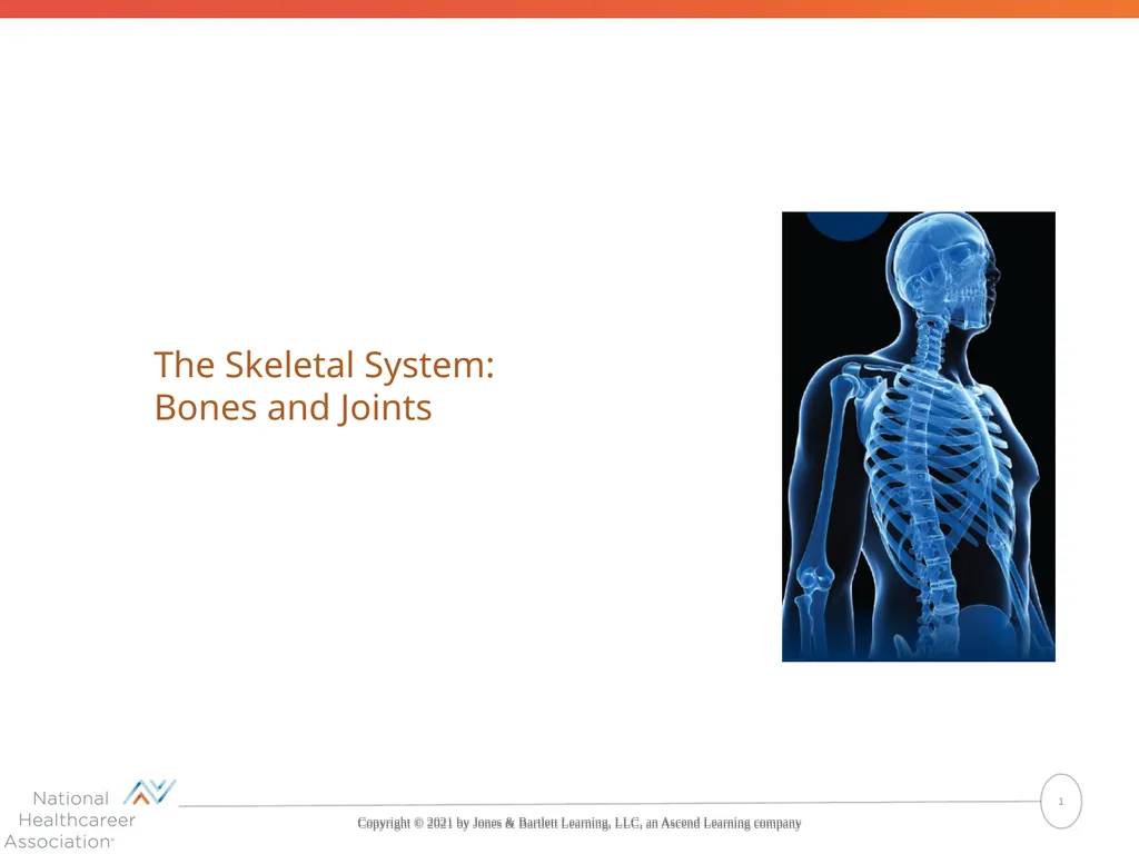
The Skeletal System: Bones and Joints Copyright © 2021 by Jones & Bartlett Learning, LLC, an Ascend Learning company 1<br>
02
Learning Objectives (1 of 1) List the functions of bone
Describe the structure of a long bone.
Name the three different types of cells in bone, and describe the functions of each.
Differentiate between compact bone and spongy bone with respect to structure and location.
Explain how a long bone grows.
Name and describe nine markings found on bones.
Name, locate, and describe the bones in the axial skeleton.
Describe the normal curves of the spine and explain their purpose. 2<br>
Describe the structure of a long bone.
Name the three different types of cells in bone, and describe the functions of each.
Differentiate between compact bone and spongy bone with respect to structure and location.
Explain how a long bone grows.
Name and describe nine markings found on bones.
Name, locate, and describe the bones in the axial skeleton.
Describe the normal curves of the spine and explain their purpose. 2<br>
03
Learning Objectives (2 of 2) Name, locate, and describe the bones in the appendicular skeleton.
Describe five types of bone disorders.
Name and describe eight types of fractures.
Describe three categories of joints based on degree of movement, and give examples of each.
Name six types of synovial joints, and demonstrate the movements that occur at each.
Describe three types of joint disorders.
Describe methods used to correct diseased joints.
Describe how the skeletal system changes with age.
Show how word parts are used to build words related to the skeleton. 3<br>
Describe five types of bone disorders.
Name and describe eight types of fractures.
Describe three categories of joints based on degree of movement, and give examples of each.
Name six types of synovial joints, and demonstrate the movements that occur at each.
Describe three types of joint disorders.
Describe methods used to correct diseased joints.
Describe how the skeletal system changes with age.
Show how word parts are used to build words related to the skeleton. 3<br>
04
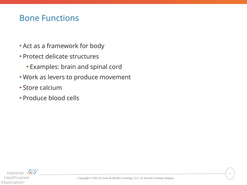
Bone Functions Act as a framework for body
Protect delicate structures
Examples: brain and spinal cord
Work as levers to produce movement
Store calcium
Produce blood cells 4<br>
Protect delicate structures
Examples: brain and spinal cord
Work as levers to produce movement
Store calcium
Produce blood cells 4<br>
05
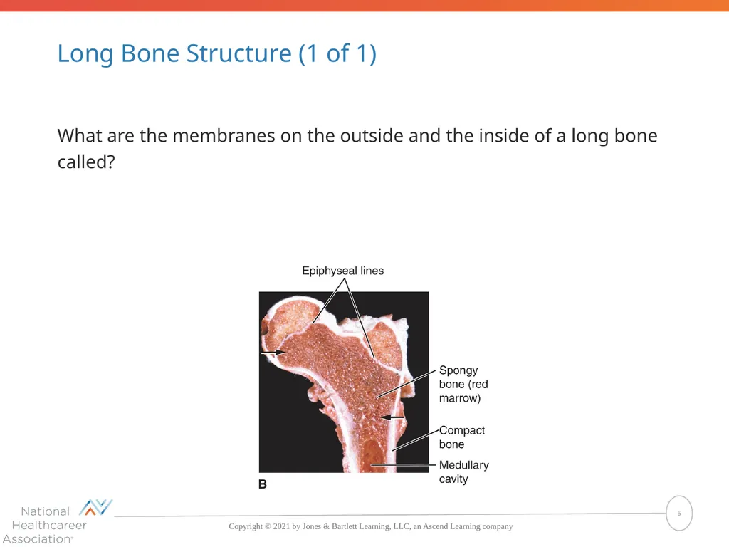
Long Bone Structure (1 of 1) What are the membranes on the outside and the inside of a long bone called? 5<br>
06
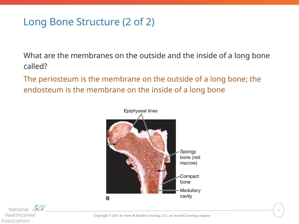
Long Bone Structure (2 of 2) What are the membranes on the outside and the inside of a long bone called?
The periosteum is the membrane on the outside of a long bone; the endosteum is the membrane on the inside of a long bone 6<br>
The periosteum is the membrane on the outside of a long bone; the endosteum is the membrane on the inside of a long bone 6<br>
07
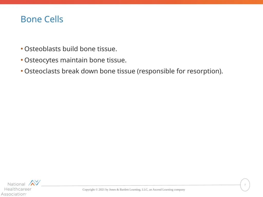
Bone Cells Osteoblasts build bone tissue.
Osteocytes maintain bone tissue.
Osteoclasts break down bone tissue (responsible for resorption). 7<br>
Osteocytes maintain bone tissue.
Osteoclasts break down bone tissue (responsible for resorption). 7<br>
08
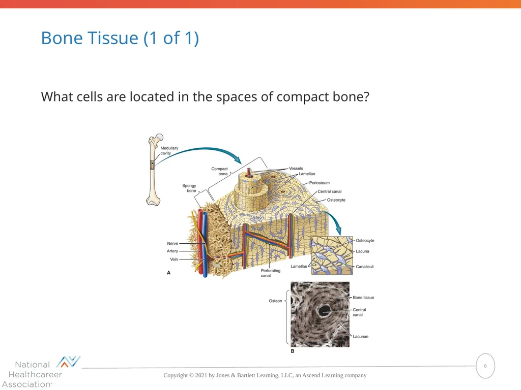
Bone Tissue (1 of 1) What cells are located in the spaces of compact bone? 8<br>
09
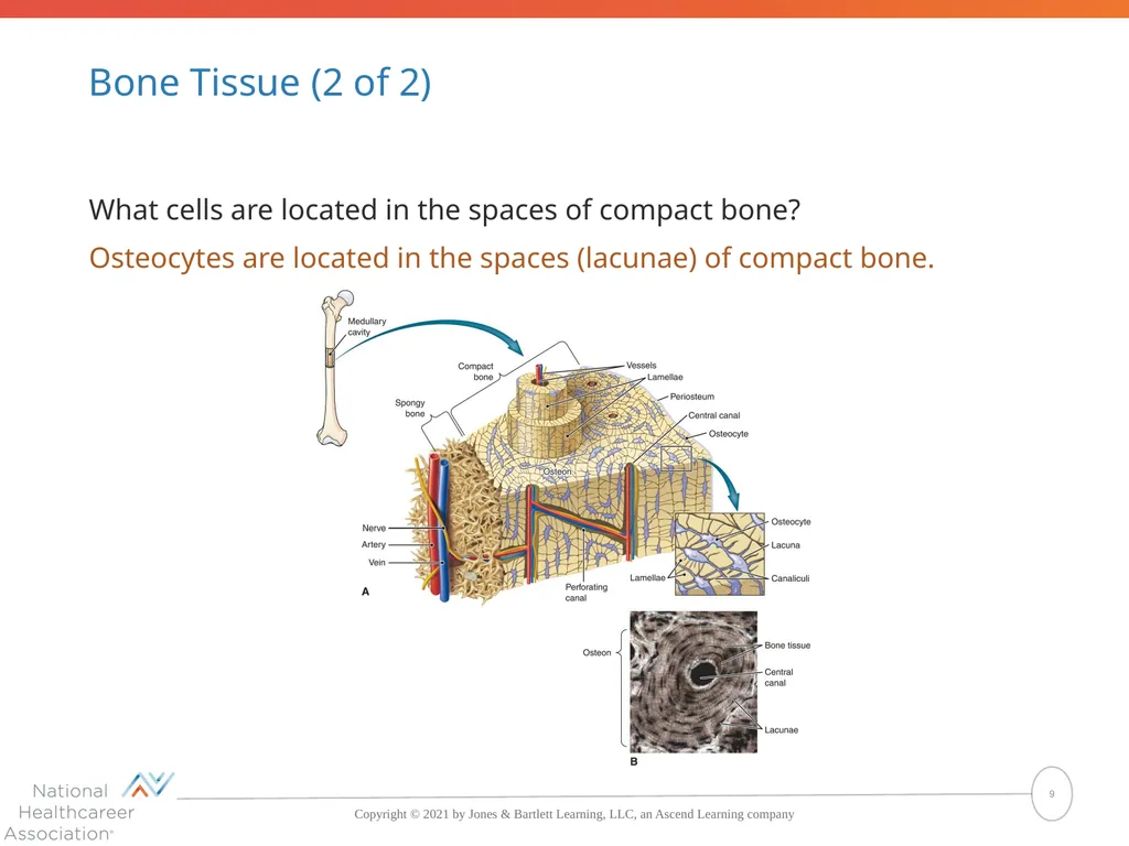
Bone Tissue (2 of 2) What cells are located in the spaces of compact bone?
Osteocytes are located in the spaces (lacunae) of compact bone. 9<br>
Osteocytes are located in the spaces (lacunae) of compact bone. 9<br>
10
Bones and Bone Tissue (1 of 1) What are the scientific names for the shaft and the ends of a long bone?
The diaphysis is the shaft of a long bone; the epiphysis is the end of a long bone.
What compounds are deposited in the intercellular matrix of the embryonic skeleton to harden it?
Calcium compounds are deposited in the intercellular matrix of the embryonic skeleton to harden it. 10<br>
The diaphysis is the shaft of a long bone; the epiphysis is the end of a long bone.
What compounds are deposited in the intercellular matrix of the embryonic skeleton to harden it?
Calcium compounds are deposited in the intercellular matrix of the embryonic skeleton to harden it. 10<br>
11
Bones and Bone Tissue (2 of 2) What are the three types of cells found in bone, and what is the role of each?
The cells found in bone are osteoblasts, which build bone tissue; osteocytes, which maintain bone; and osteoclasts, which break down (resorb) bone.
What are the two types of osseous (bone) tissue, and where is each type found?
Compact bone makes up the main shaft of a long bone and the outer layer of other bones; spongy (cancellous) bone makes up the ends of the long bones and the center of other bones. It also lines the medullary cavity of the long bones. 11<br>
The cells found in bone are osteoblasts, which build bone tissue; osteocytes, which maintain bone; and osteoclasts, which break down (resorb) bone.
What are the two types of osseous (bone) tissue, and where is each type found?
Compact bone makes up the main shaft of a long bone and the outer layer of other bones; spongy (cancellous) bone makes up the ends of the long bones and the center of other bones. It also lines the medullary cavity of the long bones. 11<br>
12
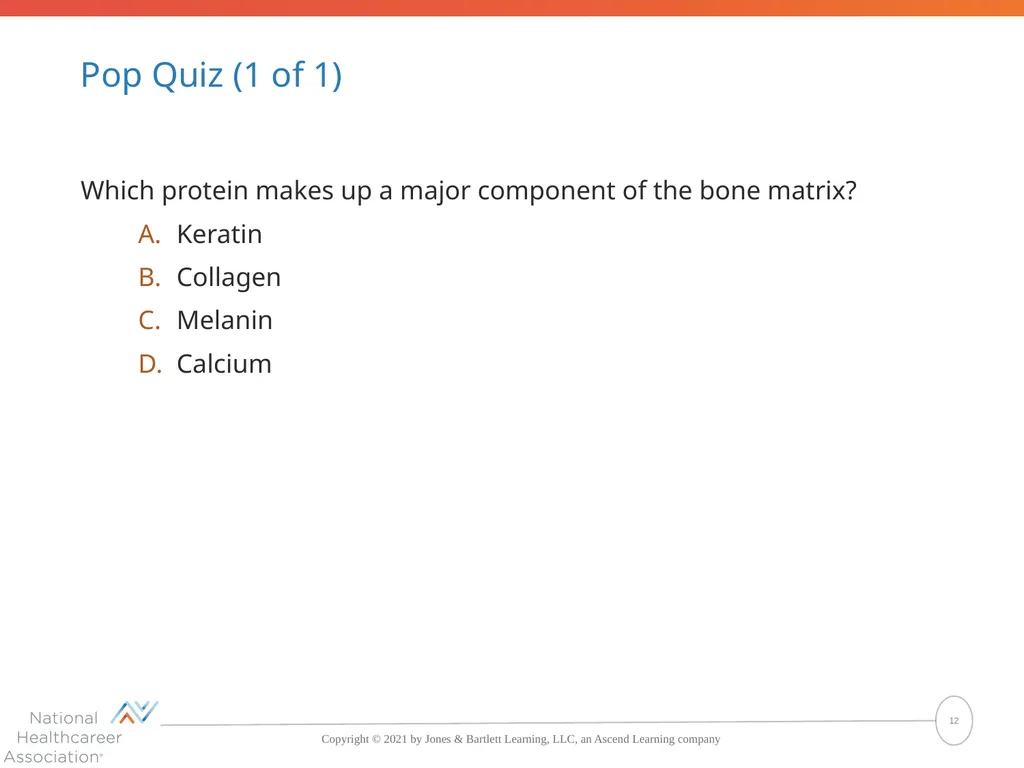
Pop Quiz (1 of 1) Which protein makes up a major component of the bone matrix?
Keratin
Collagen
Melanin
Calcium 12<br>
Keratin
Collagen
Melanin
Calcium 12<br>
13
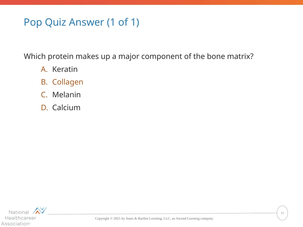
Pop Quiz Answer (1 of 1) Which protein makes up a major component of the bone matrix?
Keratin
Collagen
Melanin
Calcium The correct answer is Collagen 13<br>
Keratin
Collagen
Melanin
Calcium The correct answer is Collagen 13<br>
14
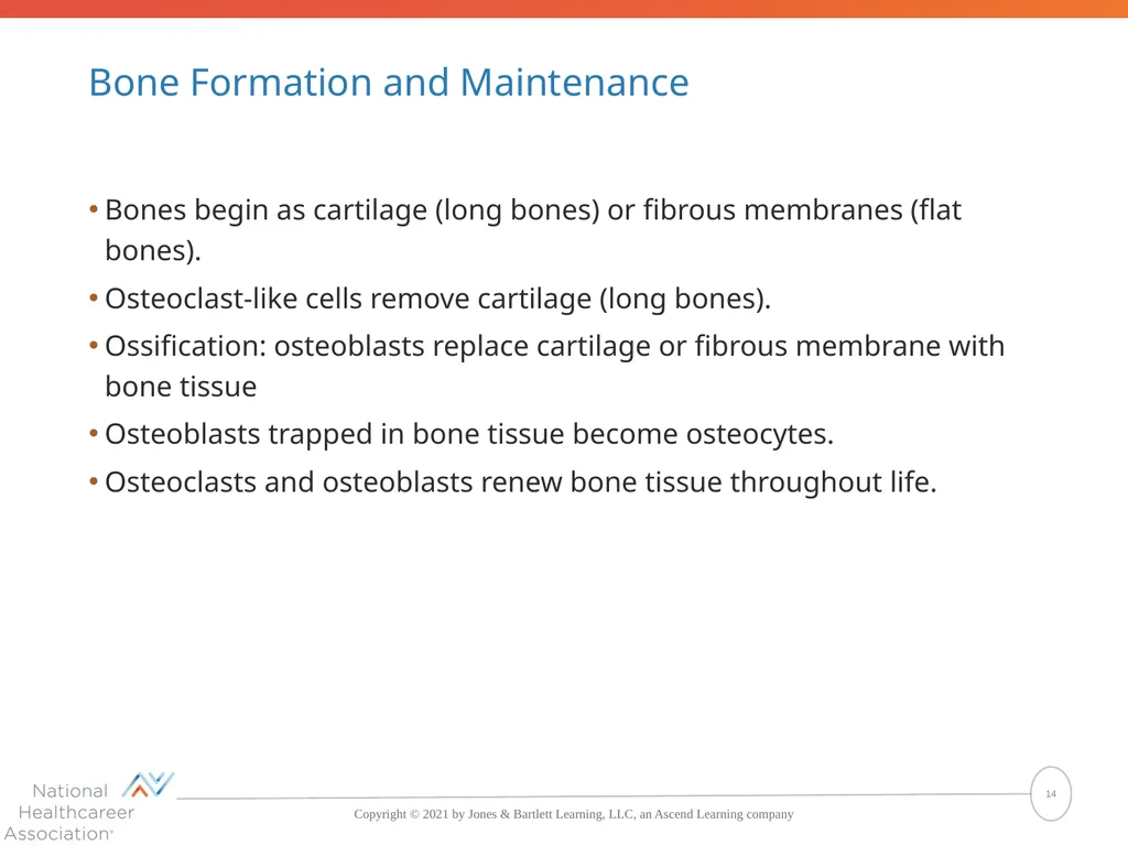
Bone Formation and Maintenance Bones begin as cartilage (long bones) or fibrous membranes (flat bones).
Osteoclast-like cells remove cartilage (long bones).
Ossification: osteoblasts replace cartilage or fibrous membrane with bone tissue
Osteoblasts trapped in bone tissue become osteocytes.
Osteoclasts and osteoblasts renew bone tissue throughout life. 14<br>
Osteoclast-like cells remove cartilage (long bones).
Ossification: osteoblasts replace cartilage or fibrous membrane with bone tissue
Osteoblasts trapped in bone tissue become osteocytes.
Osteoclasts and osteoblasts renew bone tissue throughout life. 14<br>
15
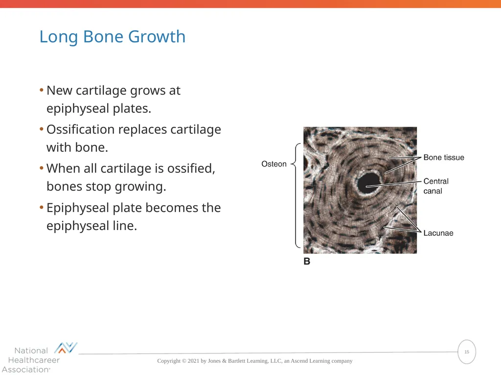
Long Bone Growth New cartilage grows at epiphyseal plates.
Ossification replaces cartilage with bone.
When all cartilage is ossified, bones stop growing.
Epiphyseal plate becomes the epiphyseal line. 15<br>
Ossification replaces cartilage with bone.
When all cartilage is ossified, bones stop growing.
Epiphyseal plate becomes the epiphyseal line. 15<br>
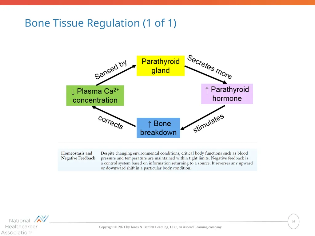
16
Bone Tissue Regulation (1 of 1) 16<br>
17
Bone Tissue Regulation (2 of 2) Bones strengthen and remodel in response to physical stresses. 17<br>
18
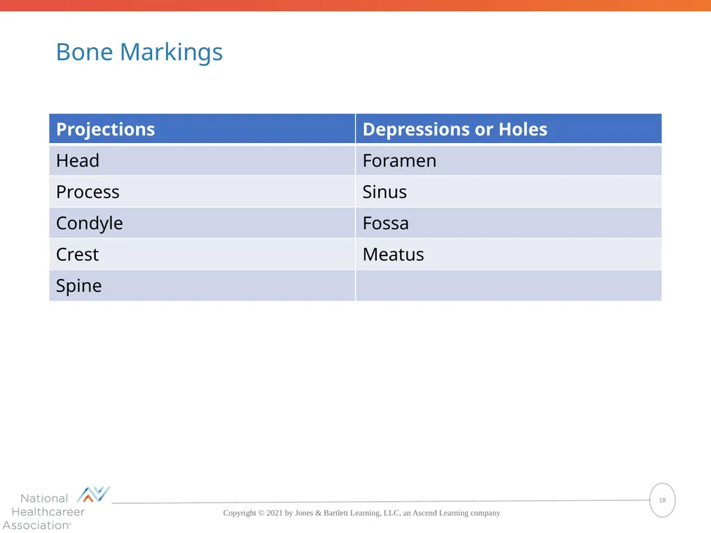
Bone Markings 18<br>
19
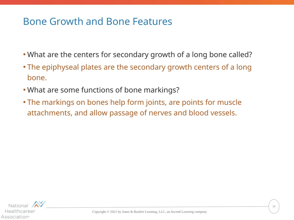
Bone Growth and Bone Features What are the centers for secondary growth of a long bone called?
The epiphyseal plates are the secondary growth centers of a long bone.
What are some functions of bone markings?
The markings on bones help form joints, are points for muscle attachments, and allow passage of nerves and blood vessels. 19<br>
The epiphyseal plates are the secondary growth centers of a long bone.
What are some functions of bone markings?
The markings on bones help form joints, are points for muscle attachments, and allow passage of nerves and blood vessels. 19<br>
20
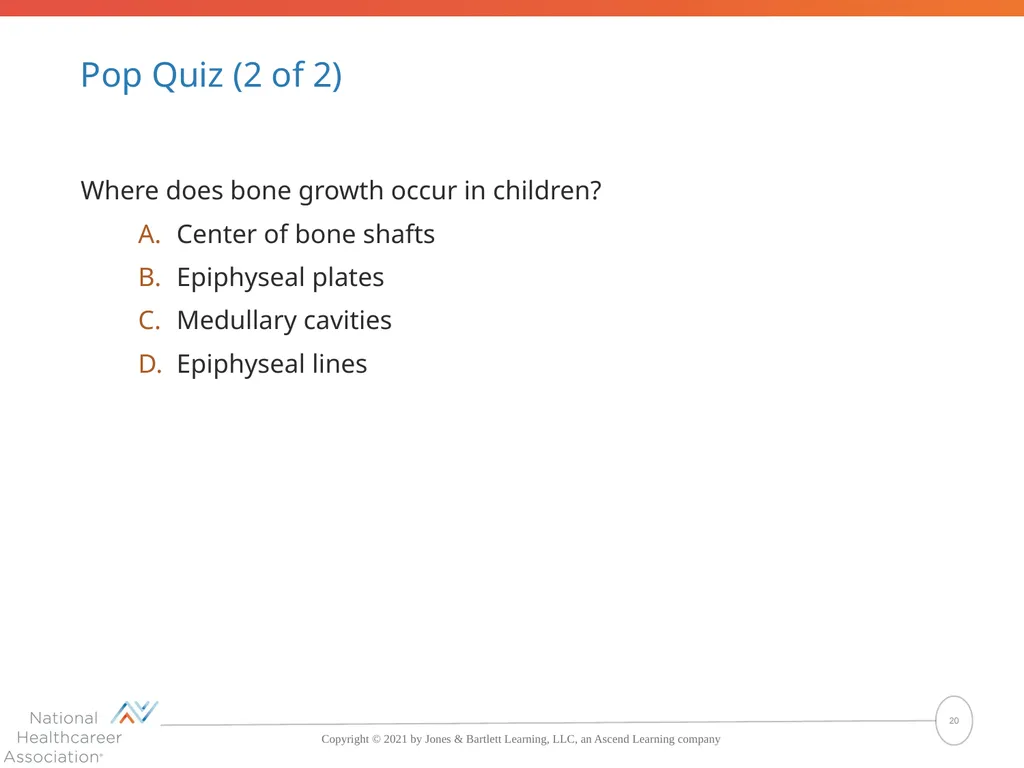
Pop Quiz (2 of 2) Where does bone growth occur in children?
Center of bone shafts
Epiphyseal plates
Medullary cavities
Epiphyseal lines 20<br>
Center of bone shafts
Epiphyseal plates
Medullary cavities
Epiphyseal lines 20<br>
21
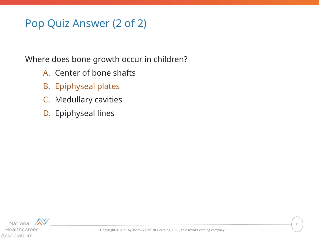
Pop Quiz Answer (2 of 2) Where does bone growth occur in children?
Center of bone shafts
Epiphyseal plates
Medullary cavities
Epiphyseal lines The correct answer is Epiphyseal plates 21<br>
Center of bone shafts
Epiphyseal plates
Medullary cavities
Epiphyseal lines The correct answer is Epiphyseal plates 21<br>
22
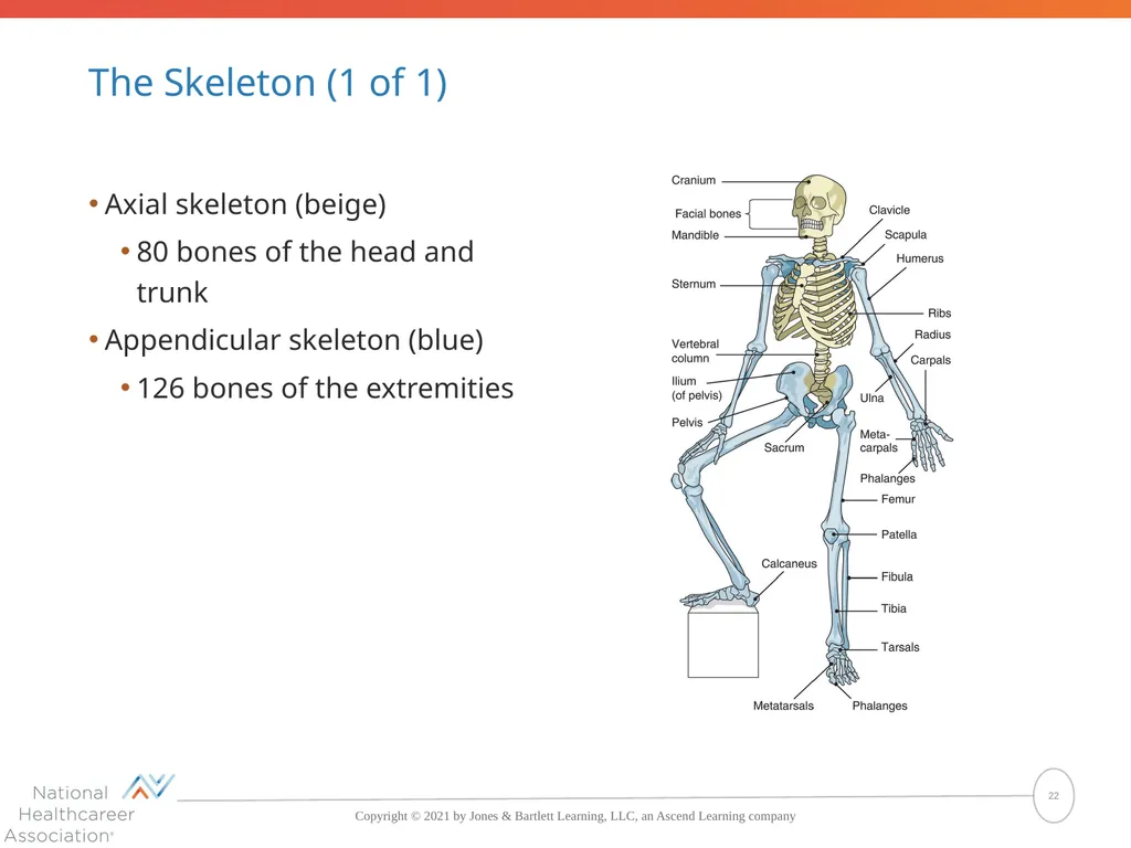
The Skeleton (1 of 1) Axial skeleton (beige)
80 bones of the head and trunk
Appendicular skeleton (blue)
126 bones of the extremities 22<br>
80 bones of the head and trunk
Appendicular skeleton (blue)
126 bones of the extremities 22<br>
23
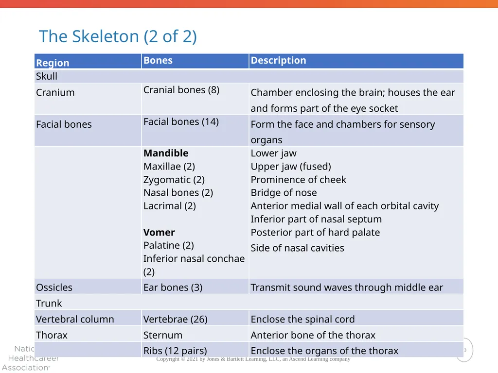
The Skeleton (2 of 2) 23<br>
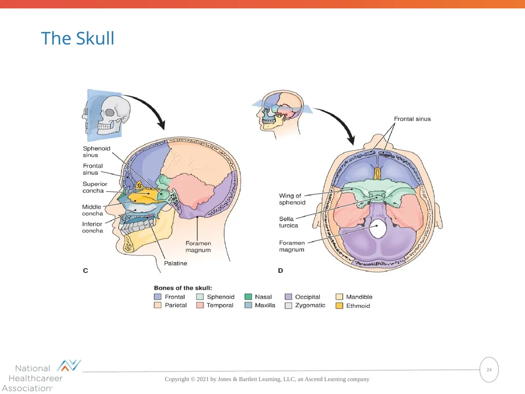
24
The Skull 24<br>
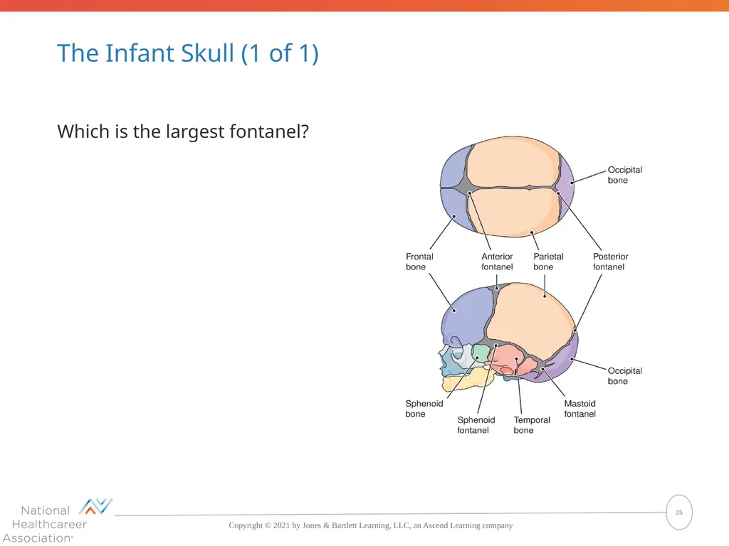
25
The Infant Skull (1 of 1) Which is the largest fontanel? 25<br>
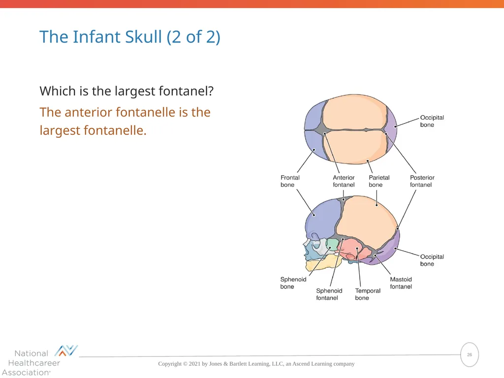
26
The Infant Skull (2 of 2) Which is the largest fontanel?
The anterior fontanelle is the largest fontanelle. 26<br>
The anterior fontanelle is the largest fontanelle. 26<br>
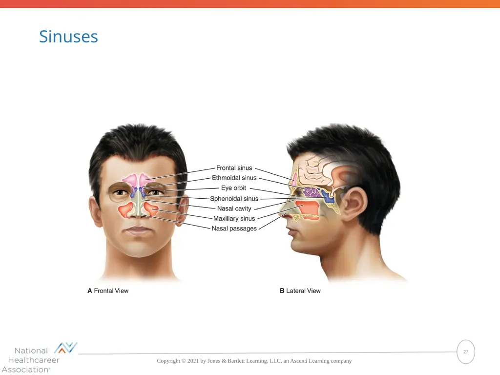
27
Sinuses 27<br>
28
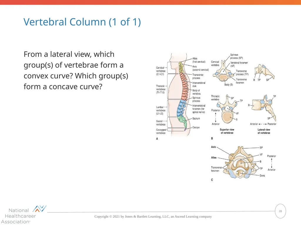
Vertebral Column (1 of 1) From a lateral view, which group(s) of vertebrae form a convex curve? Which group(s) form a concave curve? 28<br>
29
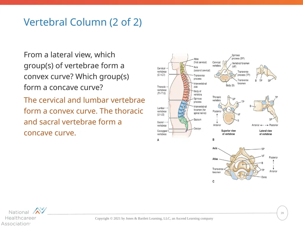
Vertebral Column (2 of 2) From a lateral view, which group(s) of vertebrae form a convex curve? Which group(s) form a concave curve?
The cervical and lumbar vertebrae form a convex curve. The thoracic and sacral vertebrae form a concave curve. 29<br>
The cervical and lumbar vertebrae form a convex curve. The thoracic and sacral vertebrae form a concave curve. 29<br>
30
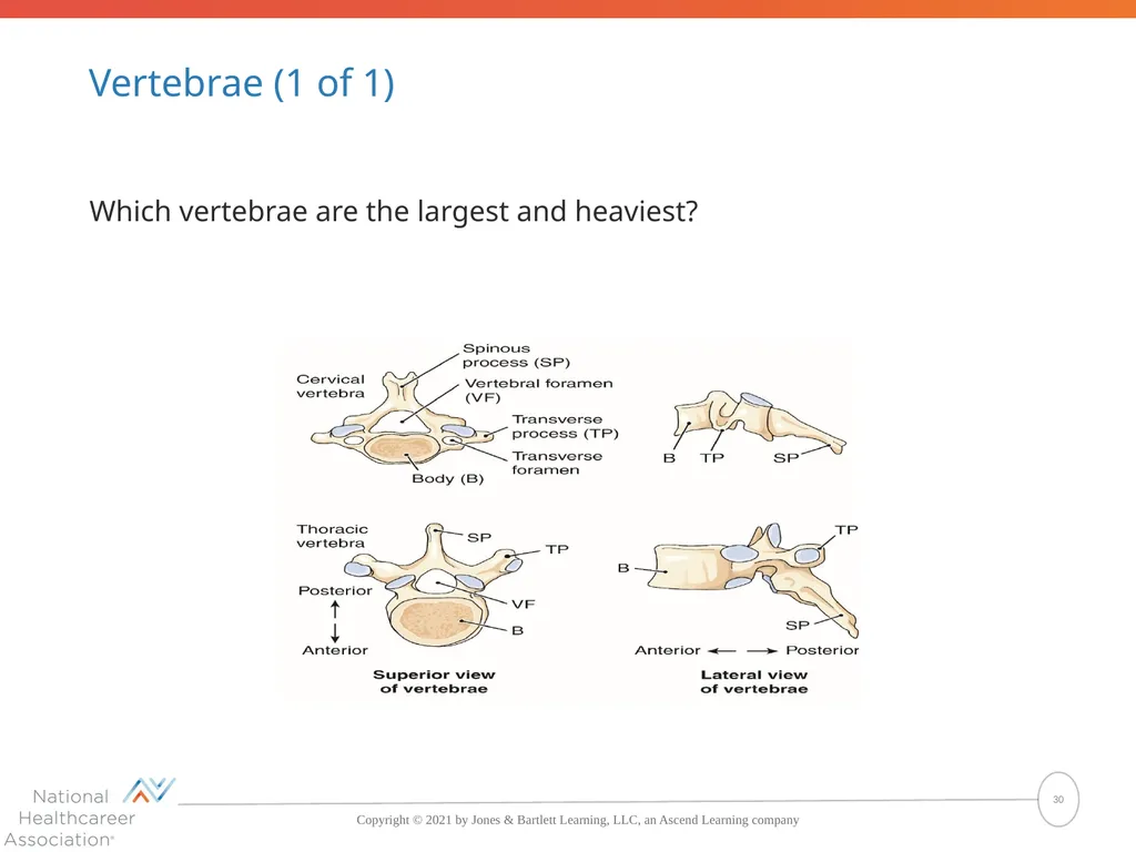
Vertebrae (1 of 1) Which vertebrae are the largest and heaviest? 30<br>
31
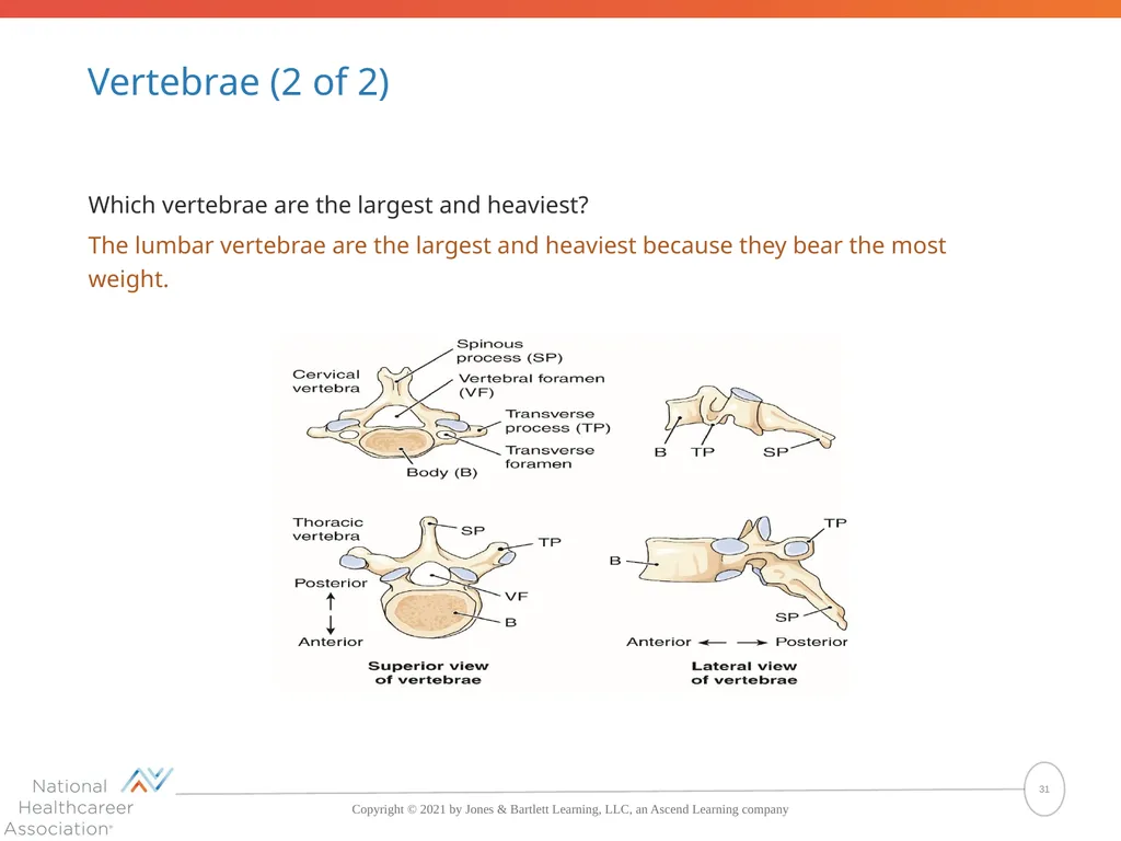
Vertebrae (2 of 2) Which vertebrae are the largest and heaviest?
The lumbar vertebrae are the largest and heaviest because they bear the most weight. 31<br>
The lumbar vertebrae are the largest and heaviest because they bear the most weight. 31<br>
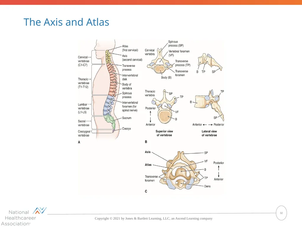
32
The Axis and Atlas 32<br>
33
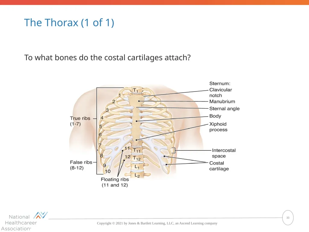
The Thorax (1 of 1) To what bones do the costal cartilages attach? 33<br>
34
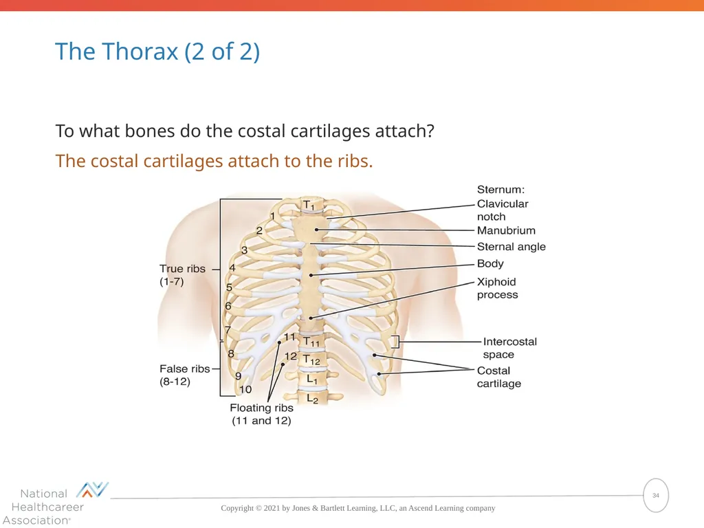
The Thorax (2 of 2) To what bones do the costal cartilages attach?
The costal cartilages attach to the ribs. 34<br>
The costal cartilages attach to the ribs. 34<br>
35
Bones of the Axial Skeleton What bones make up the skeleton of the trunk?
The skeleton of the trunk consists of the vertebral column and the bones of the thorax, which are the ribs and sternum.
What are the five regions of the vertebral column?
The five regions of the vertebral column are the cervical vertebra, thoracic vertebrae, lumbar vertebrae, sacrum, and coccyx. 35<br>
The skeleton of the trunk consists of the vertebral column and the bones of the thorax, which are the ribs and sternum.
What are the five regions of the vertebral column?
The five regions of the vertebral column are the cervical vertebra, thoracic vertebrae, lumbar vertebrae, sacrum, and coccyx. 35<br>
36
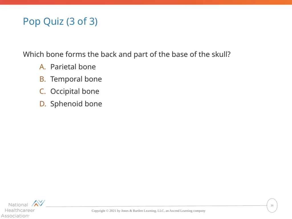
Pop Quiz (3 of 3) Which bone forms the back and part of the base of the skull?
Parietal bone
Temporal bone
Occipital bone
Sphenoid bone 36<br>
Parietal bone
Temporal bone
Occipital bone
Sphenoid bone 36<br>
37
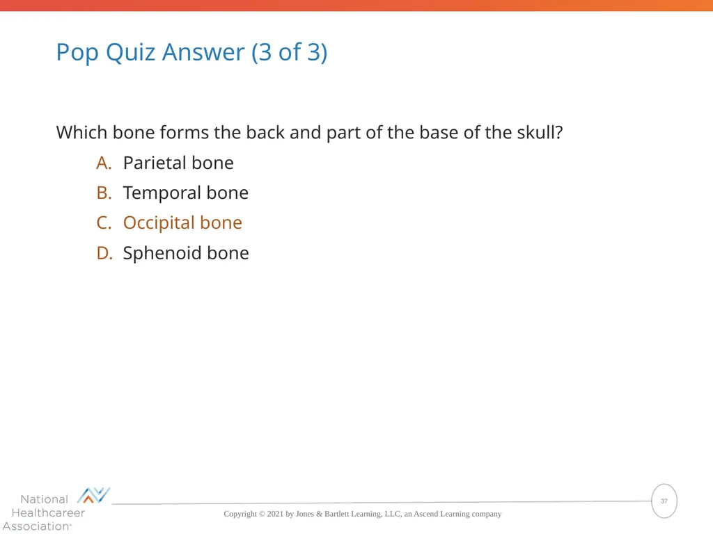
Pop Quiz Answer (3 of 3) Which bone forms the back and part of the base of the skull?
Parietal bone
Temporal bone
Occipital bone
Sphenoid bone The correct answer is Occipital bone 37<br>
Parietal bone
Temporal bone
Occipital bone
Sphenoid bone The correct answer is Occipital bone 37<br>
38
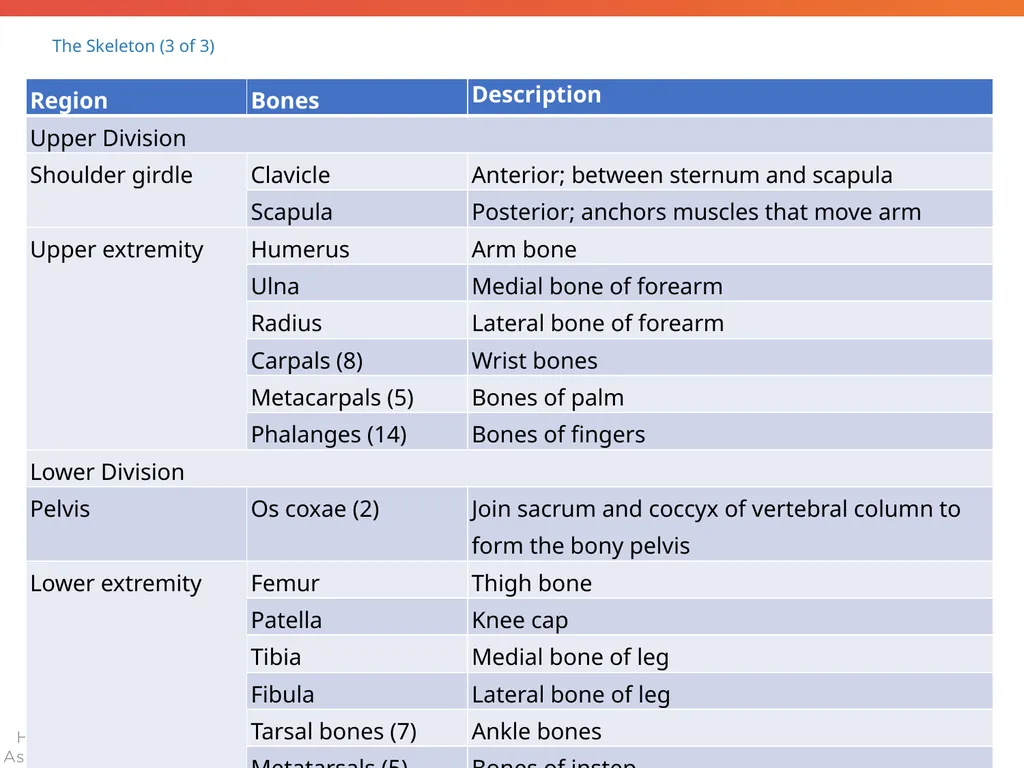
The Skeleton (3 of 3) 38<br>
39
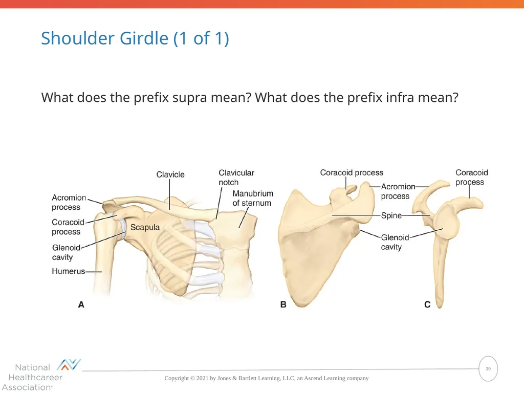
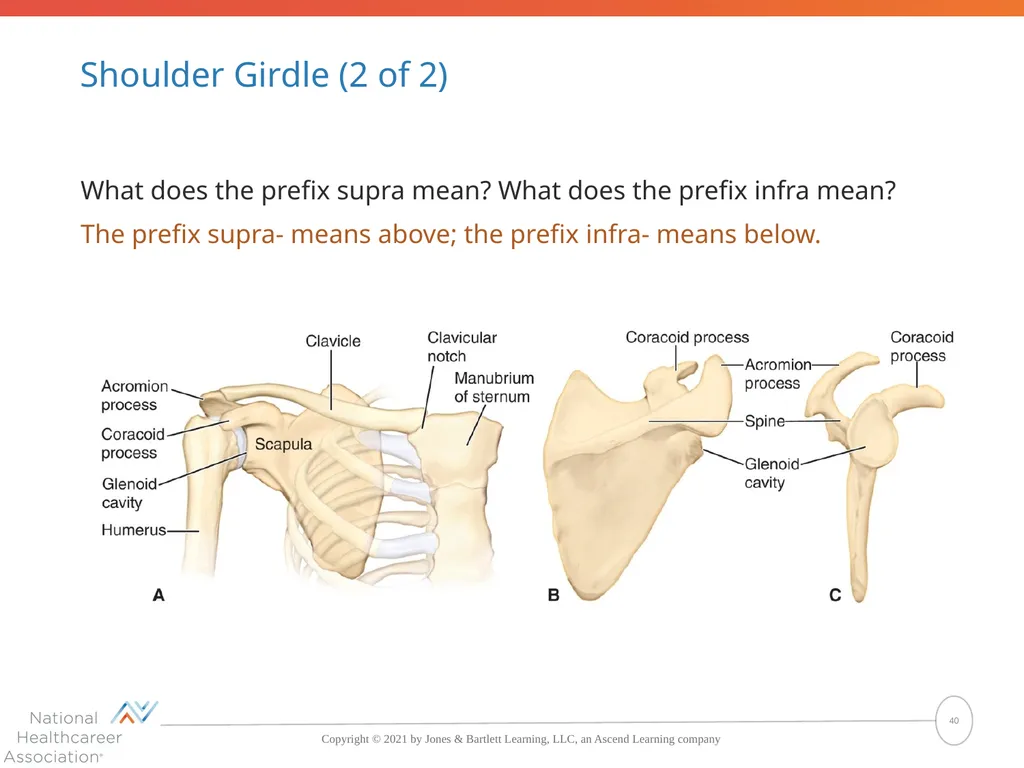
Shoulder Girdle (1 of 1) What does the prefix supra mean? What does the prefix infra mean? 39<br>
40
Shoulder Girdle (2 of 2) What does the prefix supra mean? What does the prefix infra mean?
The prefix supra- means above; the prefix infra- means below. 40<br>
The prefix supra- means above; the prefix infra- means below. 40<br>
41
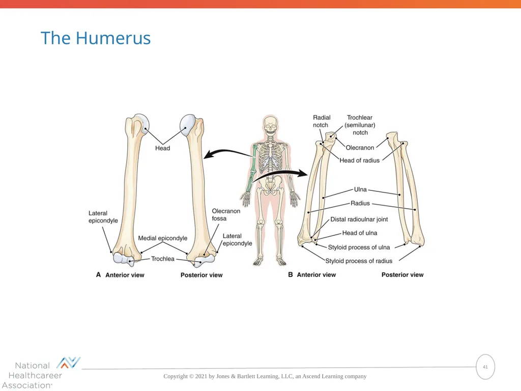
The Humerus 41<br>
42
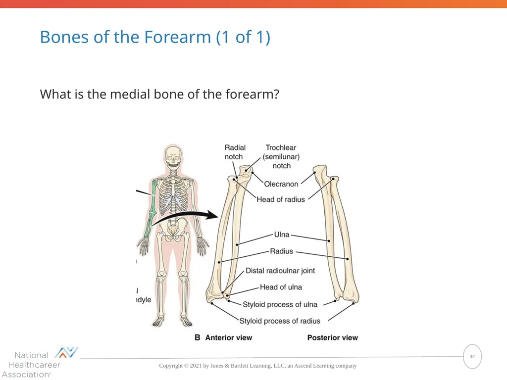
Bones of the Forearm (1 of 1) What is the medial bone of the forearm? 42<br>
43
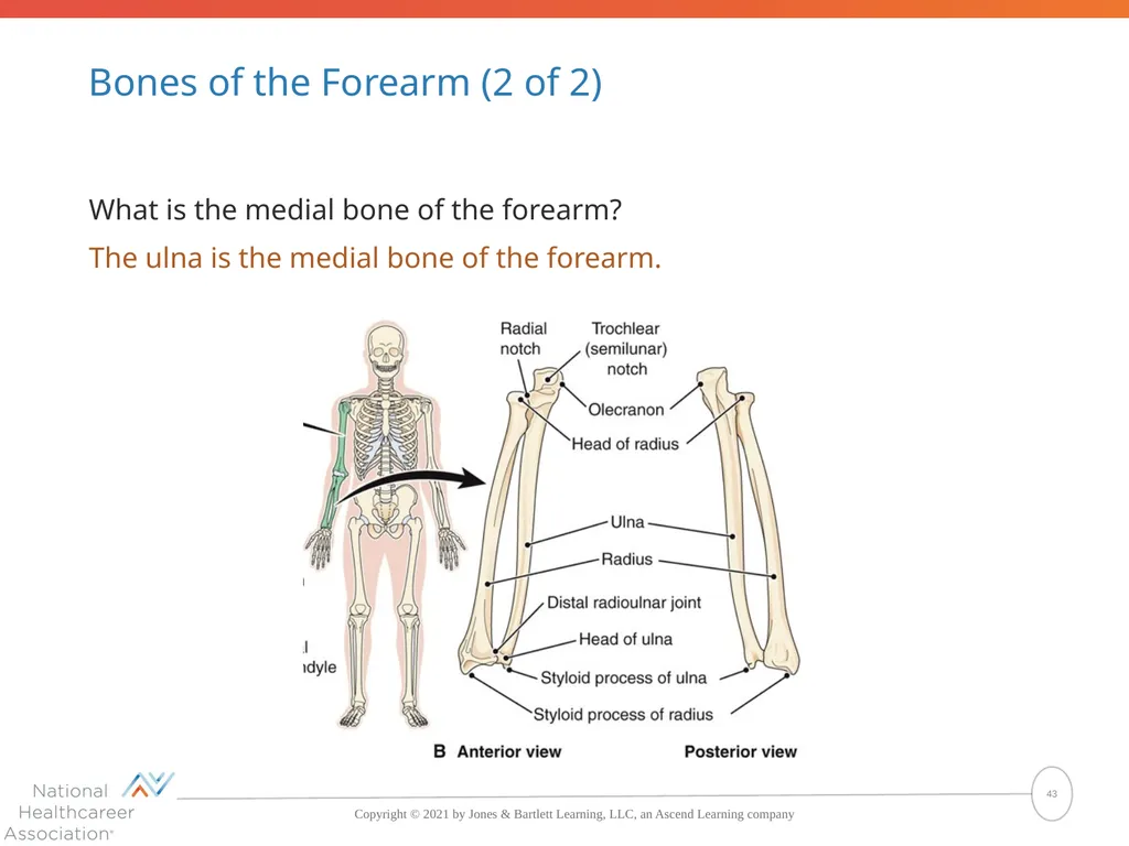
Bones of the Forearm (2 of 2) What is the medial bone of the forearm?
The ulna is the medial bone of the forearm. 43<br>
The ulna is the medial bone of the forearm. 43<br>
44
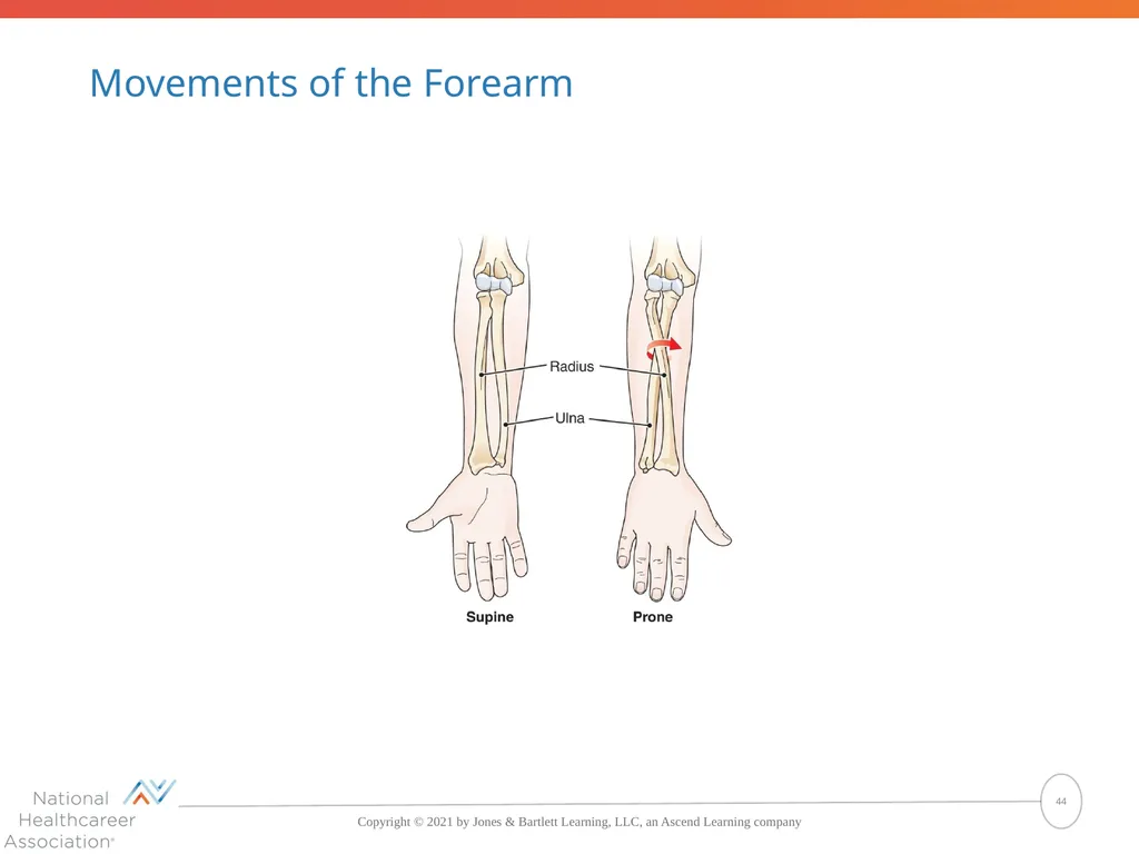
Movements of the Forearm 44<br>
45
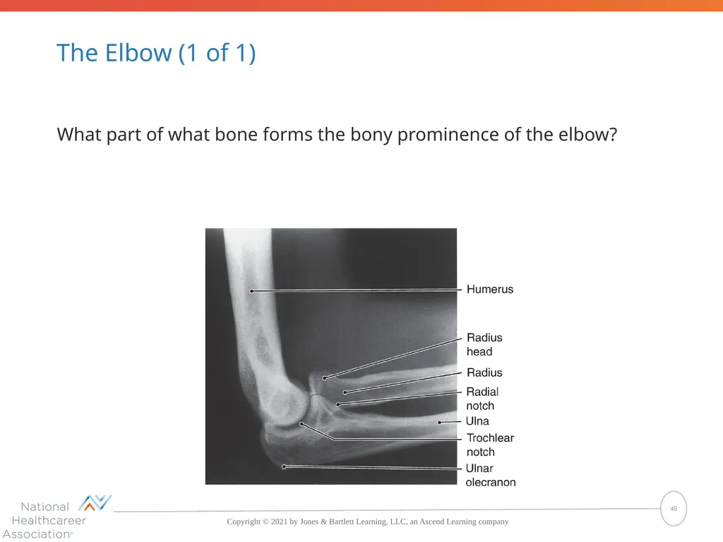
The Elbow (1 of 1) What part of what bone forms the bony prominence of the elbow? 45<br>
46
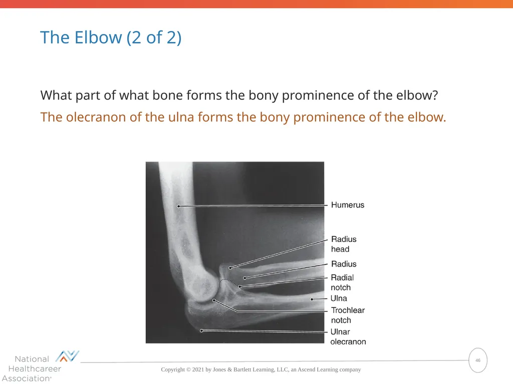
The Elbow (2 of 2) What part of what bone forms the bony prominence of the elbow?
The olecranon of the ulna forms the bony prominence of the elbow. 46<br>
The olecranon of the ulna forms the bony prominence of the elbow. 46<br>
47
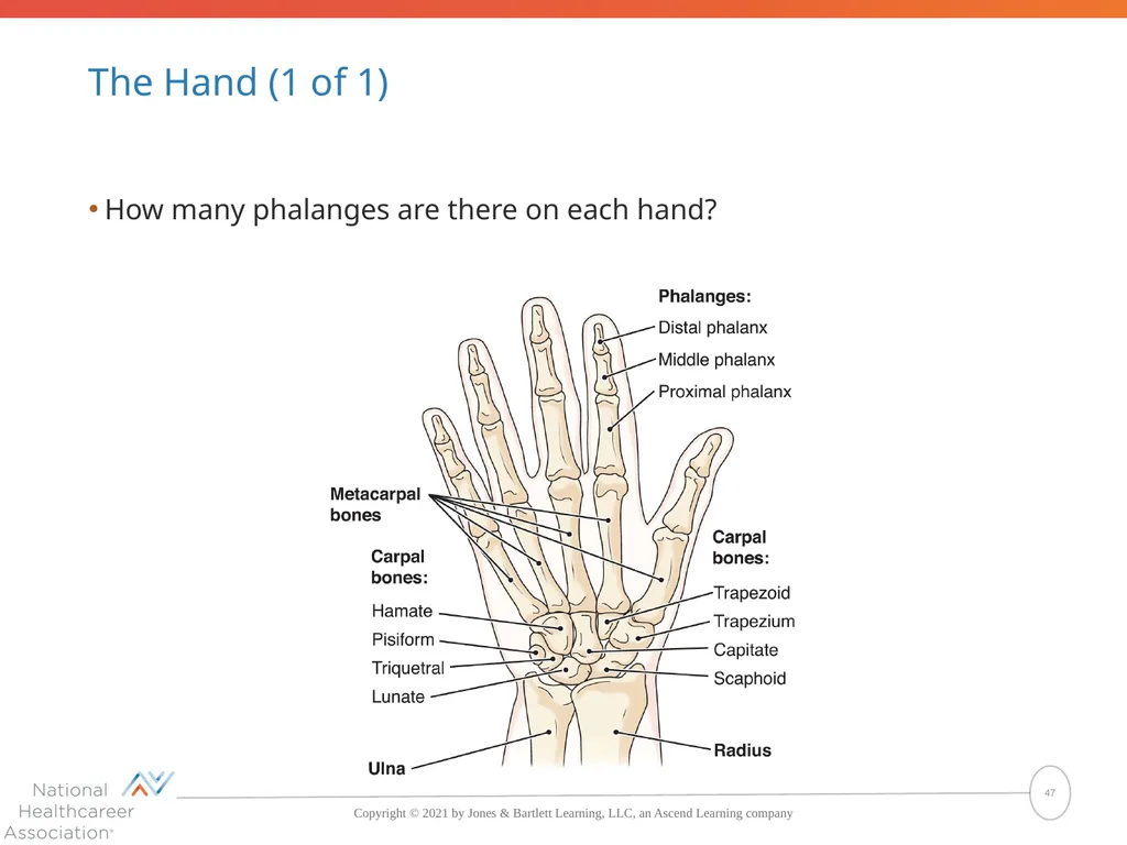
The Hand (1 of 1) How many phalanges are there on each hand? 47<br>
48
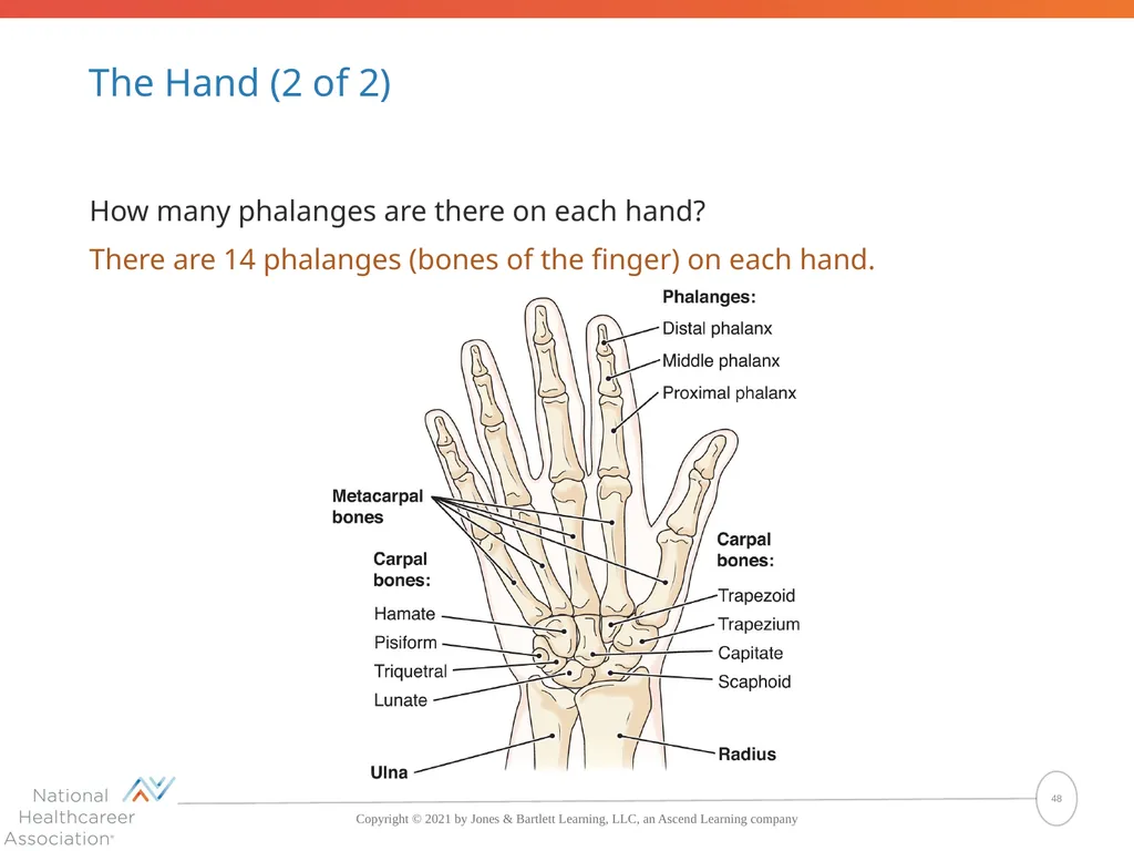
The Hand (2 of 2) How many phalanges are there on each hand?
There are 14 phalanges (bones of the finger) on each hand. 48<br>
There are 14 phalanges (bones of the finger) on each hand. 48<br>
49
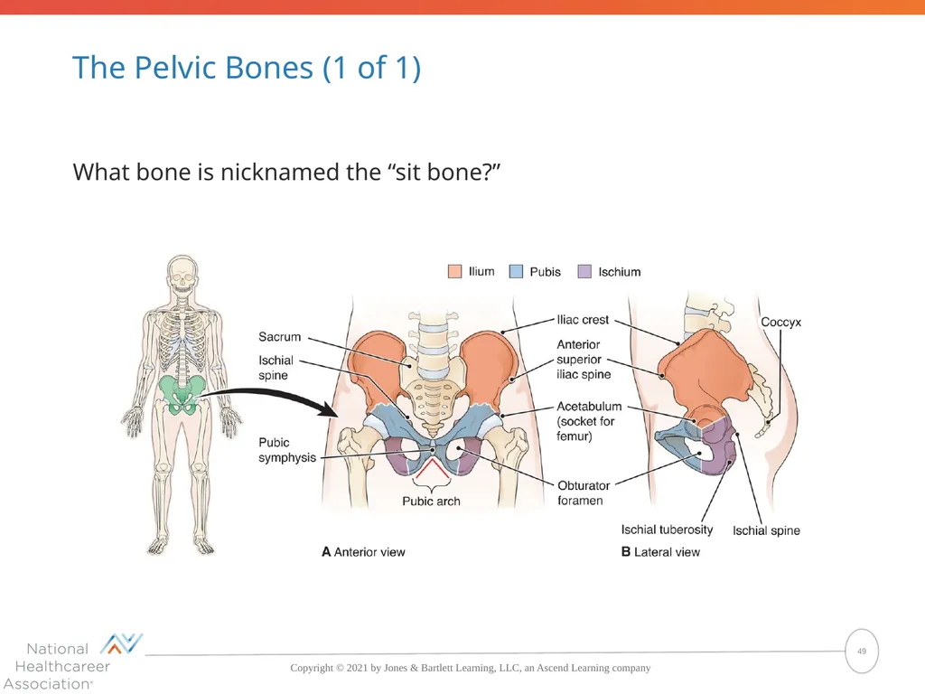
The Pelvic Bones (1 of 1) What bone is nicknamed the “sit bone?” 49<br>
50
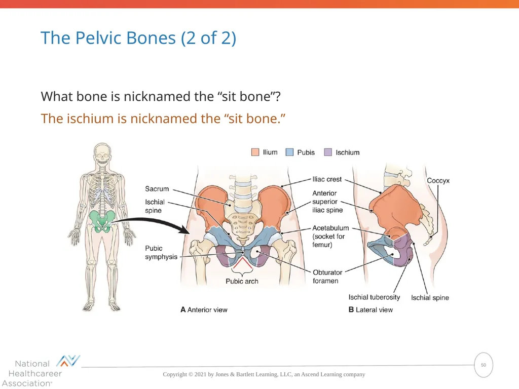
The Pelvic Bones (2 of 2) What bone is nicknamed the “sit bone”?
The ischium is nicknamed the “sit bone.” 50<br>
The ischium is nicknamed the “sit bone.” 50<br>
51
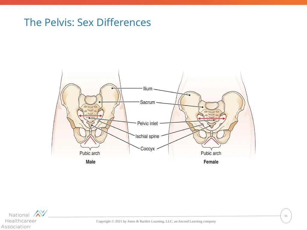
The Pelvis: Sex Differences 51<br>
52
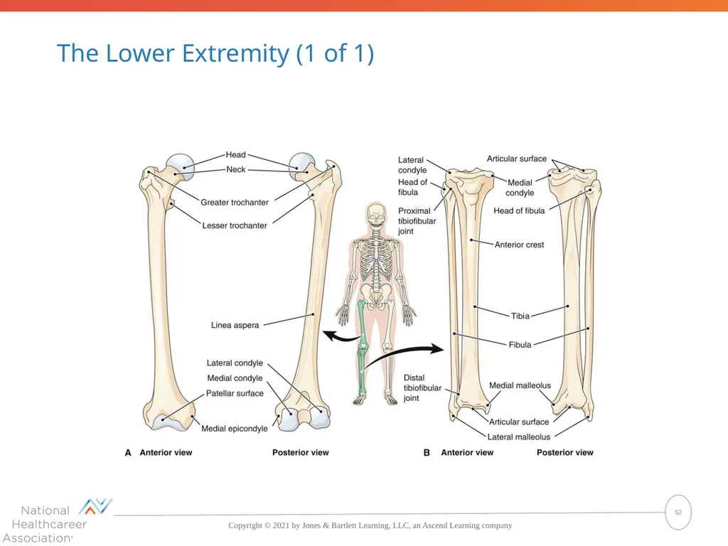
The Lower Extremity (1 of 1) 52<br>
53
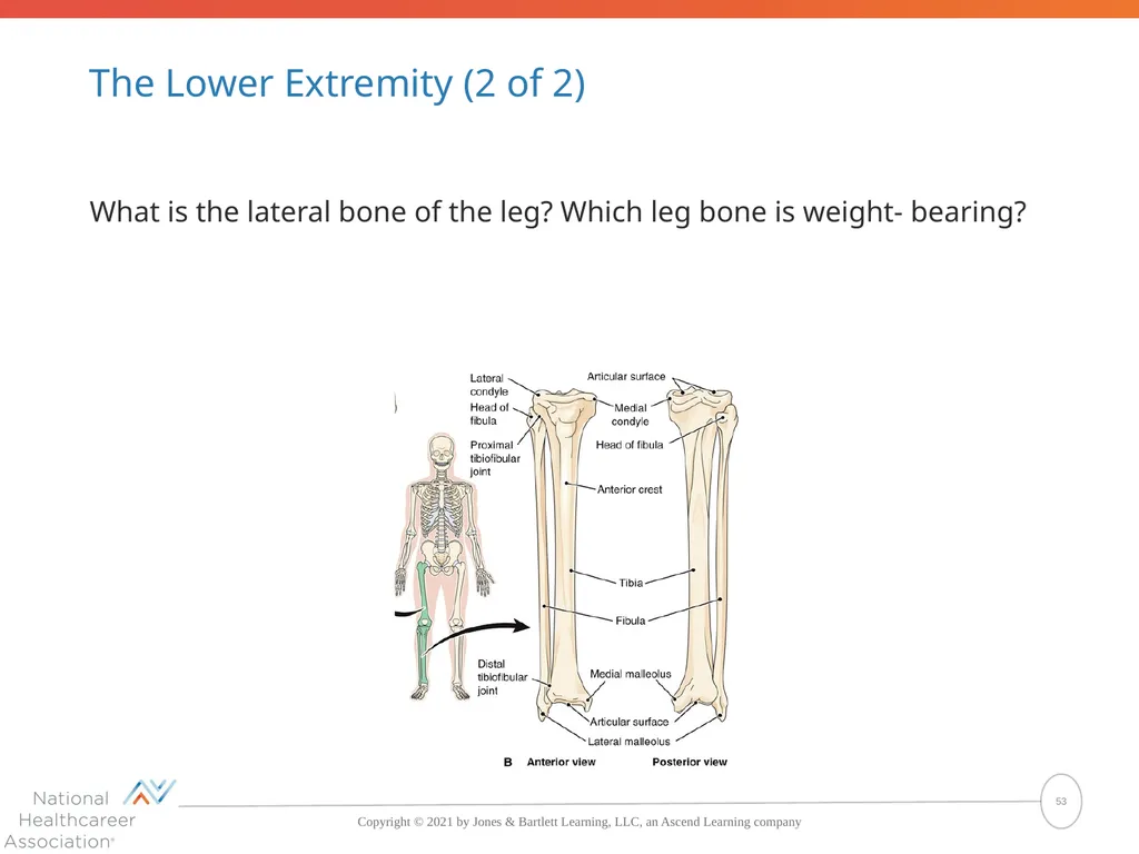
The Lower Extremity (2 of 2) What is the lateral bone of the leg? Which leg bone is weight- bearing? 53<br>
54
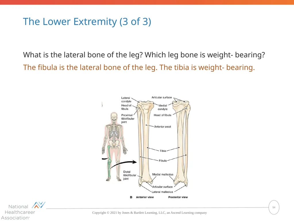
The Lower Extremity (3 of 3) What is the lateral bone of the leg? Which leg bone is weight- bearing?
The fibula is the lateral bone of the leg. The tibia is weight- bearing. 54<br>
The fibula is the lateral bone of the leg. The tibia is weight- bearing. 54<br>
55
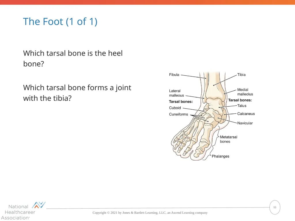
The Foot (1 of 1) Which tarsal bone is the heel bone?
Which tarsal bone forms a joint with the tibia? 55<br>
Which tarsal bone forms a joint with the tibia? 55<br>
56
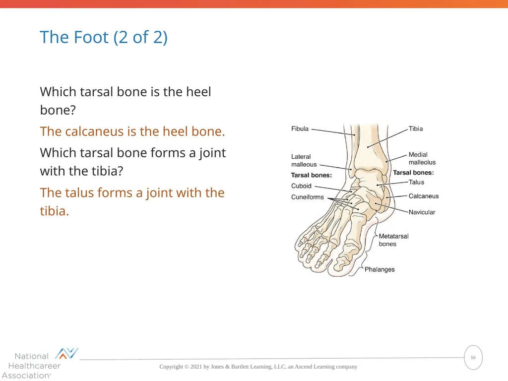
The Foot (2 of 2) Which tarsal bone is the heel bone?
The calcaneus is the heel bone.
Which tarsal bone forms a joint with the tibia?
The talus forms a joint with the tibia. 56<br>
The calcaneus is the heel bone.
Which tarsal bone forms a joint with the tibia?
The talus forms a joint with the tibia. 56<br>
57
Appendicular Skeleton What are the four regions of the appendicular skeleton?
The four regions of the appendicular skeleton are the shoulder girdle, upper extremity, pelvic bones, and lower extremity. 57<br>
The four regions of the appendicular skeleton are the shoulder girdle, upper extremity, pelvic bones, and lower extremity. 57<br>
58
Pop Quiz (4 of 4) Which of the following bones is found in the shoulder girdle?
Sternum
Humerus
Scapula
Ulna 58<br>
Sternum
Humerus
Scapula
Ulna 58<br>
59
Pop Quiz Answer (4 of 4) Which of the following bones is found in the shoulder girdle?
Sternum
Humerus
Scapula
Ulna The correct answer is Scapula 59<br>
Sternum
Humerus
Scapula
Ulna The correct answer is Scapula 59<br>
60
Disorders of Bone Metabolic diseases
Tumors
Infections
Structural problems 60<br>
Tumors
Infections
Structural problems 60<br>
61
Metabolic Disorders of Bone Characterized by a lack of normal bone formation or excess loss of bone tissue
Osteopenia
Osteoporosis
Osteitis deformans
Osteomalacia 61<br>
Osteopenia
Osteoporosis
Osteitis deformans
Osteomalacia 61<br>
62
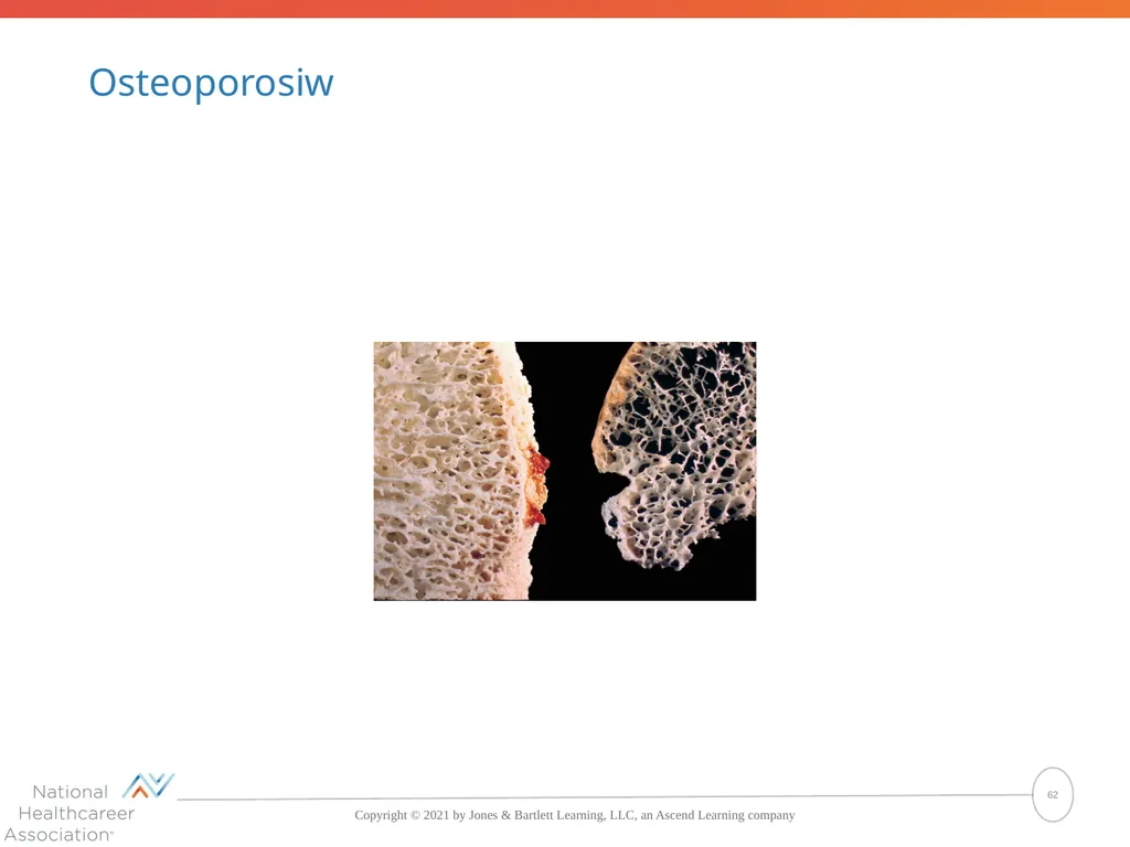
Osteoporosiw 62<br>
63
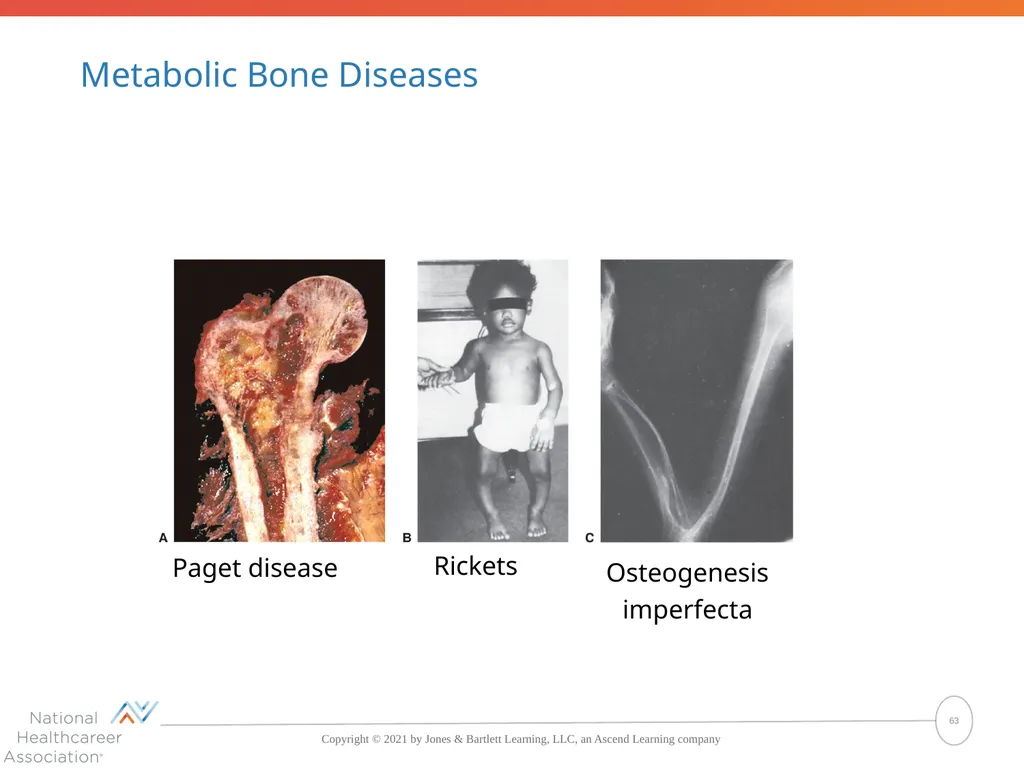
Metabolic Bone Diseases Paget disease Rickets Osteogenesis imperfecta 63<br>
64
Bone Tumors Benign: osteoma
Malignant
Osteosarcoma (bone cells)
Chondrosarcoma (cartilage cells) 64<br>
Malignant
Osteosarcoma (bone cells)
Chondrosarcoma (cartilage cells) 64<br>
65
Infections of Bone Osteomyelitis
Inflammation of the bone caused by pyogenic bacteria
Tuberculosis
Pott disease (tuberculosis of the spine) 65<br>
Inflammation of the bone caused by pyogenic bacteria
Tuberculosis
Pott disease (tuberculosis of the spine) 65<br>
66
Structural Disorders of Bone Spinal curve abnormalities
Kyphosis (hunchback)
Lordosis (swayback)
Scoliosis
Cleft palate
Flatfoot 66<br>
Kyphosis (hunchback)
Lordosis (swayback)
Scoliosis
Cleft palate
Flatfoot 66<br>
67
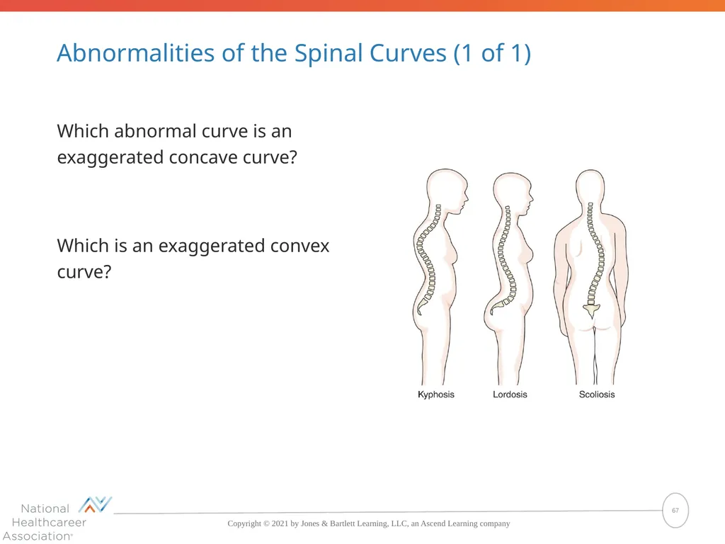
Abnormalities of the Spinal Curves (1 of 1) Which abnormal curve is an exaggerated concave curve?
Which is an exaggerated convex curve? 67<br>
Which is an exaggerated convex curve? 67<br>
68
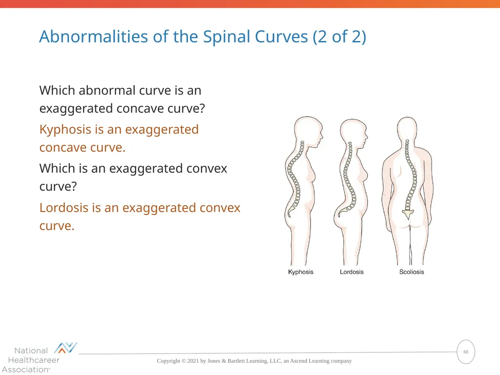
Abnormalities of the Spinal Curves (2 of 2) Which abnormal curve is an exaggerated concave curve?
Kyphosis is an exaggerated concave curve.
Which is an exaggerated convex curve?
Lordosis is an exaggerated convex curve. 68<br>
Kyphosis is an exaggerated concave curve.
Which is an exaggerated convex curve?
Lordosis is an exaggerated convex curve. 68<br>
69
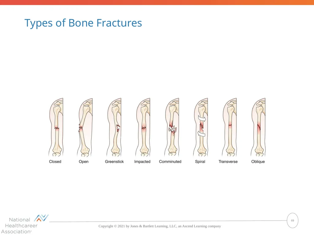
Types of Bone Fractures 69<br>
70
Disorders of Bone (1 of 1) What are five categories of bone disorders?
The five categories of bone disorders are metabolic disorders, tumors, infections, structural disorders, and fractures. 70<br>
The five categories of bone disorders are metabolic disorders, tumors, infections, structural disorders, and fractures. 70<br>
71
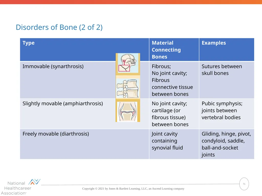
Disorders of Bone (2 of 2) 71<br>
72
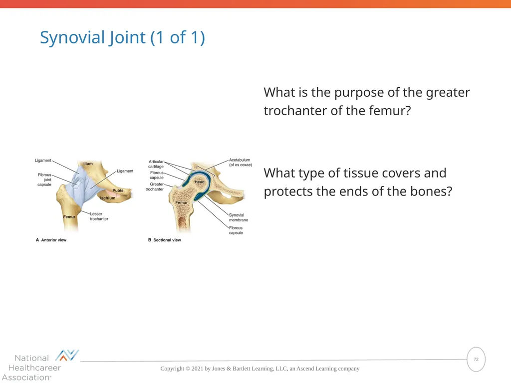
Synovial Joint (1 of 1) What is the purpose of the greater trochanter of the femur?
What type of tissue covers and protects the ends of the bones? 72<br>
What type of tissue covers and protects the ends of the bones? 72<br>
73
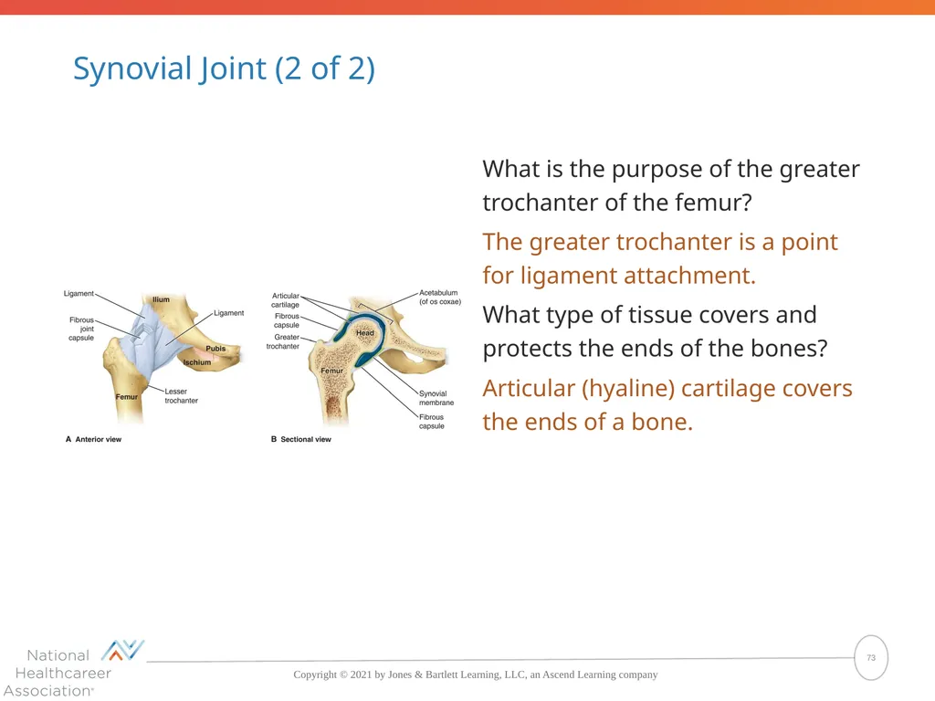
Synovial Joint (2 of 2) What is the purpose of the greater trochanter of the femur?
The greater trochanter is a point for ligament attachment.
What type of tissue covers and protects the ends of the bones?
Articular (hyaline) cartilage covers the ends of a bone. 73<br>
The greater trochanter is a point for ligament attachment.
What type of tissue covers and protects the ends of the bones?
Articular (hyaline) cartilage covers the ends of a bone. 73<br>
74
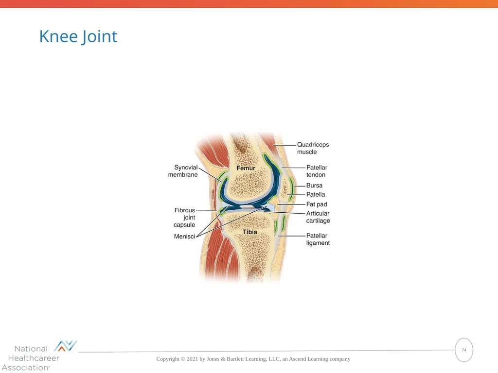
Knee Joint 74<br>
75
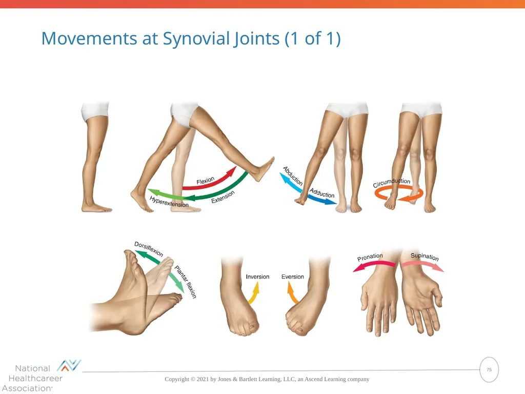
Movements at Synovial Joints (1 of 1) 75<br>
76
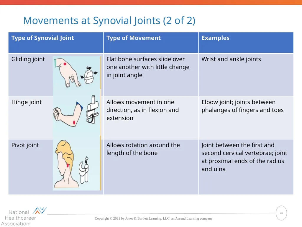
Movements at Synovial Joints (2 of 2) 76<br>
77
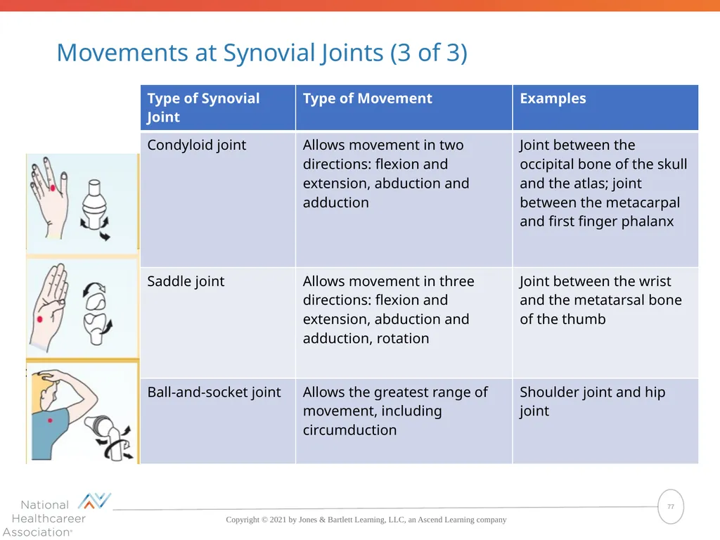
Movements at Synovial Joints (3 of 3) 77<br>
78
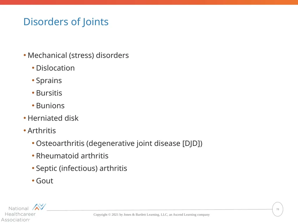
Disorders of Joints Mechanical (stress) disorders
Dislocation
Sprains
Bursitis
Bunions
Herniated disk
Arthritis
Osteoarthritis (degenerative joint disease [DJD])
Rheumatoid arthritis
Septic (infectious) arthritis
Gout 78<br>
Dislocation
Sprains
Bursitis
Bunions
Herniated disk
Arthritis
Osteoarthritis (degenerative joint disease [DJD])
Rheumatoid arthritis
Septic (infectious) arthritis
Gout 78<br>
79
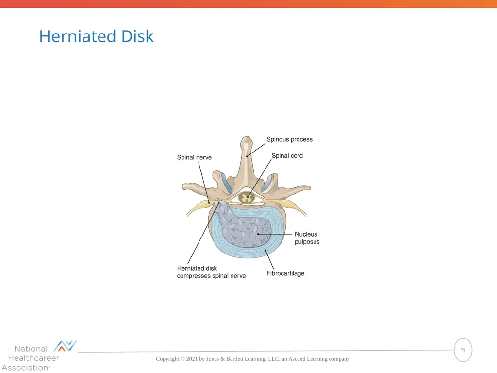
Herniated Disk 79<br>
80
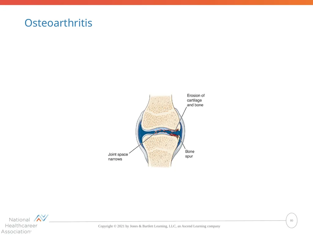
Osteoarthritis 80<br>
81
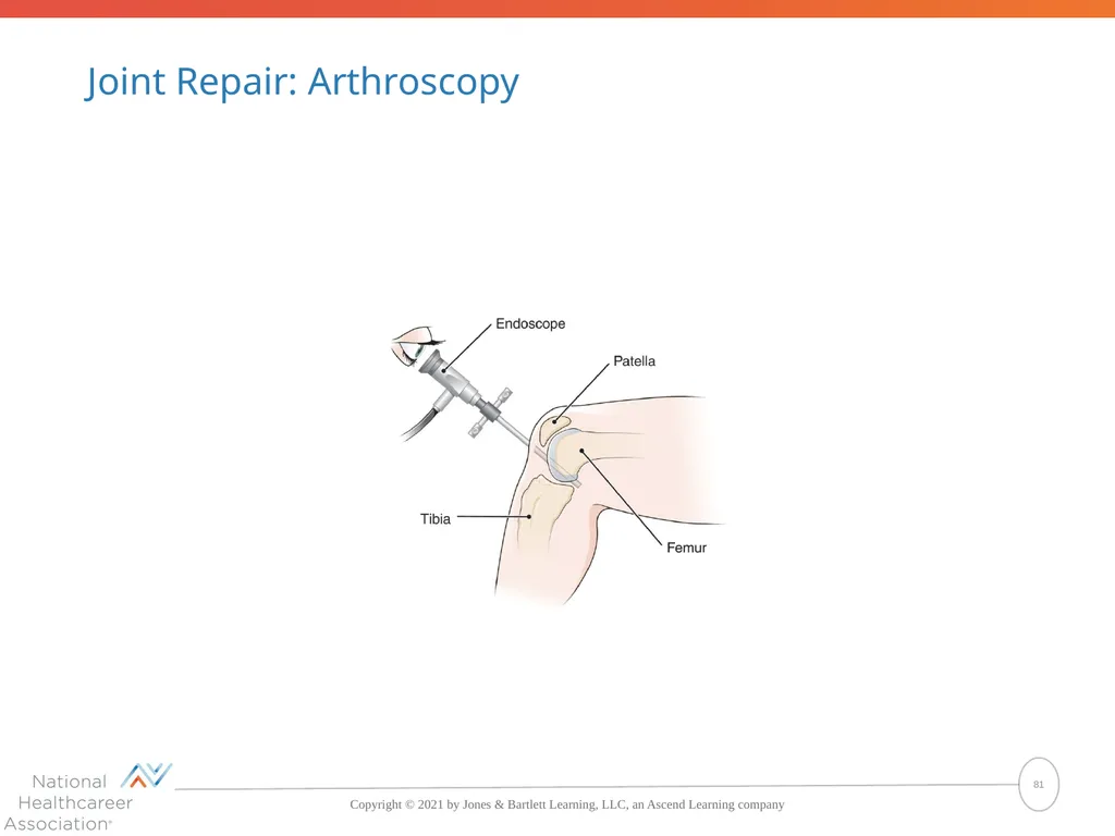
Joint Repair: Arthroscopy 81<br>
82
Joints What are three types of joints based on the degree of movement they allow?
The three types of joins based on the degree of movement allowed are synarthrosis, amphiarthrosis, and diarthrosis.
What is the most freely movable type of joint?
A synovial joint or diarthrosis
What is the most common type of joint disorder?
Arthritis is the most common type of joint disorder
What type of endoscope is used to examine and repair joints?
An arthroscope is used to examine and repair joints. 82<br>
The three types of joins based on the degree of movement allowed are synarthrosis, amphiarthrosis, and diarthrosis.
What is the most freely movable type of joint?
A synovial joint or diarthrosis
What is the most common type of joint disorder?
Arthritis is the most common type of joint disorder
What type of endoscope is used to examine and repair joints?
An arthroscope is used to examine and repair joints. 82<br>
83
Pop Quiz (5 of 5) Freely movable joints are also called which of the following?
Fibrous joints
Cartilaginous joints
Diarthroses
Amphiarthroses 83<br>
Fibrous joints
Cartilaginous joints
Diarthroses
Amphiarthroses 83<br>
84
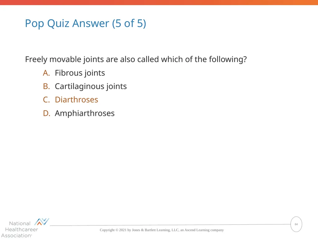
Pop Quiz Answer (5 of 5) Freely movable joints are also called which of the following?
Fibrous joints
Cartilaginous joints
Diarthroses
Amphiarthroses The correct answer is Diarthroses 84<br>
Fibrous joints
Cartilaginous joints
Diarthroses
Amphiarthroses The correct answer is Diarthroses 84<br>
85
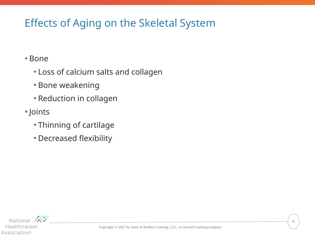
Effects of Aging on the Skeletal System Bone
Loss of calcium salts and collagen
Bone weakening
Reduction in collagen
Joints
Thinning of cartilage
Decreased flexibility 85<br>
Loss of calcium salts and collagen
Bone weakening
Reduction in collagen
Joints
Thinning of cartilage
Decreased flexibility 85<br>
86
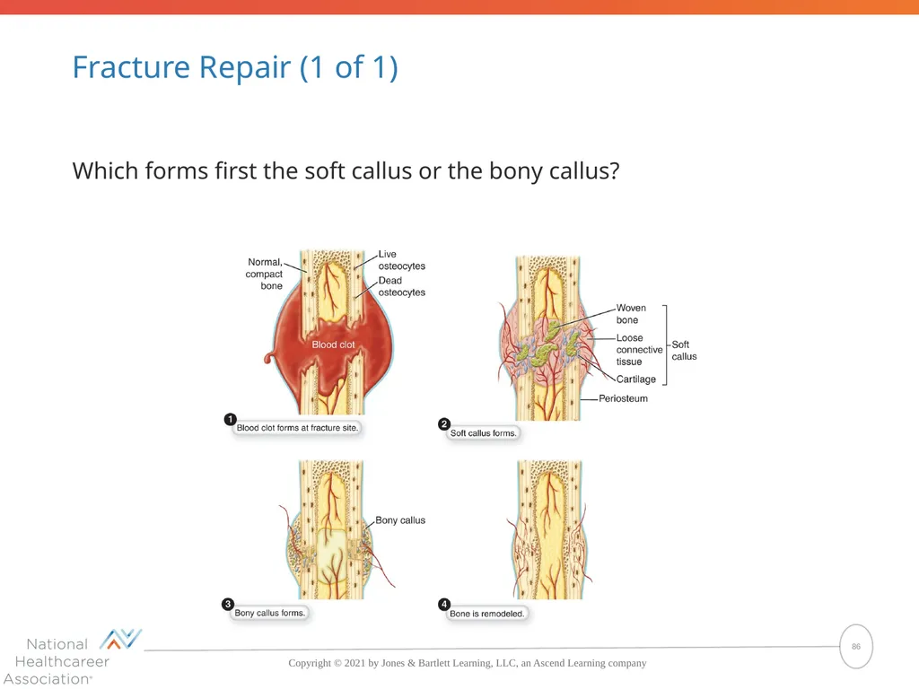
Fracture Repair (1 of 1) Which forms first the soft callus or the bony callus? 86<br>
87
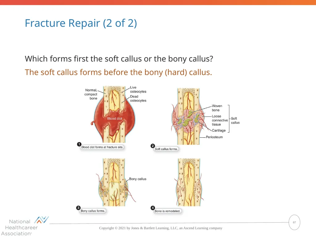
Fracture Repair (2 of 2) Which forms first the soft callus or the bony callus?
The soft callus forms before the bony (hard) callus. 87<br>
The soft callus forms before the bony (hard) callus. 87<br>
88
How Fractures Heal Blood clot forms around fracture.
Blood vessels grow into clot, pulling connective tissue with them.
Fibroblasts and chondroblasts convert clot into a soft callus.
Macrophages remove remains of blood clot, and osteoclasts resorb dead bone tissue.
Osteoblasts convert soft callus into a hard callus of spongy bone.
Osteoclasts and osteoblasts remodel hard callus into compact bone. 88<br>
Blood vessels grow into clot, pulling connective tissue with them.
Fibroblasts and chondroblasts convert clot into a soft callus.
Macrophages remove remains of blood clot, and osteoclasts resorb dead bone tissue.
Osteoblasts convert soft callus into a hard callus of spongy bone.
Osteoclasts and osteoblasts remodel hard callus into compact bone. 88<br>