
Tissue Processing Dr. Rucha Gore HISTOLOGY : It is




Description: Tissue Processing Dr. Rucha Gore HISTOLOGY : It is the branch of science which deals with the gross microscopic study of normal tissue . HISTOPATHOLOGY : It is the branch of science which deals with the gross microscopic study of tissue
Related Topics
Download Presentation
"Tissue Processing Dr. Rucha Gore HISTOLOGY : It is" is the property of its rightful owner. Permission is granted to download and print the materials on this website for personal, non-commercial use only, and to display it on your personal computer provided you do not modify the materials and that you retain all copyright notices contained in the materials. By downloading content from our website, you accept the terms of this agreement.
Presentation Transcript
slide1. Tissue Processing Dr. Rucha Gore<br>
slide2. HISTOLOGY :
It is the branch of science which deals with the gross & microscopic study of normal tissue .
HISTOPATHOLOGY :
It is the branch of science which deals with the gross & microscopic study of tissue affected by disease.
Tissue for study can be obtained from:
Biopsies
Autopsies<br>
slide3. HISTOTECHNIQUES:
The techniques for processing the tissues, whether biopsies, larger specimen removed at surgery, or tissues from autopsy so as to enable the pathologist to study them under the microscope.<br>
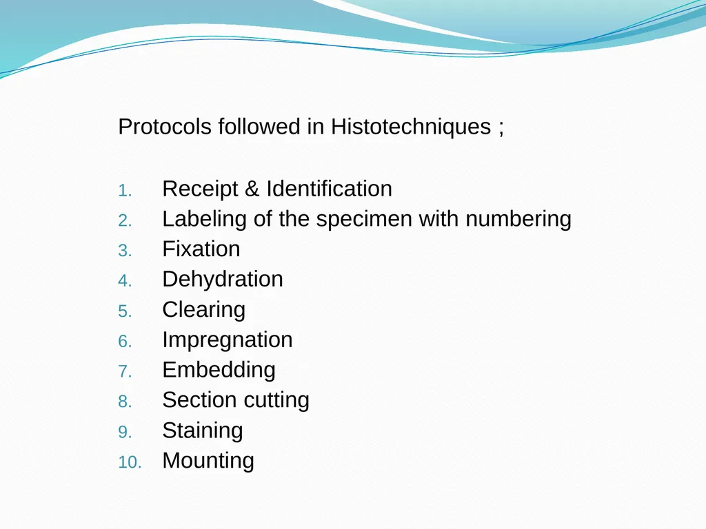
slide8. Protocols followed in Histotechniques ;
Receipt & Identification
Labeling of the specimen with numbering
Fixation
Dehydration
Clearing
Impregnation
Embedding
Section cutting
Staining
Mounting<br>
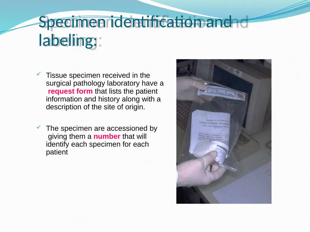
slide9. Specimen identification and labeling: Tissue specimen received in the surgical pathology laboratory have a request form that lists the patient information and history along with a description of the site of origin.
The specimen are accessioned by giving them a number that will identify each specimen for each patient<br>
slide10. Fixation It is a process in which a specimen is treated by exposing it to a fixative for a particular period of time in order to facilitate the succeeding steps.
The purpose of fixation is to preserve tissues permanently in as life-like a state as possible.
The fixative should be 15 – 20 times more in volume then the specimen.<br>
slide11. Aims of Fixation :
It should prevent autolysis & putrefaction of the cell.
It should penetrate evenly and rapidly.
It should harden the tissues
Increase the optical density
Should not cause shrinkage or swelling of the cells
Must not react with the receptor sites & thus must not
interefere with the staining procedure.
It must be cheap and easily available.<br>
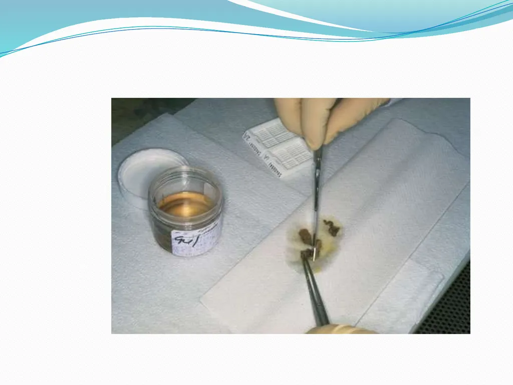
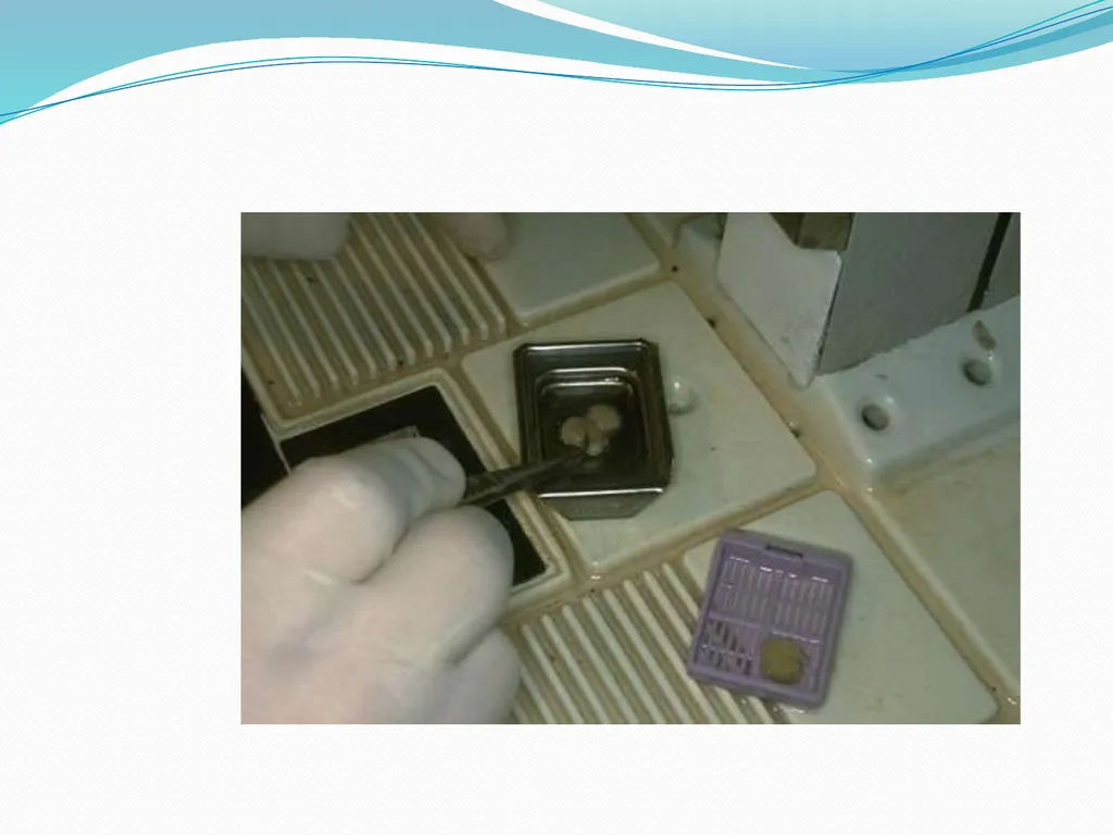
slide12. The bits should of the size of approximately 2 x 2 cm & 4- 6 micrometer in thickness for optimum fixation to take place.
These bits are then placed in metal cassettes or capsules which are then placed in the fixative.
Tiny biopsies or small specimen can be wrapped in a filter paper and then put in a cassette & fixed.<br>
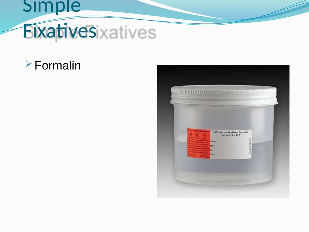
slide15. Simple Fixatives Formalin<br>
slide16. The most commonly used fixative is Formalin .
It is prepared by mixing 40 % Formaldehyde gas in 100 w/v of
distilled water.
The resultant mixture is 100 % Formalin.
Routinely, 10 % formalin is used which is prepared by mixing 10
ml of 100 % formalin in 90 ml of distilled water.
MECHANISM OF ACTION:
It forms cross links between amino acids of proteins thereby making them insoluble.
It fixes 4 mm thick tissue in 8 hours .<br>
slide17. ADVANTAGES : 1.
2.
3.
4.
5. Rapid penetration
Easy availability & cheap
Does not overharden the tissue Fixes lipids for frozen sections Ideal for mailing DISADVANTAGES: 1.
2. 3. Irritant to the nose,the eyes and mucous membranes
Formation of precipitate of paraformaldehyde which can be prevented by adding 11- 16 % methanol.
Formation of black formalin pigment , Acid formaldehyde hematin.<br>
slide18. Other Simple Fixatives Glutaraldehyde
Osmium Tetraoxide
Pottasium Dichromate
Mercuric Chloride<br>
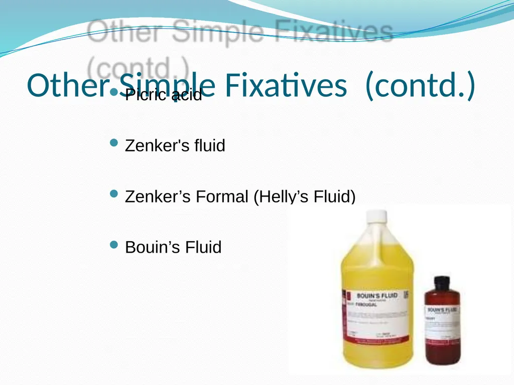
slide19. Other Simple Fixatives (contd.) Picric acid
Zenker's fluid
Zenker’s Formal (Helly’s Fluid)
Bouin’s Fluid<br>
slide20. Decalcification: It is the process of removal of the calcium salts from the specimen.
The various agents used for decalcifying are;
Nitric acid
Hydrochloric acid
Formic acid
Picric acid
Acetic acid
Citric acid<br>
slide21. Dehydration: It is the process in which the water content in the tissue to be processed is completely reduced by passing the tissue through increasing concentrations of dehydrating agents.
The various dehydrating agents used are ;
Ethyl alcohol
Acetone
Isopropyl alcohol
Dioxane<br>
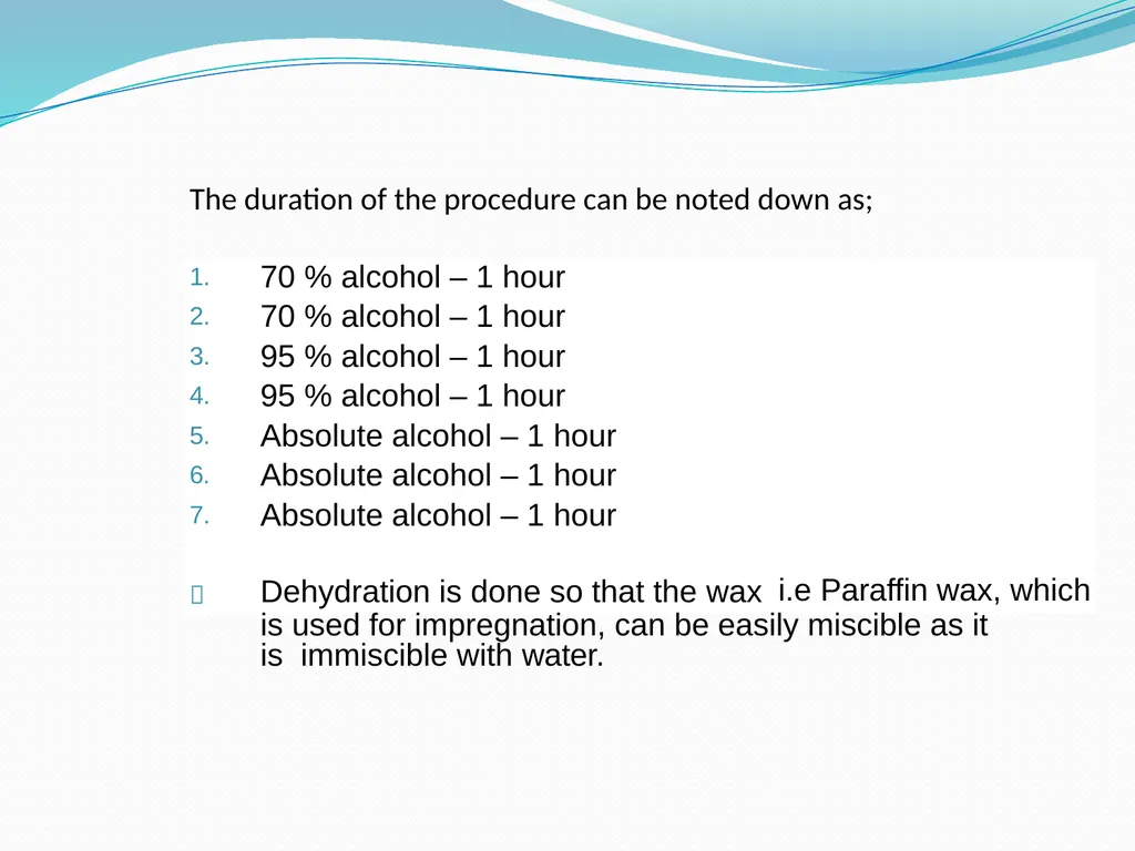
slide22. The duration of the procedure can be noted down as; is used for impregnation, can be easily miscible as it is immiscible with water.<br>
slide23. CLEARING ( DEALCOHOLIZATION): It is the procedure where in the alcohol in the tissue is
replaced by a fluid which will dissolve the wax used for
impregnating the tissues .
The various clearing agents used are ;
Cedar wood oil : The best agent but is expensive.
Benzene : It is carcinogenic.
Xylene : It is most commonly used.
Chloroform: Toxic and expensive.<br>
slide24. Impregnation: In this the tissue is kept in a wax bath containing molten paraffin wax for 6 – 8 hours .
The wax is infiltrated in the interices of the tissue which increases the optical differentiation & hardens the tissue & helps in easy sectioning of the tissue.<br>
slide25. The various waxes which are used are,
Paraffin wax
Paraplast
Paraplast plus
Gelatin
Celloidin<br>
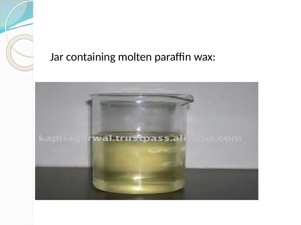
slide26. Jar containing molten paraffin wax:<br>
slide27. Embedding : It is done by transfering the tissue which has been cleared of the alcohol to a mould filled with molten wax & is allowed to cool & solidify.
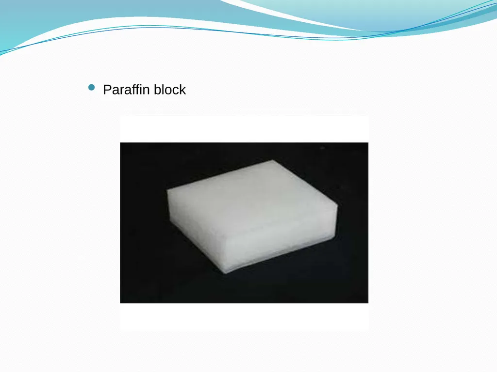
After solidification, a wax block is obtained which is then sectioned to obtain ribbons.<br>
slide28. Types of Moulds:
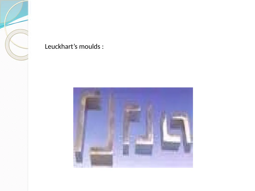
Leuckhart’s Moulds:
L- shaped brass pieces which is placed in opposing positions & can be manipulated to increase or decrease the size of the block to be prepared.
Glass or Metal petri dishes :
Watch glass
Paper boats .<br>
slide29. Leuckhart’s moulds :<br>
slide30. Paraffin block<br>
slide31. Section Cutting : It is the procedure in which the blocks which have been prepared are cut or sectioned and thin strips of varying thickness are prepared.
The instrument by which this is done is called as a Microtome.
TYPES OF MICROTOMES:
Sliding
Rotary
Rocking
Freezing
Base sledge<br>
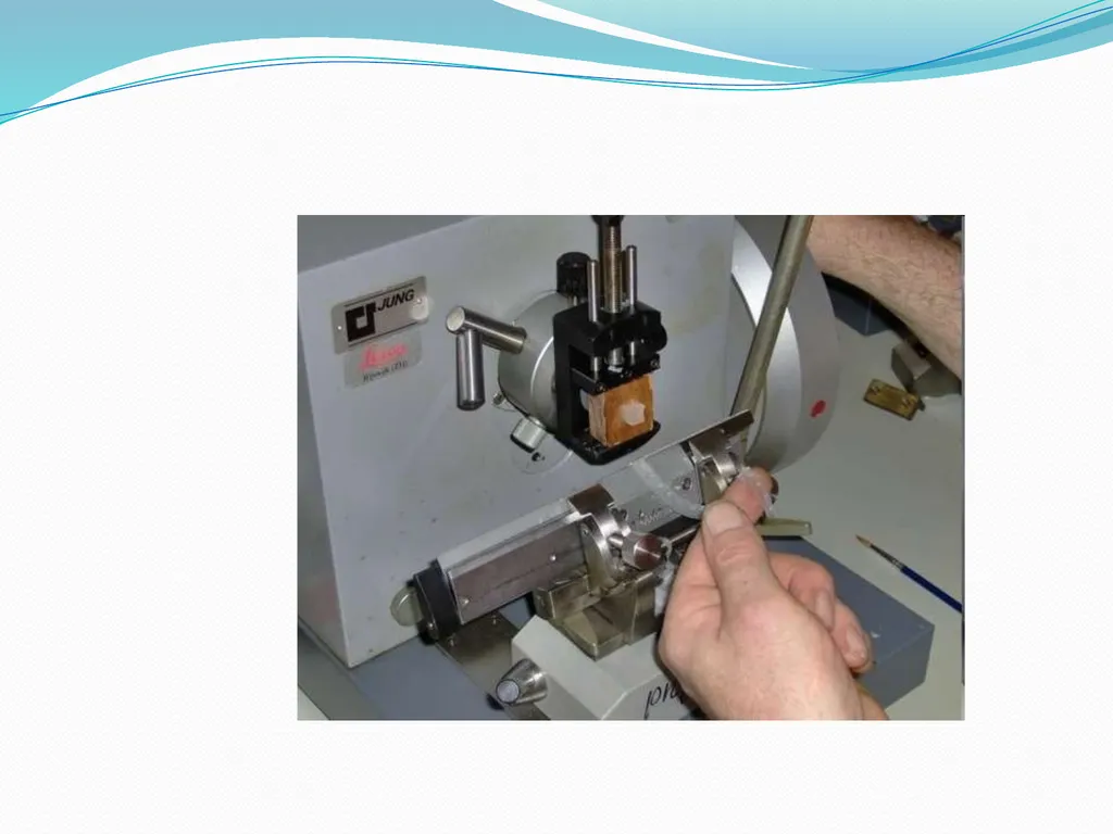
slide32. Rotary Microtome:
It is the most commonly used.
Also known as Minnot’s Rotary microtome.
In this the Block holder moves up and down while the knife
remains fixed.
It is suitable for cutting of small tissues & serial sections can be taken on it.<br>
slide33. Parts of a Microtome ( Rotary ) :
Block holder
Knife clamp screws
Knife clamps
Block adjustment
Thickness gauge
Angle of tilt adjustment
Operating handle.<br>
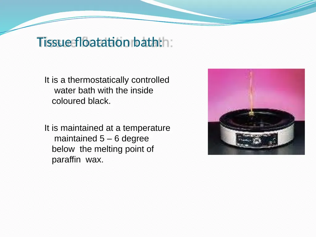
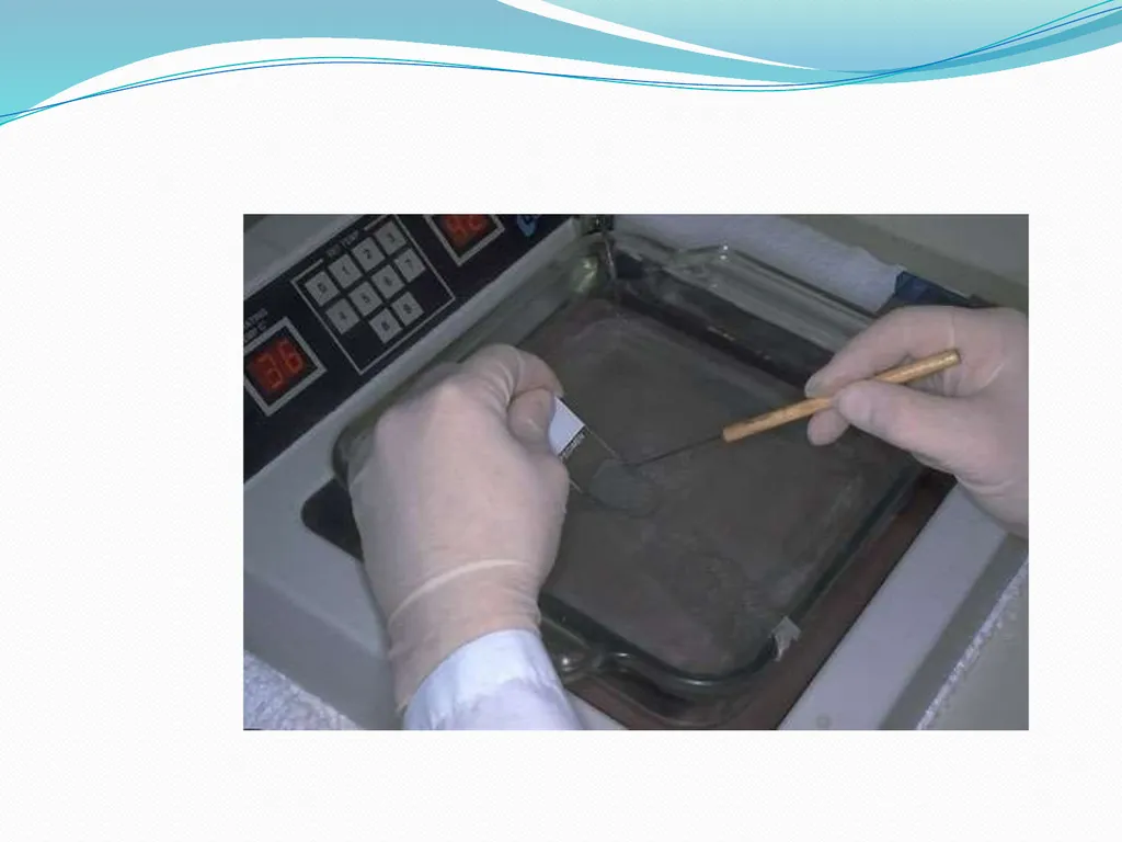
slide35. Tissue floatation bath: It is a thermostatically controlled water bath with the inside coloured black.
It is maintained at a temperature maintained 5 – 6 degree below the melting point of paraffin wax.<br>
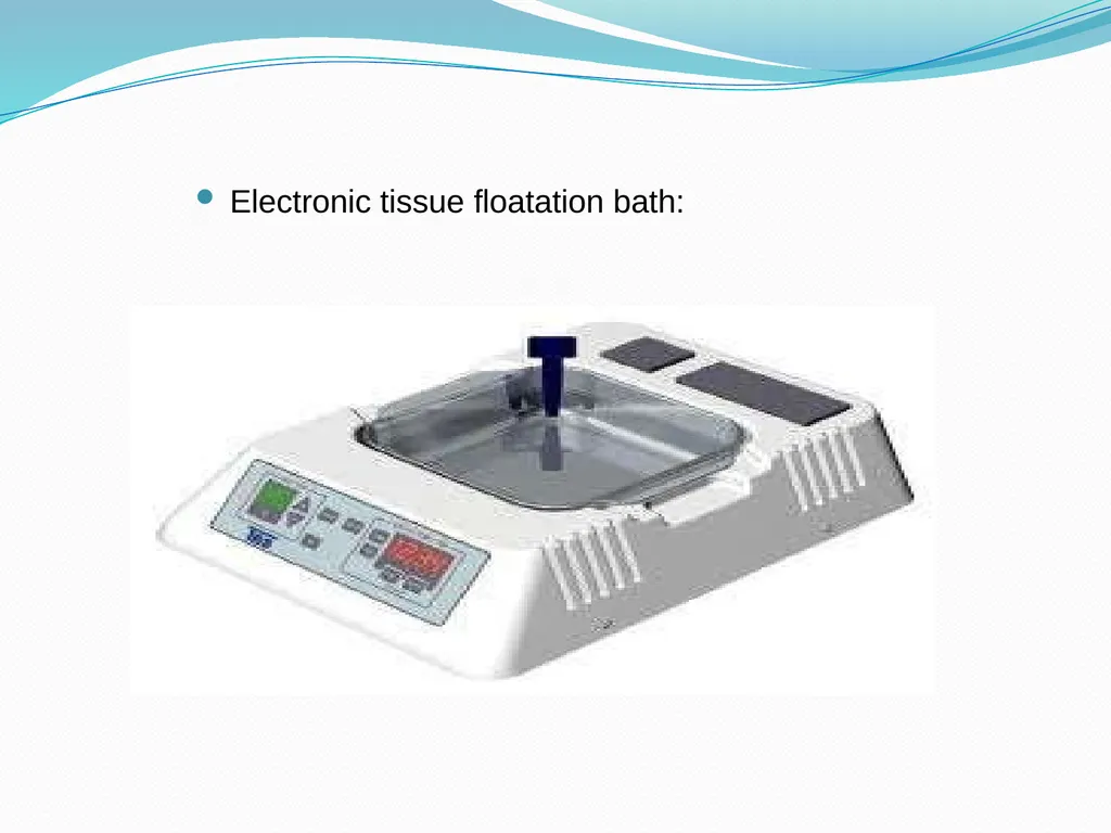
slide36. Electronic tissue floatation bath:<br>
slide39. Staining : Staining of the section is done to bring out the particular details
in the tissue under study .
The most commonly used stain in routine practice is Haematoxylin & eosin stain.<br>
slide40. Procedure :
Deparaffinization with xylene.
Hydration
Wash under water
Stain with Haematoxylin for 15 min
Wash with water
Differentiate with 1 % acid alcohol
Wash with water for 10 min
Stain with 1% Eosin for 2 min
Wash with water.
Dehydration
Clearing with xylene
Dry
Mount<br>
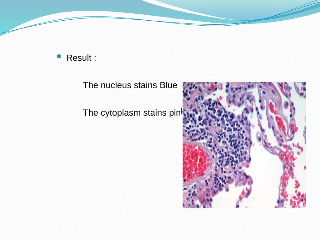
slide41. Result :
The nucleus stains Blue
The cytoplasm stains pin k.<br>
slide42. Mounting: Adhesives used for fixing the sections on the slides :
Albumin solution ( Mayor’s egg albumin)
Starch paste
Gelatin<br>
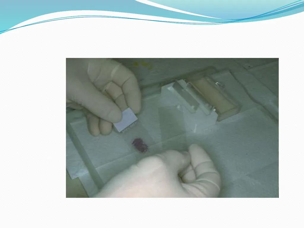
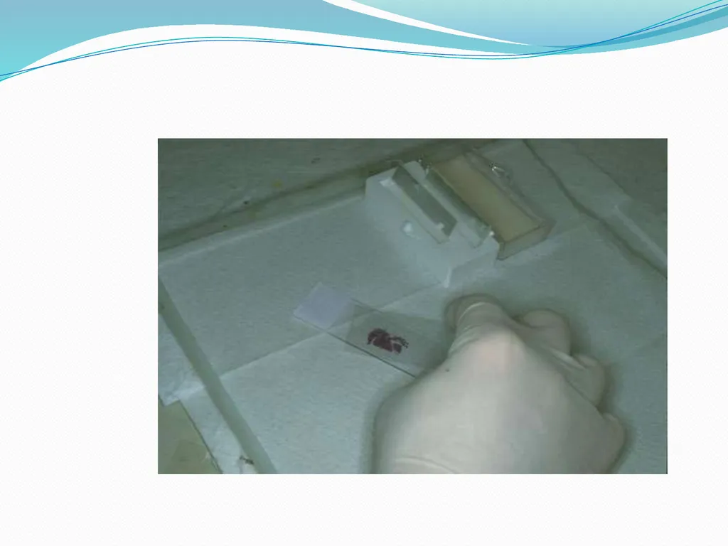
slide43. Mountants :
DPX ( Distrene Dibutyl phthalate Xylene ).
Canada Balsam
Colophonium resin
Terpene resin<br>
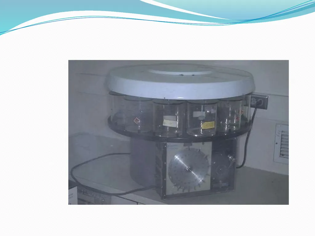
slide46. Automation: Automated tissue processor:
All the before mentioned procedures upto the impregnation step can be done automatically in a single, unmanned instrument , which is the Automated Tissue processor.
Advantages :
It provides constant agitation during every step which
ensures better fixation & processing.
It reduces the work load & in turns improves the overall output of the laboratory.<br>
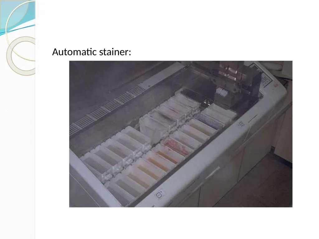
slide48. Automatic stainer:<br>
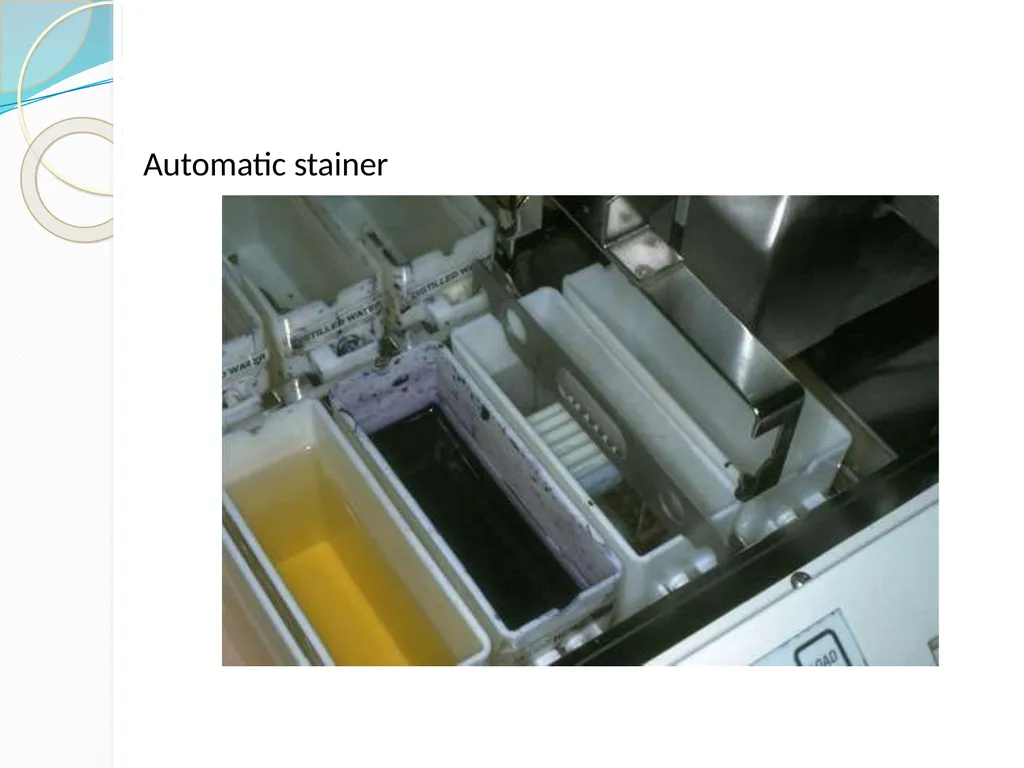
slide49. Automatic stainer<br>
slide50. Thank You !<br>
slide2. HISTOLOGY :
It is the branch of science which deals with the gross & microscopic study of normal tissue .
HISTOPATHOLOGY :
It is the branch of science which deals with the gross & microscopic study of tissue affected by disease.
Tissue for study can be obtained from:
Biopsies
Autopsies<br>
slide3. HISTOTECHNIQUES:
The techniques for processing the tissues, whether biopsies, larger specimen removed at surgery, or tissues from autopsy so as to enable the pathologist to study them under the microscope.<br>
slide8. Protocols followed in Histotechniques ;
Receipt & Identification
Labeling of the specimen with numbering
Fixation
Dehydration
Clearing
Impregnation
Embedding
Section cutting
Staining
Mounting<br>
slide9. Specimen identification and labeling: Tissue specimen received in the surgical pathology laboratory have a request form that lists the patient information and history along with a description of the site of origin.
The specimen are accessioned by giving them a number that will identify each specimen for each patient<br>
slide10. Fixation It is a process in which a specimen is treated by exposing it to a fixative for a particular period of time in order to facilitate the succeeding steps.
The purpose of fixation is to preserve tissues permanently in as life-like a state as possible.
The fixative should be 15 – 20 times more in volume then the specimen.<br>
slide11. Aims of Fixation :
It should prevent autolysis & putrefaction of the cell.
It should penetrate evenly and rapidly.
It should harden the tissues
Increase the optical density
Should not cause shrinkage or swelling of the cells
Must not react with the receptor sites & thus must not
interefere with the staining procedure.
It must be cheap and easily available.<br>
slide12. The bits should of the size of approximately 2 x 2 cm & 4- 6 micrometer in thickness for optimum fixation to take place.
These bits are then placed in metal cassettes or capsules which are then placed in the fixative.
Tiny biopsies or small specimen can be wrapped in a filter paper and then put in a cassette & fixed.<br>
slide15. Simple Fixatives Formalin<br>
slide16. The most commonly used fixative is Formalin .
It is prepared by mixing 40 % Formaldehyde gas in 100 w/v of
distilled water.
The resultant mixture is 100 % Formalin.
Routinely, 10 % formalin is used which is prepared by mixing 10
ml of 100 % formalin in 90 ml of distilled water.
MECHANISM OF ACTION:
It forms cross links between amino acids of proteins thereby making them insoluble.
It fixes 4 mm thick tissue in 8 hours .<br>
slide17. ADVANTAGES : 1.
2.
3.
4.
5. Rapid penetration
Easy availability & cheap
Does not overharden the tissue Fixes lipids for frozen sections Ideal for mailing DISADVANTAGES: 1.
2. 3. Irritant to the nose,the eyes and mucous membranes
Formation of precipitate of paraformaldehyde which can be prevented by adding 11- 16 % methanol.
Formation of black formalin pigment , Acid formaldehyde hematin.<br>
slide18. Other Simple Fixatives Glutaraldehyde
Osmium Tetraoxide
Pottasium Dichromate
Mercuric Chloride<br>
slide19. Other Simple Fixatives (contd.) Picric acid
Zenker's fluid
Zenker’s Formal (Helly’s Fluid)
Bouin’s Fluid<br>
slide20. Decalcification: It is the process of removal of the calcium salts from the specimen.
The various agents used for decalcifying are;
Nitric acid
Hydrochloric acid
Formic acid
Picric acid
Acetic acid
Citric acid<br>
slide21. Dehydration: It is the process in which the water content in the tissue to be processed is completely reduced by passing the tissue through increasing concentrations of dehydrating agents.
The various dehydrating agents used are ;
Ethyl alcohol
Acetone
Isopropyl alcohol
Dioxane<br>
slide22. The duration of the procedure can be noted down as; is used for impregnation, can be easily miscible as it is immiscible with water.<br>
slide23. CLEARING ( DEALCOHOLIZATION): It is the procedure where in the alcohol in the tissue is
replaced by a fluid which will dissolve the wax used for
impregnating the tissues .
The various clearing agents used are ;
Cedar wood oil : The best agent but is expensive.
Benzene : It is carcinogenic.
Xylene : It is most commonly used.
Chloroform: Toxic and expensive.<br>
slide24. Impregnation: In this the tissue is kept in a wax bath containing molten paraffin wax for 6 – 8 hours .
The wax is infiltrated in the interices of the tissue which increases the optical differentiation & hardens the tissue & helps in easy sectioning of the tissue.<br>
slide25. The various waxes which are used are,
Paraffin wax
Paraplast
Paraplast plus
Gelatin
Celloidin<br>
slide26. Jar containing molten paraffin wax:<br>
slide27. Embedding : It is done by transfering the tissue which has been cleared of the alcohol to a mould filled with molten wax & is allowed to cool & solidify.
After solidification, a wax block is obtained which is then sectioned to obtain ribbons.<br>
slide28. Types of Moulds:
Leuckhart’s Moulds:
L- shaped brass pieces which is placed in opposing positions & can be manipulated to increase or decrease the size of the block to be prepared.
Glass or Metal petri dishes :
Watch glass
Paper boats .<br>
slide29. Leuckhart’s moulds :<br>
slide30. Paraffin block<br>
slide31. Section Cutting : It is the procedure in which the blocks which have been prepared are cut or sectioned and thin strips of varying thickness are prepared.
The instrument by which this is done is called as a Microtome.
TYPES OF MICROTOMES:
Sliding
Rotary
Rocking
Freezing
Base sledge<br>
slide32. Rotary Microtome:
It is the most commonly used.
Also known as Minnot’s Rotary microtome.
In this the Block holder moves up and down while the knife
remains fixed.
It is suitable for cutting of small tissues & serial sections can be taken on it.<br>
slide33. Parts of a Microtome ( Rotary ) :
Block holder
Knife clamp screws
Knife clamps
Block adjustment
Thickness gauge
Angle of tilt adjustment
Operating handle.<br>
slide35. Tissue floatation bath: It is a thermostatically controlled water bath with the inside coloured black.
It is maintained at a temperature maintained 5 – 6 degree below the melting point of paraffin wax.<br>
slide36. Electronic tissue floatation bath:<br>
slide39. Staining : Staining of the section is done to bring out the particular details
in the tissue under study .
The most commonly used stain in routine practice is Haematoxylin & eosin stain.<br>
slide40. Procedure :
Deparaffinization with xylene.
Hydration
Wash under water
Stain with Haematoxylin for 15 min
Wash with water
Differentiate with 1 % acid alcohol
Wash with water for 10 min
Stain with 1% Eosin for 2 min
Wash with water.
Dehydration
Clearing with xylene
Dry
Mount<br>
slide41. Result :
The nucleus stains Blue
The cytoplasm stains pin k.<br>
slide42. Mounting: Adhesives used for fixing the sections on the slides :
Albumin solution ( Mayor’s egg albumin)
Starch paste
Gelatin<br>
slide43. Mountants :
DPX ( Distrene Dibutyl phthalate Xylene ).
Canada Balsam
Colophonium resin
Terpene resin<br>
slide46. Automation: Automated tissue processor:
All the before mentioned procedures upto the impregnation step can be done automatically in a single, unmanned instrument , which is the Automated Tissue processor.
Advantages :
It provides constant agitation during every step which
ensures better fixation & processing.
It reduces the work load & in turns improves the overall output of the laboratory.<br>
slide48. Automatic stainer:<br>
slide49. Automatic stainer<br>
slide50. Thank You !<br>