To: Pharm. D II yr. Students Sub: Pharmaceutical Microbiology By J. SIVA JYOTHI Asst. Professor HINDU COLLEGE OF PHARMACY GUNTUR RICKETTSIA INTRODUCTION Obligate intracellular parasites. Gram- negative pleomorphic rods. Parasite of
"To: Pharm. D II yr. Students Sub: Pharmaceutical" is the property of its rightful owner. Permission is granted to
download and print the materials on this website for personal, non-commercial use only, and to display it
on your personal computer provided you do not modify the materials and that you retain all copyright
notices contained in the materials. By downloading content from our website, you accept the terms of this
agreement.
Presentation Transcript
01
To: Pharm. D II yr. Students
Sub: Pharmaceutical Microbiology
By
J. SIVA JYOTHI
Asst. Professor
HINDU COLLEGE OF PHARMACY
GUNTUR RICKETTSIA<br>
02
INTRODUCTION Obligate intracellular parasites.
Gram- negative pleomorphic rods.
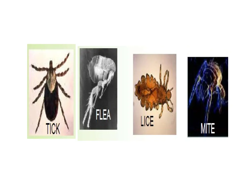
Parasite of arthropods – fleas, lice, ticks and mites.
No Human to human transmission.
Despite the similar name, Rickettsia bacteria do not cause rickets, which is a result of vitamin D deficiency.
In the past, positioned somewhere between viruses and true bacteria.<br>
03
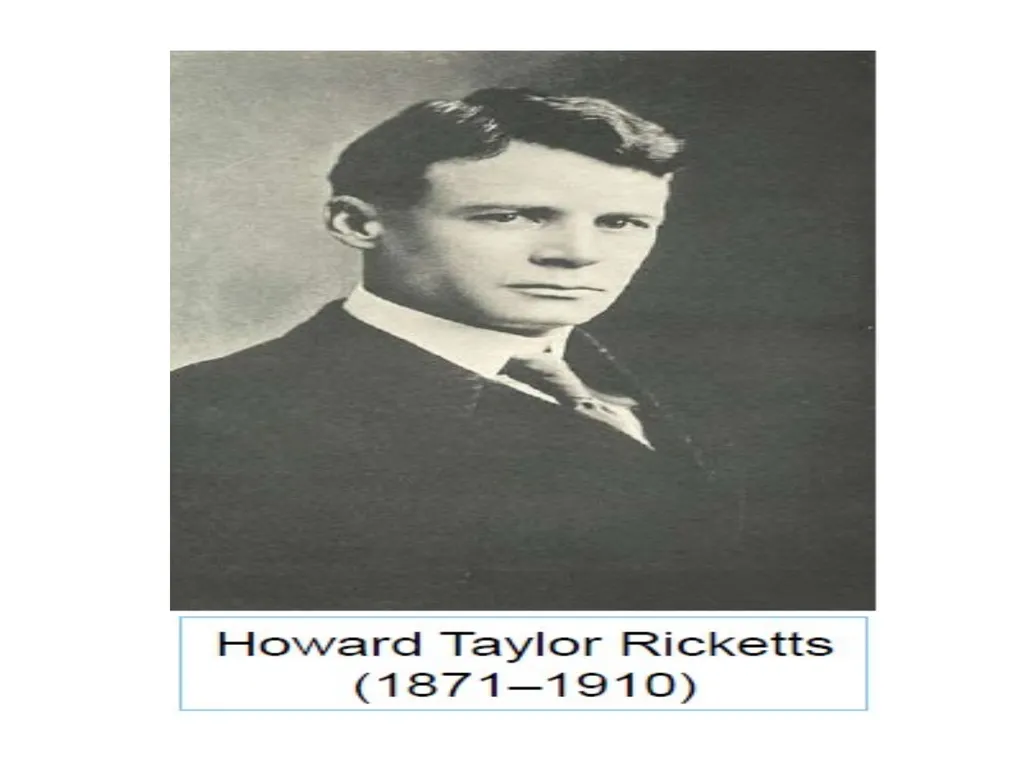
NOMENCLATURE The genus Rickettsia is named after Howard Taylor Ricketts (1871–1910), who studied Rocky Mountain spotted fever in the Bitterroot Valley of Montana.
Ricketts was devoted to his research and, on several occasions, injected himself with pathogens to study their effects.
In 1910, Ricketts became interested in a strain of typhus known as tabardillo, due to a major outbreak in Mexico City.
Days after isolating the organism that he believed caused typhus, he himself died of the disease.<br>
04
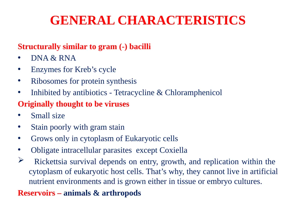
GENERAL CHARACTERISTICS Structurally similar to gram (-) bacilli
DNA & RNA
Enzymes for Kreb’s cycle
Ribosomes for protein synthesis
Inhibited by antibiotics - Tetracycline & Chloramphenicol
Originally thought to be viruses
Small size
Stain poorly with gram stain
Grows only in cytoplasm of Eukaryotic cells
Obligate intracellular parasites except Coxiella
Rickettsia survival depends on entry, growth, and replication within the cytoplasm of eukaryotic host cells. That’s why, they cannot live in artificial nutrient environments and is grown either in tissue or embryo cultures.
Reservoirs – animals & arthropods<br>
05
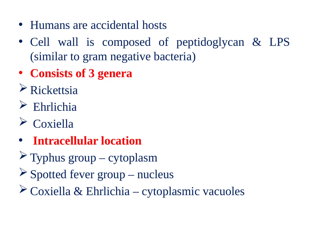
Humans are accidental hosts
Cell wall is composed of peptidoglycan & LPS (similar to gram negative bacteria)
Consists of 3 genera
Rickettsia
Ehrlichia
Coxiella
Intracellular location
Typhus group – cytoplasm
Spotted fever group – nucleus
Coxiella & Ehrlichia – cytoplasmic vacuoles<br>
06
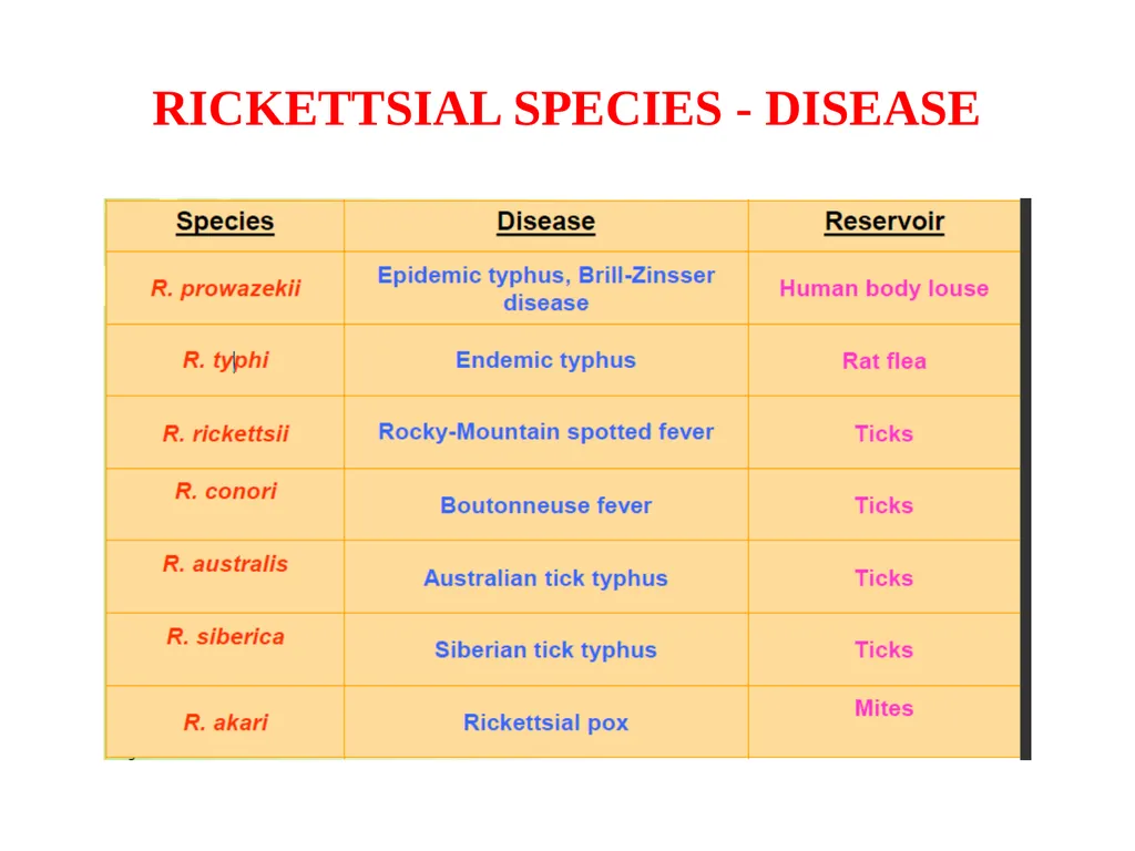
RICKETTSIAL SPECIES - DISEASE<br>
07
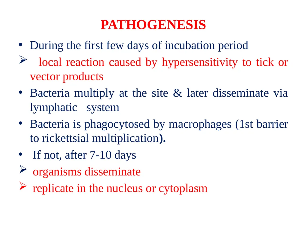
PATHOGENESIS During the first few days of incubation period
local reaction caused by hypersensitivity to tick or vector products
Bacteria multiply at the site & later disseminate via lymphatic system
Bacteria is phagocytosed by macrophages (1st barrier to rickettsial multiplication).
If not, after 7-10 days
organisms disseminate
replicate in the nucleus or cytoplasm<br>
08
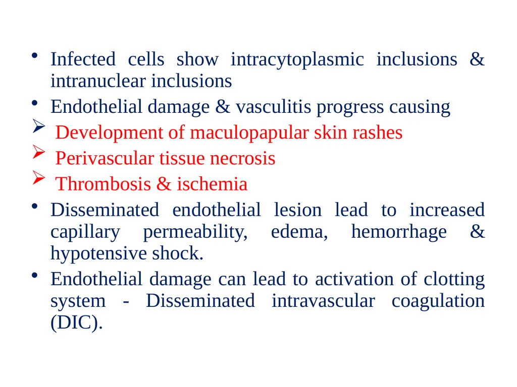
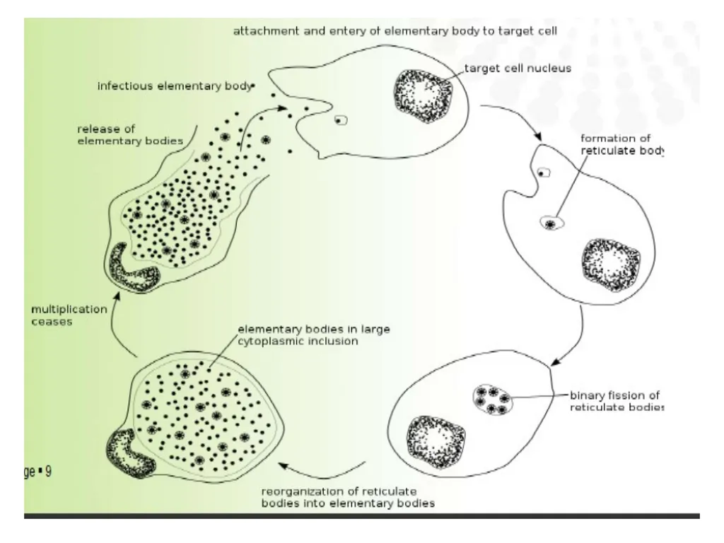
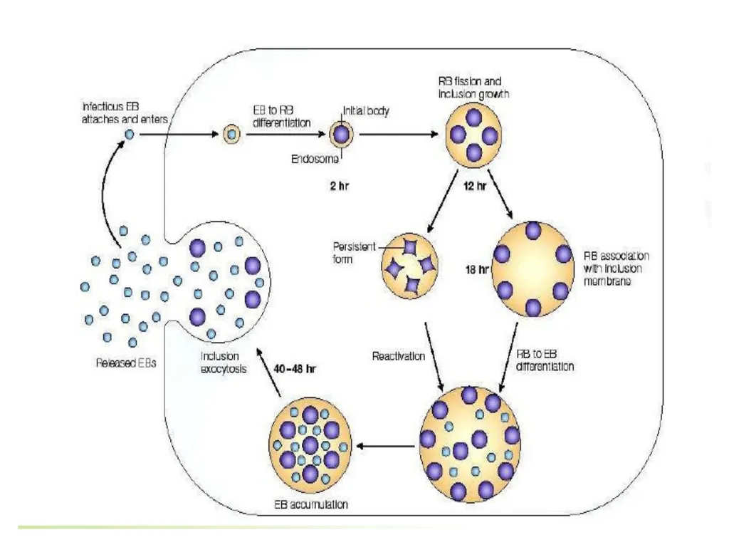
Infected cells show intracytoplasmic inclusions & intranuclear inclusions
Endothelial damage & vasculitis progress causing
Development of maculopapular skin rashes
Perivascular tissue necrosis
Thrombosis & ischemia
Disseminated endothelial lesion lead to increased capillary permeability, edema, hemorrhage & hypotensive shock.
Endothelial damage can lead to activation of clotting system - Disseminated intravascular coagulation (DIC).<br>
09
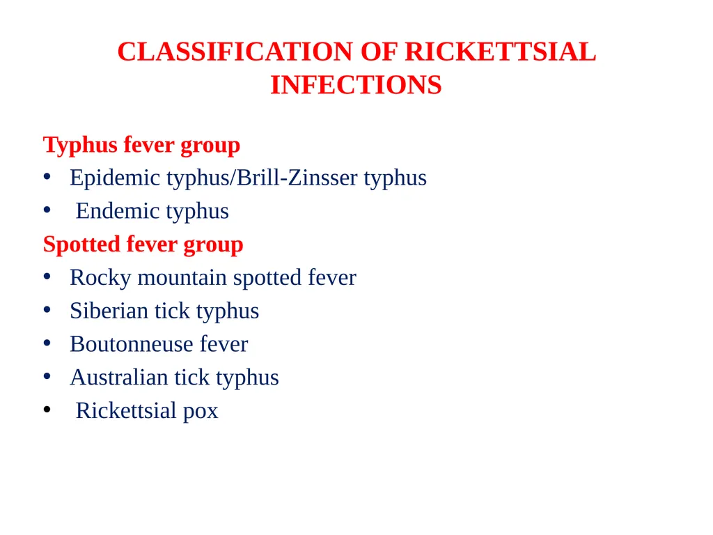
CLASSIFICATION OF RICKETTSIAL INFECTIONS Typhus fever group
Epidemic typhus/Brill-Zinsser typhus
Endemic typhus
Spotted fever group
Rocky mountain spotted fever
Siberian tick typhus
Boutonneuse fever
Australian tick typhus
Rickettsial pox<br>
10
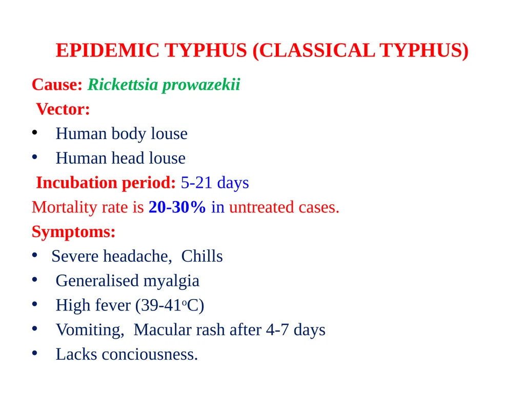
EPIDEMIC TYPHUS (CLASSICAL TYPHUS) Cause: Rickettsia prowazekii
Vector:
Human body louse
Human head louse
Incubation period: 5-21 days
Mortality rate is 20-30% in untreated cases.
Symptoms:
Severe headache, Chills
Generalised myalgia
High fever (39-41oC)
Vomiting, Macular rash after 4-7 days
Lacks conciousness.<br>
11
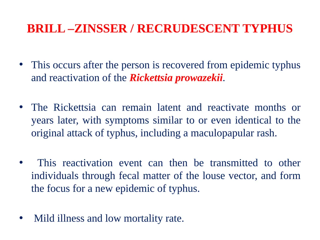
BRILL –ZINSSER / RECRUDESCENT TYPHUS This occurs after the person is recovered from epidemic typhus and reactivation of the Rickettsia prowazekii.
The Rickettsia can remain latent and reactivate months or years later, with symptoms similar to or even identical to the original attack of typhus, including a maculopapular rash.
This reactivation event can then be transmitted to other individuals through fecal matter of the louse vector, and form the focus for a new epidemic of typhus.
Mild illness and low mortality rate.<br>
12
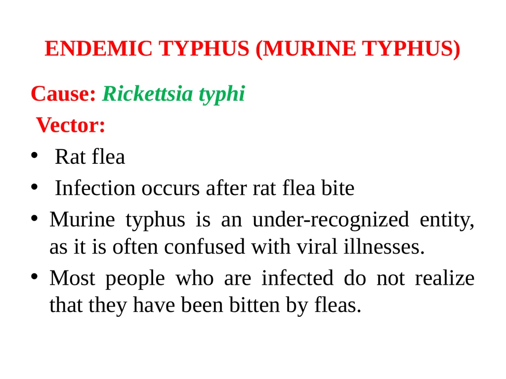
ENDEMIC TYPHUS (MURINE TYPHUS) Cause: Rickettsia typhi
Vector:
Rat flea
Infection occurs after rat flea bite
Murine typhus is an under-recognized entity, as it is often confused with viral illnesses.
Most people who are infected do not realize that they have been bitten by fleas.<br>
13
ENDEMIC TYPHUS (MURINE TYPHUS) Symptoms
Headache
Fever
Muscle pain
Joint pain
Nausea
Vomiting
40–50% of patients will develop a discrete rash six days after the onset of signs.
Up to 45% will develop neurological signs such as confusion, stupor, seizures or imbalance.<br>
14
ROCKY MOUNTAIN SPOTTED FEVER Cause: R. rickettsii
Infection occurs after tick bite
Incubation period: 1 week
Most serious form
More similar to typhus fever but the rash appears earlier and is more prominent.<br>
15
Initial symptoms:
Fever
Nausea
Emesis (vomiting)
Severe headache
Muscle pain
Lack of appetite
Parotitis
Later signs and symptoms:
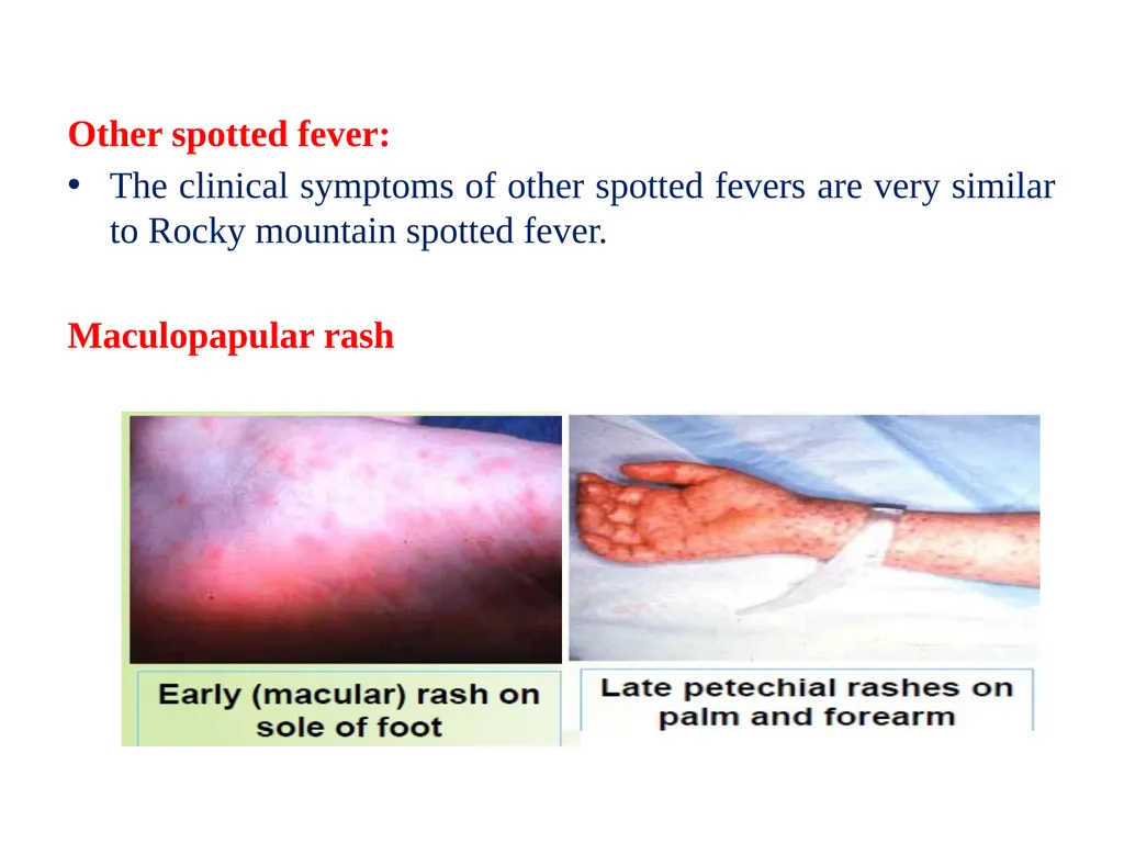
Maculopapular rash
Petechial rash
Abdominal pain
Joint pain
Forgetfulness<br>
16
RICKETTSIAL POX Cause: R. akari
Vector: Mite
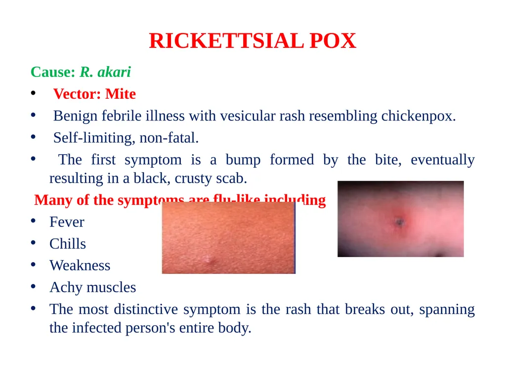
Benign febrile illness with vesicular rash resembling chickenpox.
Self-limiting, non-fatal.
The first symptom is a bump formed by the bite, eventually resulting in a black, crusty scab.
Many of the symptoms are flu-like including
Fever
Chills
Weakness
Achy muscles
The most distinctive symptom is the rash that breaks out, spanning the infected person's entire body.<br>
17
Other spotted fever:
The clinical symptoms of other spotted fevers are very similar to Rocky mountain spotted fever.
Maculopapular rash<br>
18
LABORATORY DIAGNOSIS Culture & isolation
Serologic test
Culture & isolation
Blood is inoculated in guinea pigs/mice.
Observed on 3rd – 4th week.
Animal responds to different rickettsial species can vary.
Difficult & dangerous because of the highly infectious nature of Rickettsiae.
Symptoms:
Rise in temperature – all species.
Scrotal inflammation, swelling, necrosis – R.typhi, R.conori, R.akari (except R.prowazekii)<br>
19
Serologic test
Weil-Felix test
Antibody detection
Based on cross-reactivity between some strains of Proteus & Rickettsia
Complement fixation
Not very sensitive & time consuming
Indirect fluorescence (EIA)
More sensitive & specific
Allows discrimination between IgM & IgG antibodies which helps in early diagnosis
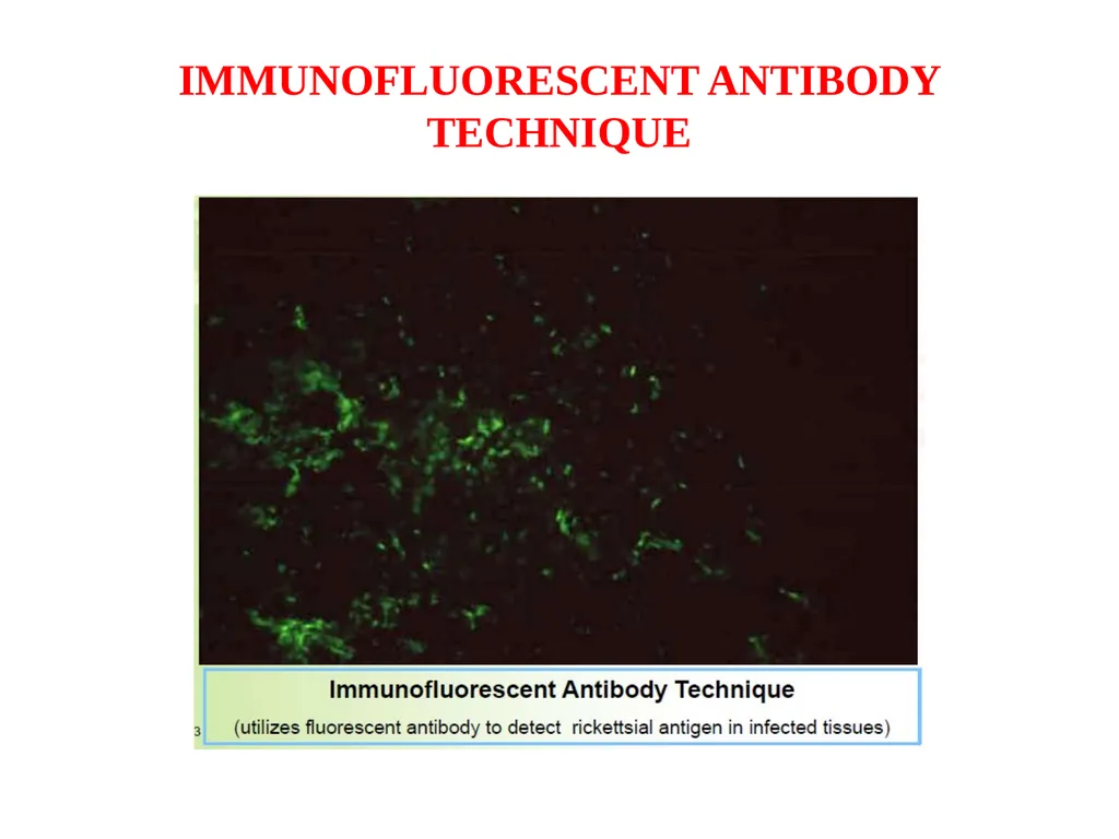
Direct immunofluorescence
The only serologic test that is useful for clinical diagnosis
100% specific & 70% sensitive allowing diagnosis in 3-4 days into the illness<br>
20
WEIL-FELIX TEST Heterophile agglutination test
Using non motile Proteus vulgaris strains (OX 19, OX 2, OX K) to find
rickettsial antibodies in patient’s serum.
Procedure:
Serum is diluted in three separate series of tubes followed by the addition of equal amount of OX 19, OX 2, OX K in 3 separate series of tubes.
Incubation at 37oC for overnight.
Observe for agglutination.
Interpretation:
Strong Agglutination with OX 19 => epidemic & endemic typhus.
Strong agglutination with OX 19 & OX 2 => Spotted fever
Strong agglutination with OX K => Scrub typhus (Scrub typhus by Orientia tsutsugamushi ).<br>
21
IMMUNOFLUORESCENT ANTIBODY TECHNIQUE<br>
22
TREATMENT Adequate antibiotic therapy initiated early in the first week of illness is highly effective and is associated with the best outcome.
Fever usually subsides within 24-72 hours after starting antibiotic therapy. If fever fails to subside with the use of a suitable antibiotic, the diagnosis of rickettsial disease should be reconsidered.
Doxycycline is the drug of choice; it is preferred over other tetracyclines for treatment of rickettsial infections.
Chloramphenicol may be used as an alternative.
Recent data from Europe suggest that fluoroquinolones, such as ciprofloxacin and ofloxacin, may be effective in the treatment of certain rickettsial disease.<br>