04
Feeding of high concentrate ration increases production VFA.
Unabsorbed VFA pass from rumen into abomasum and decrease the contractility.
Results accumulation of ingesta in the abomasum with production of large volumes of gases causing distention and displacement of the organ.
A linear relationship has been reported between the amounts of grains fed and amount of gases produced.
Hypocalcemia, metritis, mastitis, ketonuria, traumatic reticulitis may decrease the motility of abomasum predispose to displacement.<br>
05
Metabolic alkalosis also reduces abomasal contractions.
During pregnancy rumen is lifted by gravid uterus.
Abomasum may slide to the left under the rumen.
Following parturition rumen will come to its normal position.
Distended and atonic abomasum get trapped between the rumen and
Left abdominal wall to cause LDA.
Clinical signs:-
LDA – Intermittent anorexia, gradual decrease in milk yield and ketosis, rapid loss of body condition, dull, listless, shifting of weight from one leg to other due to abdominal pain, Scanty faeces diarrhoeic constipated.<br>
06
Sluggish and weak rumen movement, T, HR, RR are normal, dehydration.
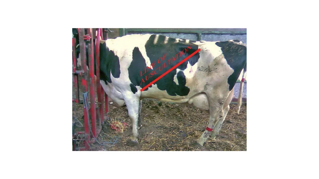
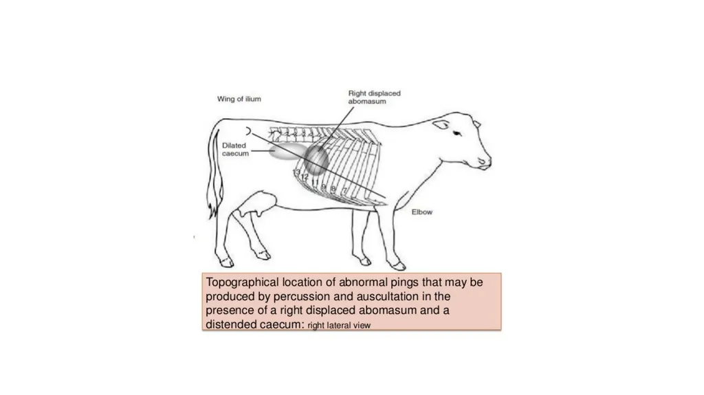
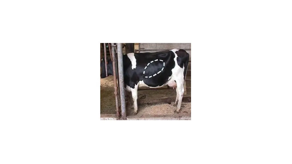
High pitched tympanic resonance (ping) heard oval area along a line from the left elbow to tuber coxae.
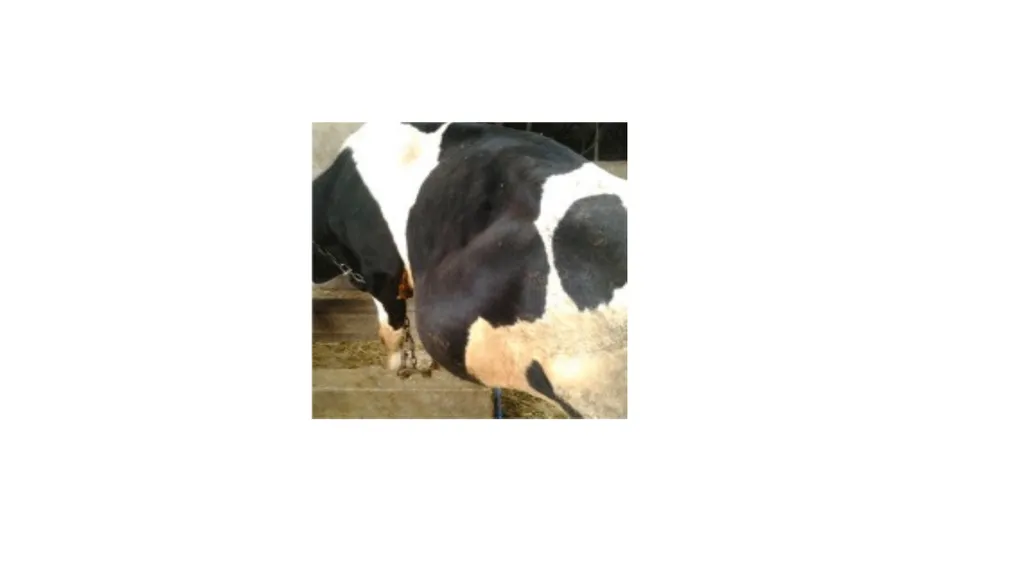
Bulging of left paralumbar fossa.
LDA rarely palpated per rectal examination.
Simple dilatation of abomasum and RDA are similar to LDA.
The distended abomasum may be palpated caudal to last rib.
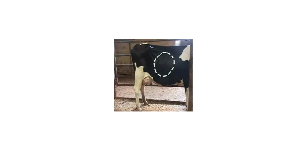
Tympanic resonance heard cranial to right para lumber fossa.
In AV signs are more pronounced.<br>
07
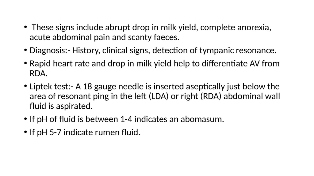
These signs include abrupt drop in milk yield, complete anorexia, acute abdominal pain and scanty faeces.
Diagnosis:- History, clinical signs, detection of tympanic resonance.
Rapid heart rate and drop in milk yield help to differentiate AV from RDA.
Liptek test:- A 18 gauge needle is inserted aseptically just below the area of resonant ping in the left (LDA) or right (RDA) abdominal wall fluid is aspirated.
If pH of fluid is between 1-4 indicates an abomasum.
If pH 5-7 indicate rumen fluid.<br>
08
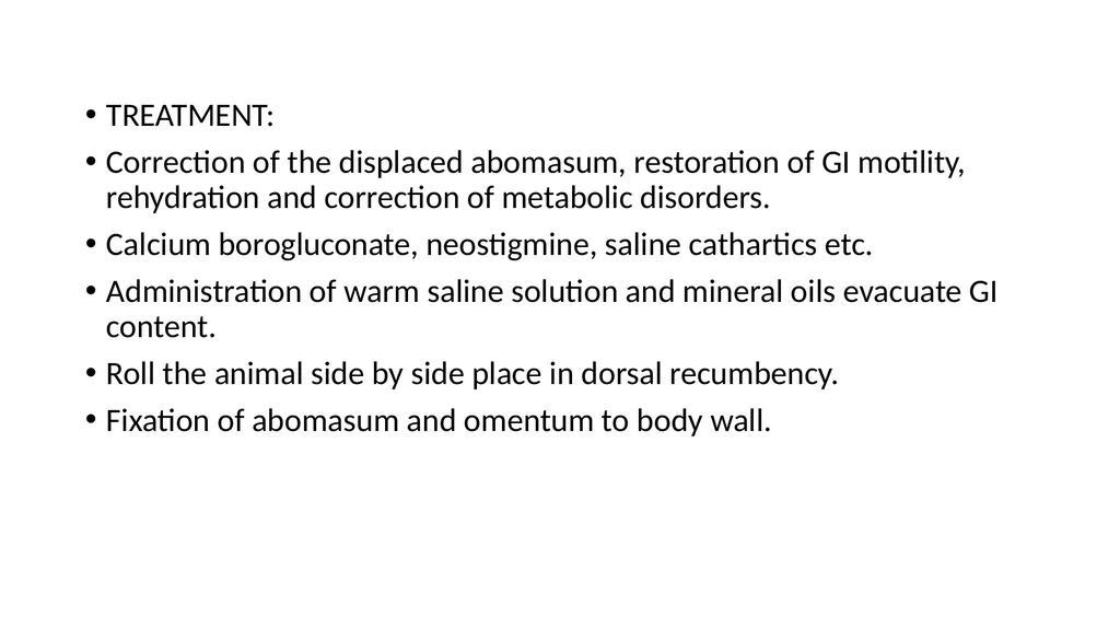
TREATMENT:
Correction of the displaced abomasum, restoration of GI motility, rehydration and correction of metabolic disorders.
Calcium borogluconate, neostigmine, saline cathartics etc.
Administration of warm saline solution and mineral oils evacuate GI content.
Roll the animal side by side place in dorsal recumbency.
Fixation of abomasum and omentum to body wall.<br>
09
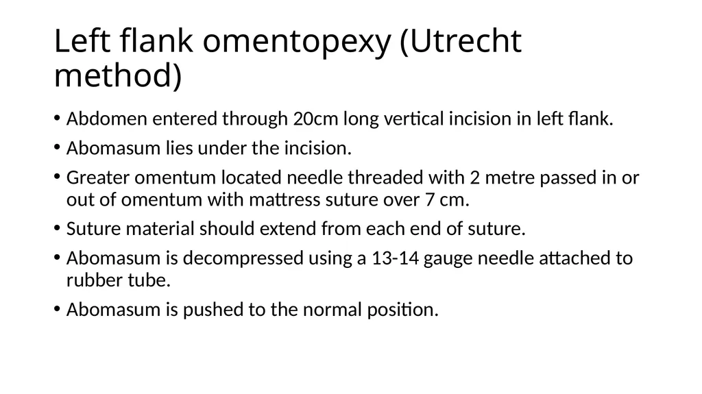
Left flank omentopexy (Utrecht method) Abdomen entered through 20cm long vertical incision in left flank.
Abomasum lies under the incision.
Greater omentum located needle threaded with 2 metre passed in or out of omentum with mattress suture over 7 cm.
Suture material should extend from each end of suture.
Abomasum is decompressed using a 13-14 gauge needle attached to rubber tube.
Abomasum is pushed to the normal position.<br>
10
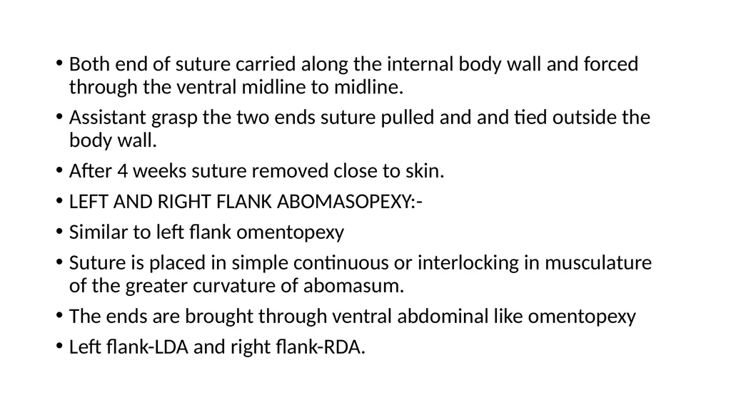
Both end of suture carried along the internal body wall and forced through the ventral midline to midline.
Assistant grasp the two ends suture pulled and and tied outside the body wall.
After 4 weeks suture removed close to skin.
LEFT AND RIGHT FLANK ABOMASOPEXY:-
Similar to left flank omentopexy
Suture is placed in simple continuous or interlocking in musculature of the greater curvature of abomasum.
The ends are brought through ventral abdominal like omentopexy
Left flank-LDA and right flank-RDA.<br>
11
Both end of suture carried along the internal body wall and forced through the ventral midline to midline.
Assistant grasp the two ends suture pulled and and tied outside the body wall.
After 4 weeks suture removed close to skin.
LEFT AND RIGHT FLANK ABOMASOPEXY:-
Similar to left flank omentopexy
Suture is placed in simple continuous or interlocking in musculature of the greater curvature of abomasum.
The ends are brought through ventral abdominal like omentopexy
Left flank-LDA and right flank-RDA.<br>
12
Right flank omentopexy:-
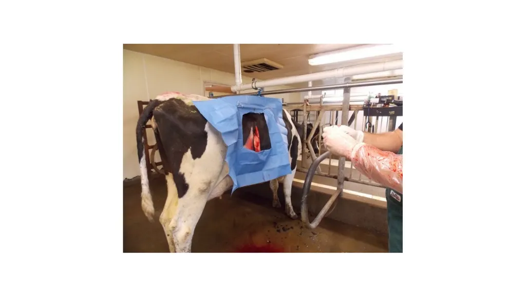
Right flank laparotomy is done in the standing animal.
Abomasum is decompressed and volvulus corrected and abomasum repositioned.
Two mattress suture of heavy catgut, one each cranial and caudal to incision are placed through peritoneum, transverse abdominal muscles and fold of omentum.
The peritoneum and transverse muscles are then sutured in simple continuous pattern incorporating omentum.
The laparotomy incision is closed in usual manner.<br>
13
The aim is to produce adhesions between parietal and visceral peritoneum covering the greater omentum to hold omasum in normal position.
Ventral paramedian abomasopexy:-
The technique is used in both LDA and RDA.
Surgery done under dorsal recumbency by sedation.
The abdominal cavity entered through long incision between the ventral midline and right subcutaneous abdominal vein starting from just caudal to xiphoid process and cranial to umbilicus.
The abomasum is pulled towards incision, decompressed and repositioned.<br>
14
The greater curvature is sutured to the peritoneum and muscles of lateral aspect of the paramedian incision.
The abdomen incision is closed routinely.
This technique is easy.<br>
15
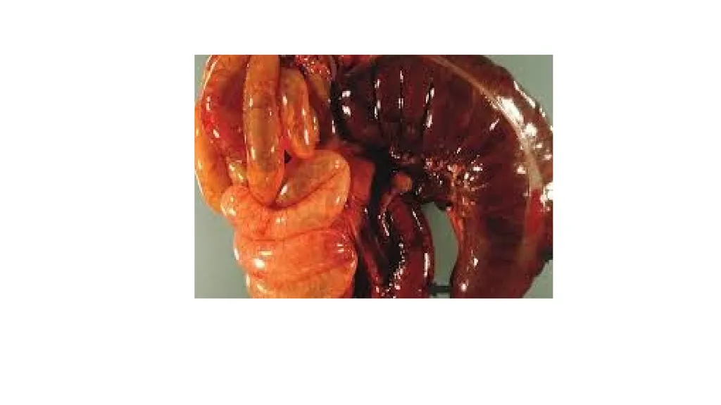
CAECAL DILATION AND TORSION It involves distension, displacement and torsion of caecum.
Free end of caecum in cattle is devoid of mesentery lead to rotation.
Dilatation may proceed to torsion.
After parturition and pregnant animal of cow, bullocks, sheep and goats may observed.
Also observed in buffalo.
Etiology not known But excessive grain feeding is animal reported.
Feeding of excessive grain – increased VFA and gas - Atony or hypomotility of caecum - dilation and torsion of the organ<br>
16
CLINICAL SIGNS :-
Simple dilatation of caecum may be acute if torsion occurs.
Symtom similar to bowel obstruction.
Early cases abdominal pain.
Rapid loss of appetite, cessation of defaecation and dehydration.
T, PR and HR are normal in simple dilatation but subnormal Temp and tachycardia in case torsion.
Hypomotility of rumen or atony of rumen present in most of the cases.
The right paralumbar fossa may be distended and tympanic resonance may be heard.<br>
17
Similar resonance is heard in case of RDA but resonant area is smaller and more caudal in caecal dilation.
The distended caecum may be rectally palpable like long cylindrical movable gas filled structure.
Rupture of caecum may cause death.
DIAGNOSIS:-
History, clinical signs, auscultation and percussion and rectal palpation.
Right flank laparotomy.
Hypochloraemic, hypokalemic metabolic alkalosis.
Hemoconcentration, azotaemia observed.<br>
18
Similar finding are also observed in bowel obstruction.
TREATMENT:-
Conservative treatment in case of dilatation without torsion.
It is parasympathomimetic drugs such as neostigmine.
It can given S/C every 3 to 4 hrs over 2 to 3 days in gradually decrease dose 12.5 mg to 2.5 mg
Alternative continuous drip of drug (200 mg in 10 litres of normal saline.
Liquid paraffin oral purgative.<br>
19
Above fails surgery indicated.
Right flank laparotomy is done standing animal and free end of caecum is exteriorized.
Packing the laparotomy wound and caecotomy done to remove the content.
Caecum is closed after cleaning with NS.
Suturing done with absorbable suture material like enterotomy.
Torsion is corrected and caecum paled in normal position.
The laparotomy wound is closed in the usual manner.
Typhlectomy is also indicated in necrosed caecum.
Blood vessels ligated and resected necrosed caecum<br>
20
Ileum and colon are anastomosed by Connell pattern using chromic catgut<br>