UNIT-5 (REGIONAL SURGERY-II) UG COURSES DR. MITHILESH KUMAR Assistant Professor cum Jr. Scientist Veterinary Surgery and Radiology Bihar Veterinary College, Patna-800014 THORACOTOMY Means opening of thoracic cavity. Techniques are (i)
"UNIT-5 (REGIONAL SURGERY-II) UG COURSES DR." is the property of its rightful owner. Permission is granted to
download and print the materials on this website for personal, non-commercial use only, and to display it
on your personal computer provided you do not modify the materials and that you retain all copyright
notices contained in the materials. By downloading content from our website, you accept the terms of this
agreement.
Presentation Transcript
01
UNIT-5 (REGIONAL SURGERY-II) UG COURSES DR. MITHILESH KUMAR
Assistant Professor cum Jr. Scientist
Veterinary Surgery and Radiology
Bihar Veterinary College, Patna-800014<br>
02
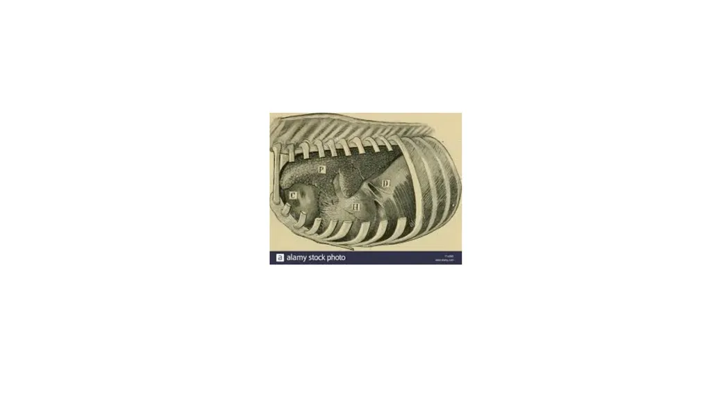
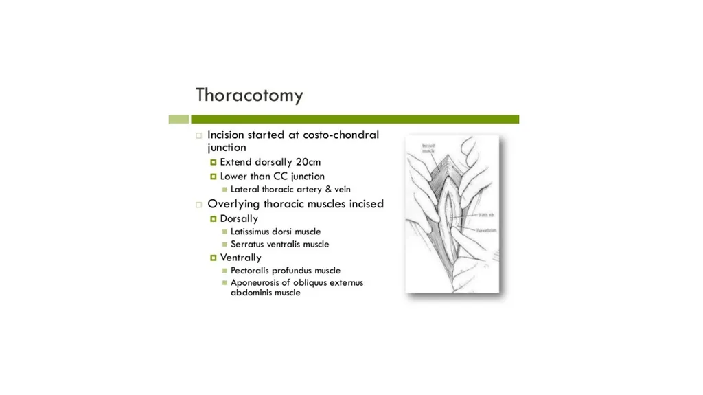
THORACOTOMY Means opening of thoracic cavity.
Techniques are
(i) Intercostal incision
(ii) Rib resection
(iii) Split – rib incision
Thoracotomy through paracostal abdominal incision is mainly indicated to repair rents in diaphragm or to drain diaphragmatic abscess.<br>
03
Pre-operative evaluation of the lesion by percussion, auscultation and radiography and ultrasonography of the chest can help to outline the exact location of the lesion.
Intercostal Incision:- Intercostal incision should be placed cranial to rib as intercostal vessels are located caudally.
The incision should be extended with scissors to the desired length.
A self retraining rib retractor is used for adequate exposure of the intrathoracic organs.
A series of interrupted sutures are placed around the adjacent ribs using nonabsorbable suture material for closure of the wound.<br>
04
A simple continuous suture in the intercostal muscles seals the incision against air leaks.
The overlying muscles and skin are in routine manner.
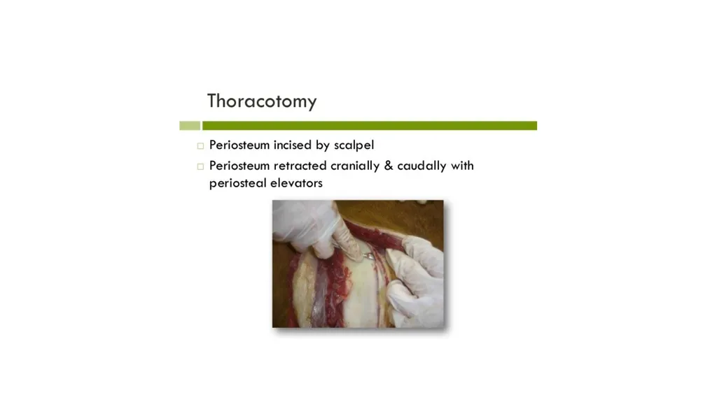
Rib resection:- After skin and muscles have been incised a longitudinal incision is made on the periosteum of the exposed rib.
The periosteum is stripped from the lateral surface and cranial and caudal borders of the rib with help of periosteal elevator.
A curved haemostat introduced between the periosteum and medial surface of the rib is moved up and down to separate the periosteum completely from the rib.
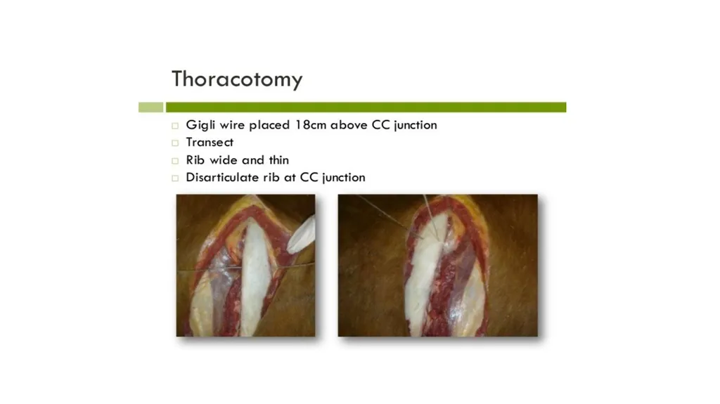
To expose the periosteal bed, the rib is resected with wire saw at the proximal end.<br>
05
The distal end is then disarticulated at the costochondral junction.
The pleural incision is extended dorsally and ventrally with scissors.
The wound is closed in layers i.e. the pleura and periosteum together, intercostal muscles and then superficial muscles and fascia together.
Continuous lock stich sutures using heavy chromic catgut.
The skin sutured routinely.
Split –rib technique :-
After exposing the rib, straight longitudinal incision is made in its center by means of an oscillating electrical bone saw.
The rib is then sectioned transversely at either ends of the primary incision.<br>
06
Closure of rib incision is accomplished by placing 4 to 5 interrupted sutures of either stainless steel wire or heavy black braided silk around the rib.
Muscles closed in layers and skin sutured as usual.
It provides maximum exposure of the pleural space without involvement of rib retractors.
Closure is simple and quick.<br>
07
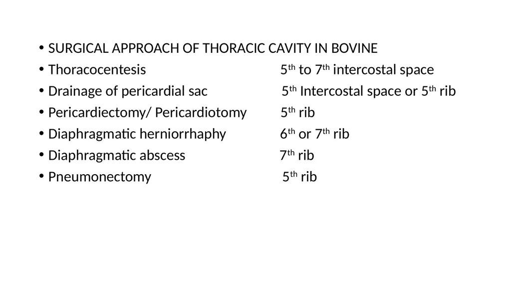
SURGICAL APPROACH OF THORACIC CAVITY IN BOVINE
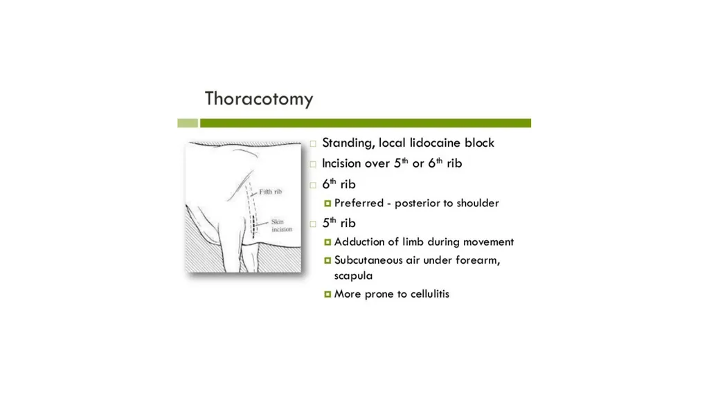
Thoracocentesis 5th to 7th intercostal space
Drainage of pericardial sac 5th Intercostal space or 5th rib
Pericardiectomy/ Pericardiotomy 5th rib
Diaphragmatic herniorrhaphy 6th or 7th rib
Diaphragmatic abscess 7th rib
Pneumonectomy 5th rib<br>
08
THORACOCENTESIS:- indicated to collect the fluid for physical, cytological, biochemical and culture and sensitivity tests.
These tests help to point the cause of the effusions.
All aseptic precautions should be used for thoracocentesis to avoid introduction of infection.
Pneumothorax:- Air accumulates in the pleural space.
THORACIC WOUNDS: Wound occur as a result of trauma.
It is necessary to prevent pneumothorax and collapse of lung.
Rib fractures due to blow to the thoracic wall cause splinters or sharp rib ends which may penetrate the lungs to cause pneumothorax.
Non penetrating wounds are common.<br>
09
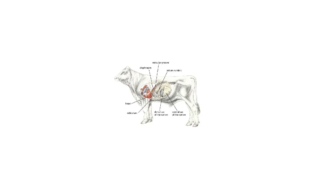
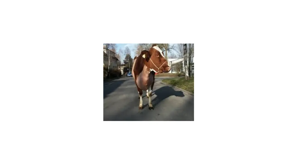
TRAUMATIC PERICARDITIS It is occurs as a result of penetration of the pericardium by sharp foreign body.
Distance between reticulum and pericardium is only few cm.
Sharp foreign body can easily pierce the diaphragm and enter the pericardium.
Exudative, suppurative or constrictive pericarditis characterized by symptoms of toxemia or congestive heart failure.
Higher incidence in pregnant or recently calved animals as increased intraabdominal pressure pushes the foreign body towards thorax.<br>
10
Pericardium may be pierced.
Sometime pierce the myocardium and come through thoracic wall.
Abscess forms on either side of chest behind the elbow.
In few cases come into reticulum and rarely come in faeces.
Trauma to pericardium initiates inflammation and exudates continue to accumulate in the pericardial sac.
Contamination spreads.
Formation of adhesions between epicardium and pericardium.
Fluid accumulates in the Pericardial sac impairs pump of heart.
Exhibits signs of congestive heart failure.<br>
11
Right sided heart failure is common.
Absorption bacterial toxins.
CLINICAL SIGNS:- Complete anorexia, drop in milk yield and reluctance to walk, walk with short steps and stiff gait, Pyrexia, increased pulse rate, abdominal respiration, arched back, abducted elbows, grunting, brisket oedema.
Oedema of jaw, dewlap and ventral abdominal region extending upto udder indicating in advance cases, indicating congestive heart failure.
The jugular veins engorged and show pulsation.
Initially pericardial friction sounds are heard on auscultation.
Later muffled heart sound in more fluid content<br>
12
Splashing heart sound if gas.
Prognosis is poor with signs of congestive failure.
DIAGNOSIS:-
Clinical signs, Pericardiocentesis and radiography.
Hematological, biochemical and electrocardiographic changes indicate severity .
Pericardiocentesis through 5th or 6th intercostal space shows offensive odour fluid.
low erythrocyte count, Hb and packed cell volume alongwith leukocytosis, neutrophilia and elevated ESR
Urine is acidic albuminuria.<br>
13
Jugular veins distended and pulsatile.
Lateral radiography of chest to diagnose the foreign body and shadow of heart.
Radiography of chest helps to differentiate pericarditis from DH.
Pericardiocentesis.
TREATMENT :-
Conservative treatment- diuretics, antimicrobial therapy.
Elevation of front feet by 15-20cm.
Surgical treatment:-
Pericardiocentesis, Pericardiotomy or Pericardiectomy with or without pericardial graft.<br>
14
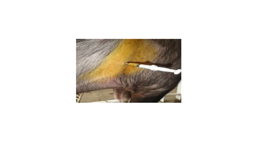
Pericardiocentesis – drain the fluid from the pericardial sac and inject antibiotics and proteolytic enzymes.
Pericardiocentesis done in standing animal.
Large bore needle is inserted between 4th and 5th intercostal space at the level of elbow region.
Pericardiotomy –
Laparoruminotomy done before pericardiotomy.
Foreign body penetrating diaphragm or pericardium should be removed.
Pericardiotomy involves incising the pericardium and draining the
fluid.<br>
15
The main objectives of pericardiocentesis are to drain fluid from pericardial sac and inject antibiotics and proteolytic enzymes.
It is temporary relief.
Pericardiocentesis done in standing animal with forelimb drawn forward.
Needle inserted between 4th and 6th intercostal space in left side.
Laparorumenotomy done before pericardiotomy to remove the FB,s.
Select the early cases.
Surgery should be done early diagnosis and has not lost compete appetite, good body condition and young.
Thoracic approach mentioned earlier.<br>
16
5% DNS given intravenously during surgery.
Following thoracotomy, pericardiotomy or pericardiectomy done.
Pericardiotomy involves incising the pericardium and draining the fluid.
Many effusions are bread and butter type remove the exudate to clear the pericardium.
Foreign body removed.
Wash the cavity with warm isotonic saline solution containing antibiotics.
Pericardial and pleural incision closed with continuous suture suture using absorbable suture material.<br>
17
Penicillin is sprinkled inside cavity.
Close the thoracic cavity as usual.
Sometime drainage tube attached inside the pericardial sac.
Flushing with mild antibiotic solution containing proteolytic enzyme daily for several days until no discharge from the sac.<br>
18
Occurs in high producing adult dairy cows in age group of 3-7 years.
Reported in bulls, calves and sheep.
RDA is relatively more frequent than LDA.
Rare in buffaloes.
In DH part of abomasum gets herniated along with reticulum.
Exhibit signs of DH without signs of abomasal involvement.
Correction of DH cures the condition.
Detected only by laparoruminotomy.
Occur at any time of gestation<br>