What is ECG? UNDERSTANDING ECG LESSON -2 By AMANDEEP KAUR ASIANM Contents 01. What is ECG Basic definition and fundamental principles of electrocardiography 02. ECG Components Understanding P waves, QRS complexes, and T waves 03. Clinical
"What is ECG? UNDERSTANDING ECG LESSON -2 By" is the property of its rightful owner. Permission is granted to
download and print the materials on this website for personal, non-commercial use only, and to display it
on your personal computer provided you do not modify the materials and that you retain all copyright
notices contained in the materials. By downloading content from our website, you accept the terms of this
agreement.
Presentation Transcript
01
What is ECG? UNDERSTANDING ECG LESSON -2 By
AMANDEEP KAUR
ASI/ANM<br>
02
Contents 01. What is ECG
Basic definition and fundamental principles of electrocardiography
02. ECG Components
Understanding P waves, QRS complexes, and T waves
03. Clinical Applications
How ECG helps diagnose cardiac conditions
04. Normal vs Abnormal
Recognizing healthy and pathological patterns<br>
03
What is ECG? Electrocardiography Overview
ECG is a non-invasive diagnostic tool that records the electrical activity of the heart over time, providing crucial insights into cardiac health and function.
Electrical Activity
Measures electrical impulses that trigger heart muscle contractions
Real-time Monitoring
Continuous recording of heart rhythm and electrical patterns<br>
04
ECG Wave Components P Wave
Atrial depolarization - represents electrical activity in the upper heart chambers
QRS Complex
Ventricular depolarization - shows electrical activity in the lower heart chamber
T Wave
Ventricular repolarization - indicates recovery phase of heart muscle<br>
05
ECG Recording Process Patient Preparation
Clean skin, position electrodes at specific anatomical landmarks
Electrode Placement
Apply 10 electrodes in standard 12-lead configuration
Recording
Capture 10-second rhythm strip for analysis
Interpretation
Analyze waveforms, intervals, and rhythm patterns<br>
06
Clinical Applications Arrhythmia Detection
Identifies irregular heartbeats, bradycardia, and tachycardia
Ischemia Diagnosis
Detects signs of reduced blood flow to heart muscle
Myocardial Infarction
Diagnoses heart attacks by identifying characteristic changes
Monitoring
Tracks treatment effectiveness and disease progression<br>
07
Normal ECG Parameters PR Interval
120-200ms - represents atrioventricular conduction time
QRS Duration
60-100ms - indicates ventricular depolarization time<br>
08
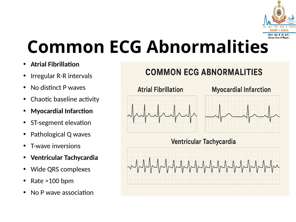
Common ECG Abnormalities Atrial Fibrillation
Irregular R-R intervals
No distinct P waves
Chaotic baseline activity
Myocardial Infarction
ST-segment elevation
Pathological Q waves
T-wave inversions
Ventricular Tachycardia
Wide QRS complexes
Rate >100 bpm
No P wave association<br>
09
ECG Interpretation Guidelines Systematic Approach
Follow a consistent method: rate, rhythm, intervals, morphology, and clinical correlation
Calculate heart rate and rhythm
Measure all intervals and segments
Assess wave morphology and axis
Clinical Integration
Always correlate ECG findings with patient symptoms and clinical presentation
Compare with previous ECGs
Consider patient medications
Evaluate for acute changes
Professional Review
Complex cases require cardiologist consultation for accurate interpretation
Seek expert opinion when uncertain
Use decision support tools
Document findings clearly<br>