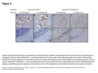
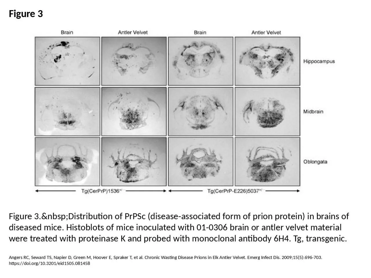
PPT-Figure 3 Figure 3. Distribution of PrPSc (disease-associated form of prion protein)
Author : vivian | Published Date : 2024-02-09
Angers RC Seward TS Napier D Green M Hoover E Spraker T et al Chronic Wasting Disease Prions in Elk Antler Velvet Emerg Infect Dis 2009155696703 httpsdoiorg103201eid1505081458
Presentation Embed Code
Download Presentation
Download Presentation The PPT/PDF document "Figure 3 Figure 3. Distribution..." is the property of its rightful owner. Permission is granted to download and print the materials on this website for personal, non-commercial use only, and to display it on your personal computer provided you do not modify the materials and that you retain all copyright notices contained in the materials. By downloading content from our website, you accept the terms of this agreement.
Figure 3 Figure 3. Distribution of PrPSc (disease-associated form of prion protein): Transcript
Angers RC Seward TS Napier D Green M Hoover E Spraker T et al Chronic Wasting Disease Prions in Elk Antler Velvet Emerg Infect Dis 2009155696703 httpsdoiorg103201eid1505081458. Creutzfeldt. -Jakob Disease. Human equivalent of mad cow . disease. Rare. , degenerative, fatal disease. Approximately 1 case per million per year. Typically people are diagnosed around age 60 . CJD belongs to a family of human and animal diseases known as the transmissible spongiform . Carlos Francisco Mendoza. Human Prion Diseases. Comparison of Prp. C. and . Prp. Sc. Evidence Supports the Prion Hypothesis. Genetic . Polymorphism. RPSA Polymorphisms have no direct influence on susceptibility to sporadic CJD. Prions. are infectious agents composed of protein in . misfolded. form.. initially thought to be viruses which replicated slowly within their hosts, but no nucleic acid found associated with infectious material.. Wadsworth J, Joiner S, Linehan JM, Balkema-Buschmann A, Spiropoulos J, Simmons MM, et al. Atypical Scrapie Prions from Sheep and Lack of Disease in Transgenic Mice Overexpressing Human Prion Protein. Emerg Infect Dis. 2013;19(11):1731-1739. https://doi.org/10.3201/eid1911.121341. Torres J, Andréoletti O, Lacroux C, Prieto I, Lorenzo P, Larska M, et al. Classical Bovine Spongiform Encephalopathy by Transmission of H-Type Prion in Homologous Prion Protein Context. Emerg Infect Dis. 2011;17(9):1636-1644. https://doi.org/10.3201/eid1709.101403. Babelhadj B, Di Bari M, Pirisinu L, Chiappini B, Gaouar S, Riccardi G, et al. Prion Disease in Dromedary Camels, Algeria. Emerg Infect Dis. 2018;24(6):1029-1036. https://doi.org/10.3201/eid2406.172007. Diack AB, Ritchie D, Peden AH, Brown D, Boyle A, Morabito L, et al. Variably Protease-Sensitive Prionopathy, a Unique Prion Variant with Inefficient Transmission Properties. Emerg Infect Dis. 2014;20(12):1969-1979. https://doi.org/10.3201/eid2012.140214. Seuberlich T, Gsponer M, Drögemüller C, Polak MP, McCutcheon S, Heim D, et al. Novel Prion Protein in BSE-affected Cattle, Switzerland. Emerg Infect Dis. 2012;18(1):158-159. https://doi.org/10.3201/eid1801.111225. Angers RC, Seward TS, Napier D, Green M, Hoover E, Spraker T, et al. Chronic Wasting Disease Prions in Elk Antler Velvet. Emerg Infect Dis. 2009;15(5):696-703. https://doi.org/10.3201/eid1505.081458. Baron T, Bencsik A, Biacabe A, Morignat E, Bessen RA. Phenotypic Similarity of Transmissible Mink Encephalopathy in Cattle and L-type Bovine Spongiform Encephalopathy in a Mouse Model. Emerg Infect Dis. 2007;13(12):1887-1894. https://doi.org/10.3201/eid1312.070635. Honkavuori KS, Shivaprasad H, Williams BL, Quan P, Hornig M, Street C, et al. Novel Borna Virus in Psittacine Birds with Proventricular Dilatation Disease. Emerg Infect Dis. 2008;14(12):1883-1886. https://doi.org/10.3201/eid1412.080984. Weissenböck H, Bakonyi T, Sekulin K, Ehrensperger F, Doneley RJ, Dürrwald R, et al. Avian Bornaviruses in Psittacine Birds from Europe and Australia with Proventricular Dilatation Disease. Emerg Infect Dis. 2009;15(9):1453-1459. https://doi.org/10.3201/eid1509.090353. 2. CNS Infection - Meninges. CNS infections: most are due to sepsis. Pathogenesis:. Hematogenous spread (most common) . Traumatic implantation . Local extension from nearby infection . Ascent of peripheral nerve. Torres J, Andréoletti O, Lacroux C, Prieto I, Lorenzo P, Larska M, et al. Classical Bovine Spongiform Encephalopathy by Transmission of H-Type Prion in Homologous Prion Protein Context. Emerg Infect Dis. 2011;17(9):1636-1644. https://doi.org/10.3201/eid1709.101403.
Download Document
Here is the link to download the presentation.
"Figure 3 Figure 3. Distribution of PrPSc (disease-associated form of prion protein)"The content belongs to its owner. You may download and print it for personal use, without modification, and keep all copyright notices. By downloading, you agree to these terms.
Related Documents