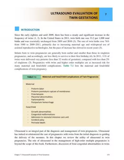
/
CAREFULL DIAGNOSIS & MANAGEMENT OF MONOCHORIONIC MONOAMNIOTIC TWINS CAREFULL DIAGNOSIS & MANAGEMENT OF MONOCHORIONIC MONOAMNIOTIC TWINS

CAREFULL DIAGNOSIS & MANAGEMENT OF MONOCHORIONIC MONOAMNIOTIC TWINS - PowerPoint Presentation

wang
wang . @wang
Follow
343 views
Uploaded On 2022-06-18

CAREFULL DIAGNOSIS & MANAGEMENT OF MONOCHORIONIC MONOAMNIOTIC TWINS - PPT Presentation

DRABINAYA VIJAYAN Sree Balaji Medical College amp Hospital Chennai INDIA MOMO TWINS Monochorionic monoamniotic twins are a subtype in monozygotic twin pregnancy DEFINITION Monoamniotic twins ID: 920792

twins twin cord monoamniotic twin twins monoamniotic cord single fetal pregnancy perinatal pregnancies weeks monochorionic momo monitoring umbilical mortality

Share:

Link:

Embed:

Download Presentation from below link

Download Presentation The PPT/PDF document "CAREFULL DIAGNOSIS & MANAGEMENT OF M..." is the property of its rightful owner. Permission is granted to download and print the materials on this web site for personal, non-commercial use only, and to display it on your personal computer provided you do not modify the materials and that you retain all copyright notices contained in the materials. By downloading content from our website, you accept the terms of this agreement.


Presentation Transcript

Slide1

CAREFULL DIAGNOSIS & MANAGEMENT OF MONOCHORIONIC MONOAMNIOTIC TWINS

DR.ABINAYA VIJAYAN

Sree

Balaji

Medical College & Hospital

Chennai, INDIA

Slide2

MOMO TWINSMonochorionic

monoamniotic

twins are a subtype in monozygotic twin pregnancy

Slide3

DEFINITION Monoamniotic twins

are identical

twins

that share the single chorionic sac, 

a single

yolk sac

 and a single

amniotic sac

.

always identical

always monochorionic and are usually termed

Monoamniotic

-Monochorionic ("

MoMo

")

twins

.

They also share the placenta, but have two separate umbilical cords.

Slide4

PATHOLOGY-

It results from a separation of a single ovum at 8-13 days following

fertilisation

(i.e. later than with an MCDA pregnancy). 

By this time a trophoblast

has already formed,

yielding a single placenta. 


Slide5

Slide6

INCIDENCERARE

1 in 35,000 to 1 in 60,000

pregnancies

Slide7

WHY INTENSIFIED MONITORING ???

associated with

Morbidity and

Mortality

.

Slide8

CASE REPORT

Mrs.X

, 30 yrs old, G2P1L1

-with previous Full term normal vaginal delivery,

-LCB- 9 years back

-Booked with our hospital from 2 months of

amenorrhoea

Slide9

Menstrual H/O:RMP, 3/30 days cycleNot associated with pain or clots

Marital H/O:

Married since 10 years

Non

consanguinous

marriage

Obstertric

H/O:

1st pregnancy :Conceived spontaneously

Boy/ FTNVD/ 9yrs/ Institutional/ Alive & Healthy

No H/O contraceptives

Slide10

2nd pregnancy :

1

ST

TRIMESTER : confirmed by UPT at 2 months of

amenorhoea

Dating scan done

USG at 11 wks revealed –

“MONOCHORIONIC MONOAMNIOTIC TWIN PREGNANCIES”

Tablet Folic acid taken

No H/O fever with rash/ irradiation exposure/ spotting or bleeding

p/v

.

Slide11

2nd TRIMESTER :

Quickening at 18 weeks of gestation.

Anomaly scan at 20weeks – one fetus had

SINGLE UMBILICAL ARTERY

- After 22 weeks

SERIAL ULTRASOUND

every 2 weeks was performed with regular Antenatal visits.

Every USG –

Full assessment of fetal growth - Amniotic fluid volume

- fetal

doppler

-2 doses of Inj. TT were given.

No H/O abdominal pain/ discharge

p/v

/ pedal edema

Slide12

3rd TRIMESTER:

Perceived fetal movements well

At 34 weeks – INJ.BETAMETHASONE 12mg IM 2 DOSES, 24 HOURS APART were given

Admitted at 34 weeks of gestation – “CLOSE MONITORING”

- DAILY NONSTRESS TEST WITH WEEKLY ULTRASOUND WITH DOPPLER

At 37 weeks she was taken up for EMERGENCY LSCS

- in view of PROM for >12 hours and non-progress of

labour

Slide13

Caesarean section was performed - I twin was delivered by vertex presentation and II twin by breech extraction.

She delivered

two live female babies weighing 2.5kgs and 2.9kgs respectively with good APGAR score.

The first twin had single umbilical artery .

Placental examination showed a

SINGLE PLACENTA WITH MONOCHORIONIC MONOAMNIOTIC MEMBRANE AND UMBILICAL CORD ENTANGLEMENT

Both infants showed good growth and development with nil complications at 6 months of age.

Slide14

SINGLE UMBILICAL ARTERY

Slide15

SINGLE PLACENTA WITH MOMO MEMBRANE & ENTANGLED CORD

Slide16

COMPLICATIONS

CORD ENTANGLEMENT

ANOMALIES

TWIN TO TWIN TRANSFUSION SYNDROME

PREMATURITY

Slide17

Slide18

CORD ENTANGLEMENT

42% - 80% of cases

traditionally related to high

perinatal

mortality

CORD COMPRESSION is another life threatening condition preventing oxygenation and vital nutrients resulting in fetal demise

Slide19

Cord entanglement is one of the main complications associated with monoamniotic twins. Because the twins have

NO AMNIOTIC MEMBRANE

separating them, their umbilical cords can easily become entangled.

Slide20

Cord compression is another life threatening condition common in monoamniotic twins.

As the twins move around in the amniotic sac

,

it is possible that one will compress the other"s umbilical cord.

This can prevent vital nutrients and blood from traveling to the other baby. resulting in fetal death.

CORD COMPRESSION

Slide21

Slide22

TWIN TO TWIN TRANSFUSION SYNDROME-Because there is no barrier separating the two fetuses from each other, there are almost always blood vessel connections in the placenta shared by two fetuses in

monochorionic twin

(MC) pregnancies.

-10-15% of monochorionic twins

-

In these instances, there may be significant transfer of blood from one twin (the so-called “donor”) to the other twin (the so-called “recipient”), resulting in twin-to-twin transfusion syndrome (TTTS).

Slide23

TWIN TO TWIN TRANSFUSION SYNDROMEone twin becomes undernourished whereas the other develops

hyperdynamic

circulation and heart failure.

In severe TTTS presenting with acute

polyhydramnios

during the second trimester, endoscopic laser coagulation of the intercommunicating placental vessels is associated with survival of at least one baby in about 70% of the pregnancies

TTTS is not as common among MoMo as in

MoDi

pregnacies

The presence of

polyhydramnios

, discordant fetal growth,

hydrops

, congestive heart failure, tricuspid regurgitation and discordant bladder fillings make the prenatal diagnosis of TTTS possible.

Slide24

TREATMENT

-FETOSCPOIC LASER INTERVENTION

-AMNIOREDUCTION IN DI AMNIOTICS

Slide25

PREMATURITYIt is known that uncomplicated twin pregnancies have a higher incidence of premature birth than singletons and that

MoMo

twins are at an even greater risk of being born before 32 weeks of gestation.

Slide26

Those born before 32 weeks of gestationhave a high incidence of perinatal

depression,

respiratory distress,

early and late onset sepsis,

patent

ductus

arteriosus

, necrotizing enterocolitis,

Intracranial hemorrhage,

prolonged hospitalization and

poor neurological outcomes.

Slide27

DIAGNOSISMOMO twins has the highest

perinatal

mortality, about 50%.

Detection of monochorionic

pregnancies at 10 to 14 weeks of gestation and monitoring by serial ultrasounds should lead to early diagnosis of TTTS

Slide28

ULTRASOUNDIst

TRIMESTER

* shows a twin pregnancy with a single gestational sac and a single yolk sac (differentiating from a DCDA and MCDA pregnancy)

* there is no inter twin membrane: theoretically this differentiates from a DCDA and MCDA pregnancy

o

however, even in a MCDA pregnancy the

intertwin

membrane may be difficult to see

o

therefore non-

visualisation

of the

intertwin

membrane is not in itself diagnostic

Slide29

MOMO TWINS

MCDA TWINS

Slide30

Second trimester * specific to a MCMA pregnancy:

-

there

can be presence of cord entanglement

-

there can be presence of cord fusion

- absent inter twin membrane: although may be difficult to see sometimes even with a MCDA pregnancy * common to both

MCMA

and

MCDA

pregnancies

-

a single placenta is seen

-

absent twin peak sign

Slide31

MOMO TWINS AT 16 WEEKS

Slide32

TWIN PEAK SIGN IN DCDA TWINS

Slide33

Slide34

TREATMENT-Unfortunately. there is no treatment that can reverse this pregnancy condition.

An experimental drug.

SULINDAC

- has been used to in some monoamniotic twins. This drug lowers the amount of fluid in the amniotic sac thereby reducing the amount of fetal movement.

This is thought to lower the chances of cord entanglement or compression. However. this drug has not been studied in a large number of pregnancies and its potential side effects are unknown.

Slide35

The best treatment for monoamniotic twins is to have regular and aggressive fetal monitoring.

twice-weekly monitoring of fetal heart rate and movement. particularly after the 26th week.

Aggressive monitoring can help to lower the risk of fetal death considerably.

Slide36

Slide37

CONCLUSION

Women with

monochorionic

monoamniotic

twins should be

counseled

immediately after the diagnosis of

MoMo

twins regarding the complications and perinatal mortality.

Slide38

With a multidisciplinary approach a good outcome can be achieved.

These antenatal women should be subjected to

intensified monitoring

as well early admission in the hospital for close monitoring; taking care and caution to prevent

perinatal

mortality, thus, progressing to deliver at term.

Slide39

REVIEW OF LITERATURE

Slide40

IMPROVED PERINATAL SURVIVAL WITH INPATIENT MONITORING

Slide41

ALL WOMEN WERE DELIVERED BY CAESAREAN SECTION

Slide42

INCIDENCE OF PERINATAL MORTALITY HAS DECREASED

Slide43

NO IUD IN ANY HOSPITALISED PATIENT

Slide44

RISK FOR CORD ENTANGLEMENT, CONGENITAL MALFORMATION, TTS & PREMATURITY

Slide45

REFERENCES1.Benirschke K. The biology of the twinning process: how placentation

influences outcome.

Semin

Perinatol

1995; 19: 342–350.

2.Carr SR, Aronson MP,

Coustan

DR. Survival rates of

monoamniotic twins do not decrease after 30 weeks’ gestation. Am J Obstet Gynecol

1990; 163: 719 – 722.

3.Bilardo CM,

Arabin

B.

Monoamniotic

twins. In:

Blickstein

I, Keith LG (

eds

) Multiple Pregnancy. Taylor & Francis: London and New York, 2005, pp 574 – 582.

4.

Rodis

JF,

McIlveen

PF,

Eagen

JF,

Borgida

AF, Turner GW, Campbell WA.

Monoamniotic

twins: improved

perinatal

survival with accurate prenatal diagnosis and antenatal fetal surveillance. Am J

Obstet

Gynecol

1997; 177: 1046 – 1049.

Slide46

5. Allen VM, Windrim R, Barrett J, Ohlsson

A. Management of

monoamniotic

twin pregnancies: a case series and systematic review of the literature. Br J Obstet

Gynecol

2001; 108: 931–936

6.Yosef Ezra, David

Shveiky, Ella OphirMicael

Nadjari

etal

. Intensive management and early delivery reduce antenatal mortality in

monoamniotic

twin pregnancies.

Acta

Obstet

Gynecol

Scand 2005:84; 432-435.

7.Obstetrics &

Gynaecology

: February 2009 – Volume 113 – issue 2, Part1 – pp 353-360

Perinatal

Outcome of

Monoamniotic

twin pregnancies.

8.Roque H, Gillen-Goldstein J,

Funai

E, Young BK, Lockwood CJ.

Perinatal

outcomes in

monoamniotic

gestations. J

Matern

-Fetal

Neonat

Med 2003; 13: 414–421.

Slide47