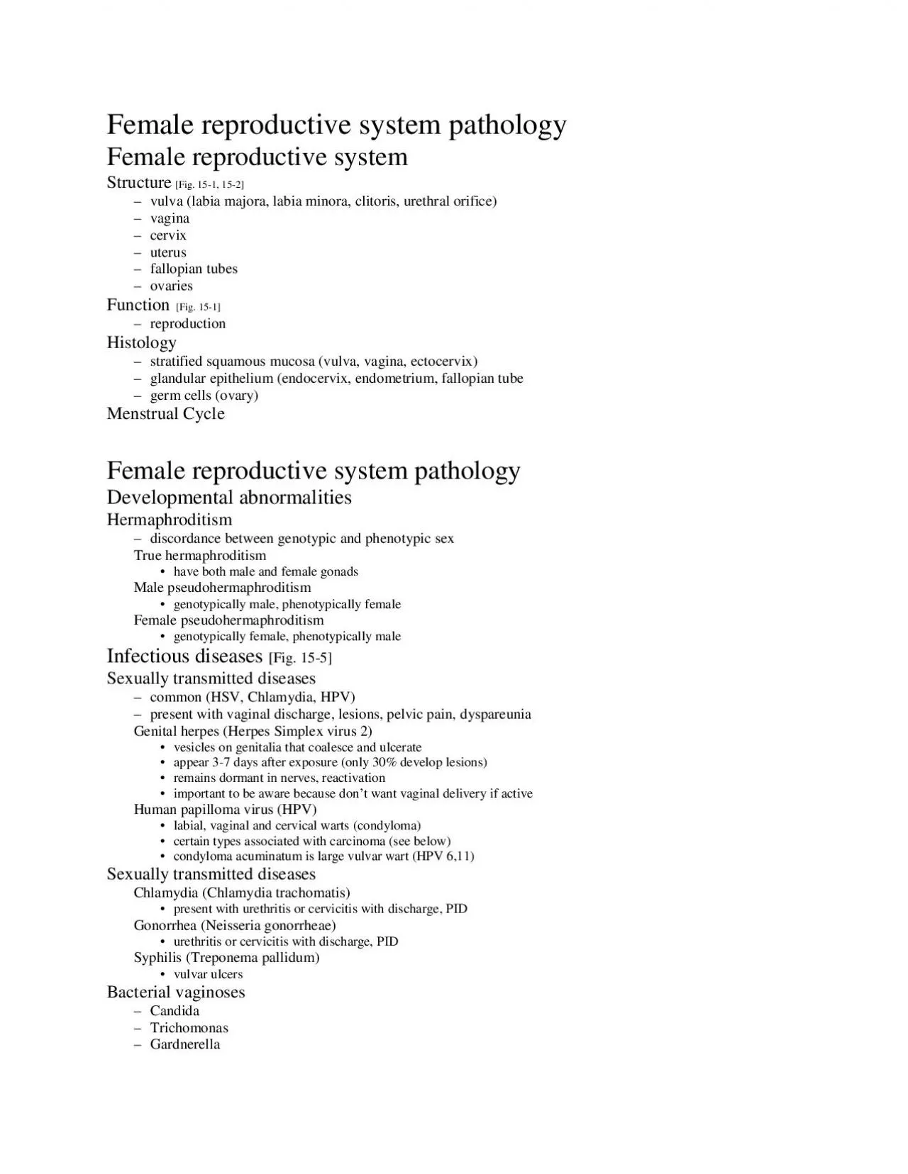
/
Female reproductive system pathologyFemale reproductive systemStructur Female reproductive system pathologyFemale reproductive systemStructur

Female reproductive system pathologyFemale reproductive systemStructur - PDF document

williams
williams . @williams
Follow
342 views
Uploaded On 2022-10-29

Female reproductive system pathologyFemale reproductive systemStructur - PPT Presentation

Female reproductive system pathology Infections Pelvic inflammatory disease chronic extensive infection of upper reproductive tract usually secondary to STD Neisseria Chlamydia salpingitis tuboo ID: 961865

malignant tumors reproductive female tumors malignant female reproductive breast benign ovarian carcinoma factors common neoplasm epithelial females estrogen cell

Share:

Link:

Embed:

Download Presentation from below link

Download Pdf The PPT/PDF document "Female reproductive system pathologyFema..." is the property of its rightful owner. Permission is granted to download and print the materials on this web site for personal, non-commercial use only, and to display it on your personal computer provided you do not modify the materials and that you retain all copyright notices contained in the materials. By downloading content from our website, you accept the terms of this agreement.


Presentation Transcript

Female reproductive system pathologyFemale reproductive systemStructure[Fig. 15-1, 15-2]vulva (labia majora, labia minora, clitoris, urethral orifice) vagina cervix uterus fallopian tubes ovaries Function [Fig. 15-1]reproduction Histology stratified squamous mucosa (vulva, vagina, ectocervix) glandular epithelium (endocervix, endometrium, fallopian tube germ cells (ovary) Menstrual Cycle Female reproductive system pathologyDevelopmental abnormalitiesHermaphroditism discordance between genotypic and phenotypic sex True hermaphroditism have both male and female gonads Male pseudohermaphroditism genotypically male, phenotypically female Female pseudohermaphroditism genotypically female, phenotypically maleInfectious diseases [Fig. 15-5] Sexually transmitted diseases common (HSV, Chlamydia, HPV) present with vaginal discharge, lesions, pelvic pain, dyspareunia Genital herpes (Herpes Simplex virus 2) vesicles on genitalia that coalesce and ulcerate appear 3-7 days after exposure (only 30% develop lesions) remains dormant in nerves, reactivation important to be aware because don’t want vaginal delivery if active Human papilloma virus (HPV) labial, vaginal and cervical warts (condyloma) certain types associated with carcinoma (see below)condyloma acuminatum is large vulvar wart (HPV 6,11)Sexually transmitted diseases Chlamydia (Chlamydia trachomatis) present with urethritis

or cervicitis with discharge, PID Gonorrhea (Neisseria gonorrheae) urethritis or cervicitis with discharge, PID Syphilis (Treponema pallidum) vulvar ulcers Bacterial vaginoses Candida Trichomonas Gardnerella Female reproductive system pathology Infections Pelvic inflammatory disease chronic, extensive infection of upper reproductive tract usually secondary to STD (Neisseria, Chlamydia) salpingitis, tubo-ovarian abscess,peritonitis complications chronic non-specific infection [fever, malaise, fatigue] infertility secondary to scarring of fallopian tubes pelvic mass with pain spread of infection Endometrial hyperplasianormal menstrual cycle requires normal functioning of the hypothalamic-pituitary-ovarian axis (figure 15-5) endometrial hyperplasia is thickening of the endometrial mucosa due to continued estrogen stimulation with inadequate progesterone anovulatory cycles (no ovulation therefore no progesterone secretion) functional causes puberty, anxiety, athlete organic excess estrogen (OCP, tumors) Complex vs. simple hyperplasia atypical hyperplasia increased risk of endometrial adenocarcinoma Neoplasms of lower reproductive tract Carcinoma of vulva squamous cell carcinoma raised or ulcerated lesion pre-neoplastic change may present as white or red patch biopsy to assess surgical excision +/- adjuvant therapy Carcinoma of vagina squamous cell carcinoma clear cell c

arcinoma women born to mothers on DES during pregnancyCarcinoma of cervix reduced mortality due to Pap test (early diagnosis)risk factors sexual intercourse at early age, multiple partners, HPV infection (certain types), other venereal diseases environmental component and other factors squamous cell carcinoma precursor lesion = dysplasia (Cervical intra-epithelial neoplasia) [Fig. 15-6] lack of normal maturation of squamous epithelium occurs at transition zone graded mild, moderate, severe cells shed into vagina (Pap smear) HPV types 16, 18, 31, 33, 34, 35 associated koilocytic change refers to characteristic changes due to HPV Female reproductive system pathology Neoplasia of the uterus Leiomyoma (fibroid) benign neoplasm derived from smooth muscle in wall of uterus most common uterine neoplasm responsive to estrogen, arise during reproductive age usually asymptomatic may produce symptoms due to mass effects, bleeding Leiomyosarcoma malignant neoplasm derived from smooth muscle in wall of uterus very rare Endometrial adenocarcinoma malignant neoplasm derived from epithelial cells in endometrium most common malignant tumor of female reproductive tract elderly females, vaginal bleed risk factors (related to increased estrogen (hyperestrinism)) estrogen secreting tumor, exogenous estrogen obesity nulliparous or early menarche, late menopause stage most important pro

gnostic feature [Fig. 15- 9] grade is also important (low, intermediate, high) diagnosis: endometrial biopsy, dilation and curettage therapy: hysterectomy +/- adjuvant therapy Ovarian cysts fluid filled cavities lined by epithelium usually arise from unruptured follicles (follicular cysts) may also represent cystic corpora lutea or inclusions of surface cells usually small, solitary, asymptomatic if large, then further investigation to rule out neoplasm Polycystic ovary syndrome multiple cysts in both ovaries due to complex hormonal disturbances of the hypothalamic-pituitary-ovarian-adrenal axis presents with menstrual irregularities cause of infertility Ovarian neoplasms Introduction [Fig. 15-12]second most common group of tumors of female reproductive tract highest mortality of female reproductive tract tumors three major groups of neoplasms based on histogenetics surface epithelial tumors germ cell tumors sex cord stromal tumors malignant ovarian tumors are uncommon in young females risk factors not well defined ovarian dysgenesis BRCA1 and BRCA2 gene mutations oral contraceptives not linked to ovarian neoplasms Female reproductive system pathology Ovarian neoplasms Surface epithelial tumors 70 % of ovarian neoplasms spectrum of histologic types serous, mucinous, endometrioid, clear cell and transitional cell types Serous epithelial tumors most common typically

cystic, filled with clear fluid benign, borderline malignant, and malignant tumors 25 % of benign tumors and 50 % of malignant tumors are bilateral distinction of benign versus malignant requires histologic examination Mucinous epithelial tumors also typically cystic, filled with viscus fluid benign, borderline malignant, and malignant tumors 25 % of benign tumors and 50 % of malignant tumors are bilateral distinction of benign versus malignant requires histologic examination Endometrioid epithelial tumors typically solid malignant Germ cell tumors 20 % of ovarian tumors, occur in young females Teratoma most common ovarian neoplasm in young females cystic, contain hair, sebaceous material (dermoid cysts) may contain teeth, bone cartilage benign may undergo malignant transformation (malignant teratoma) Immature teratoma teratoma that contains immature neural tissue may behave malignantly Fibroma benign neoplasm of fibroblasts Thecoma benign, solid and firm neoplasm of spindle cells (theca cells) produce estrogens Granulosa cell tumor neoplasm of granulosa cells benign or malignant,may produce estrogen Metastases Female reproductive system pathology Infertility ovum related sperm related genital organ factors PID Asherman’s syndrome systemic factors Diseases of pregnancy Ectopic pregnancy implantation of fertilized ovum outside the uterine cavity usually occurs in f

allopian tube trophoblast cells of placenta invade wall of tube, begins enlarging may rupture surgical emergency Placenta accreta abnormally deep penetration of placental villi into wall of uterus Placenta previa abnorma placental implantation site in lower uterine segment Toxemia of pregnancy disease of pregnancy of unknown pathogenesis resulting in characteristic symptom complex in the motherPreeclampsia presents with hypertension, edema, and proteinuria occurs in third trimester may progress to eclampsia Eclampsia hypertension, edema, proteinuria and seizures life threatening, must treat seizures, deliver babyGestational trophoblastic disease abnormalities of placentation resulting in tumor-like changes or malignant transformation Hydatidiform mole developmental abnormality of placenta trophoblastic proliferation, hydropic degeneration of chorionic villi enlarged uterus with no fetal movement, high HCG Complete mole no identifiable fetus, abnormal fertilization (46XX, all paternal) Incomplete mole usually some fetal parts, abnormal fertilization (69 chromosomes) Choriocarcinoma rare highly malignant tumor of placental origin, treat with methotrexate Female reproductive system pathology Abortion interruption of pregnancy prior to fetal viability ( 500 g, 20 wks) Spontaneous abortions no identifiable cause (1/3 of all pregnancies) Complete abortion fetus and plac

enta expelled, normal function returns Incomplete abortion retention of some fetal or placental material Missed abortion death of fetus in utero, passed several weeks laterThreatened abortion cervical os closed, spotting of blood Endometriosis endometrial tissue (uterine glands + stroma)located outside the uterus various locations, typically ovary, peritoneum cycle in response to hormonal influences pathogenesis retrograde flow traumatic implantation common, may cause pain may cause infertility benign condition chocolate cyst of ovary Breast pathology Normal BreastFunction function is to produce milk (nourish newborn) Structure [Figs. 16-1, 16-3] modified apocrine sweat gland lobules (ducts + terminal buds) drain into larger duct system hormonally influenced changes males, pre-pubertal females have nipple + ducts post-pubertal female proliferation of ducts and early acini pregnant female terminal buds develop into acini prolactin released in response to infant’s suck milk produced Breast pathology Inflammation Acute mastitis acute inflammation of the breast lactating female bacterial infection abscess may develop Chronic mastitis rare disease of unknown etiology may mimic breast cancer Fibrocystic change benign changes in breast tissue due to various factors including hormonal influences and age females of reproductive age fibrosis of intralobular stroma cystic di

lation of epithelial ducts epithelial hyperplasia various symptoms Gynecomastia increased proliferation of male breast due to various factors Breast pathologyBreast neoplasms Fibroadenoma benign neoplasm of breast epithelial and stromal elements well circumscribed, firm, mobile mass young females Breast cancer most common cancer in females second most common cause of cancer related deaths in females hormonal, environmental and genetic influences familial breast cancers BRCA-1, BRCA-2 tumor suppressor genes increased incidence of other cancersrisk factors female sex (100x males) genetic predisposition hormonal factors prolonged estrogen exposure early menarche, late menopause nulliparous other malignancies contralateral breast carcinoma endometrial carcinoma premalignant changes carcinoma in situ, atypical hyperplasia Age Race there are different forms of breast cancer most common breast cancer is infiltrating ductal carcinoma adenocarcinoma desmoplastic response of stroma lymphatic spread (axillary nodes drain most of the breast) presents as mass early detection breast self-examination mammography [Fig. 16-10] fine needle aspiration incisional biopsy therapy Surgical resection lumpectomy mastectomy axillary dissection radiation chemotherapy tamoxifen herceptin Prognosis staging most important [Fig. 16-8] histologic subtypes histological grading estrogen receptor stat