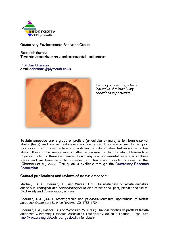
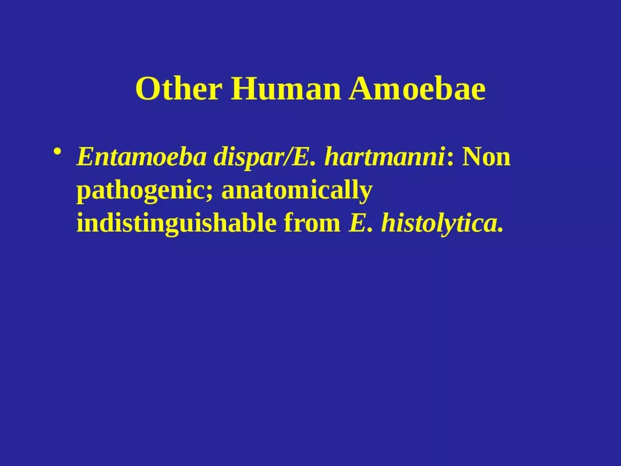
/
Other Human Amoebae Entamoeba dispar/E. hartmanni Other Human Amoebae Entamoeba dispar/E. hartmanni

Other Human Amoebae Entamoeba dispar/E. hartmanni - PowerPoint Presentation

ButterflyHeart
ButterflyHeart . @ButterflyHeart
Follow
352 views
Uploaded On 2022-07-28

Other Human Amoebae Entamoeba dispar/E. hartmanni - PPT Presentation

Non pathogenic anatomically indistinguishable from E histolytica Entamoeba coli Trophozoite Cyst Nucleus Entamoeba coli Life cycle and location identical to ID: 929906

trophozoites common trophozoite amebic common trophozoites amebic trophozoite endocommensal entamoeba naegleria acanthamoeba fowleri cyst cysts lives symptoms distribution worldwide

Share:

Link:

Embed:

Download Presentation from below link

Download Presentation The PPT/PDF document "Other Human Amoebae Entamoeba dispar/E. ..." is the property of its rightful owner. Permission is granted to download and print the materials on this web site for personal, non-commercial use only, and to display it on your personal computer provided you do not modify the materials and that you retain all copyright notices contained in the materials. By downloading content from our website, you accept the terms of this agreement.


Presentation Transcript

Slide1

Other Human Amoebae

Entamoeba dispar/E. hartmanni

: Non pathogenic; anatomically indistinguishable from

E. histolytica.

Slide2

Entamoeba coli

Trophozoite Cyst Nucleus

Slide3

Entamoeba coli

Life cycle and location identical to

E. histolytica

.

Slide4

Entamoeba coli

Life cycle and location identical to

E. histolytica

.

Most common endocommensal in people; has a worldwide distribution and 10-50% of the population can be infected in different parts of the world.

Slide5

Entamoeba coli

Life cycle and location identical to

E. histolytica

.

Most common endocommensal in people; has a worldwide distribution and 10-50% of the population can be infected in different parts of the world.

Not pathogenic.

Slide6

Entamoeba coli

Life cycle and location identical to

E. histolytica

.

Most common endocommensal in people; has a worldwide distribution and 10-50% of the population can be infected in different parts of the world.

Not pathogenic.

Feeds on bacteria and any other cells available to it;

does not invade tissue

.

Slide7

Other Hosts

Monkeys

Apes

Pigs

Slide8

10

m

m

Trophozoites 20-30

μ

m in diameter (15-50 µm)

Slide9

10

m

m

A

D

C

B

Slide10

10

m

m

A

D

C

B

E. coli

E. histolytica

Slide11

C

10

m

m

Cysts 10-30

μ

m

Slide12

Entamoeba gingivalis


Habitat: Mouth

Hosts: Humans, other primates, dogs and cats.

Prevalence is from 50 to 95%.


Stage: Trophozoite, no cyst.


Slide13

Entamoeba gingivalis


Trophozoite lives on the surface of teeth and gums. Feed on epithelial cells of the mouth, bacteria, food debris, and other cells available to them.


Slide14

Entamoeba gingivalis


Trophozoite lives on the surface of teeth and gums. Feed on epithelial cells of the mouth, bacteria, food debris, and other cells available to them.

Organisms are more common in persons with pyorrhea (gum disease) but they are not the cause of the condition.

Slide15

Entamoeba gingivalis


Trophozoite lives on the surface of teeth and gums. Feed on epithelial cells of the mouth, bacteria, food debris, and other cells available to them.

Organisms are more common in persons with pyorrhea (gum disease) but they are not the cause of the condition.

Transmission mouth to mouth, droplet spray, or sharing eating utensils.


Slide16

Endolimax nana

“The dwarf internal slug”

Trophozoite Cyst Nucleus

Slide17

Endolimax nana

Second most common endocommensal of humans, worldwide distribution 30%.

Slide18

Endolimax nana

Second most common endocommensal of humans, worldwide distribution 30%.

Lives in the large intestine mainly near the cecum and feed on bacteria; non pathogenic.

Slide19

Endolimax nana

Second most common endocommensal of humans, worldwide distribution 30%.

Lives in the large intestine mainly near the cecum and feed on bacteria; non pathogenic.

Also occurs in monkeys.

Slide20

10

m

m

Trophozoites are tiny 6-15

μ

m in diameter.

Large glycogen vacuoles are often present.

Slide21

10

m

m

Mature cyst is 5 – 14

μ

m in diameter; contains 4 nuclei; shape is round to elliptical

Slide22

Iodamoeba buetschlii

Trophozoite Cyst Nucleus

Slide23

Iodamoeba buetschlii

Not very common endocommensal in people.

Slide24

Iodamoeba buetschlii

Not very common endocommensal in people.

Lives in the large intestine, predominantly in the cecal areas.

Slide25

Iodamoeba buetschlii

Not very common endocommensal in people.

Lives in the large intestine, predominantly in the cecal areas.

Has a very high prevalence in

pigs

; 50% of pigs are infected with this ameba in France and Egypt; pigs are probably its normal host.

Slide26

10

m

m

Trophozoites are 9-14

μ

m long but may be as large as 20

μ

m

Slide27

10

m

m

Slide28

10

m

m

E. nana

I. buetschlii

Slide29

10

m

m

Cysts are 6-15

µm long and have a large glycogen vacuole.

Slide30

Dientamoeba fragilis

Trophozoite No Cyst Nucleus

Slide31

Dientamoeba fragilis

LIFE CYCLE - it does not form cysts and trophozoites cannot survive passage through the small intestine.

Slide32

Dientamoeba

fragilis

LIFE CYCLE - it does not form cysts and

trophozoites

cannot survive passage through the small intestine.

Humans probably get infected by this

endocommensal

when they ingest pinworm eggs!

Slide33

10

m

m

Trophozoites small 6-12

µm long; binucleated.

Slide34

Slide35

Slide36

free-living amoebae causing human infections

N

ormal

inhabitants of soil and water where they feed on bacteria.

A

few members have the ability to become

facultative

parasites

when an

opportunity

to enter a vertebrate exists

.

There

are able to infect humans

Slide37

Naegleria

fowleri

primary

amebic

meningoencephalitis

(PAM)

Acanthamoeba

spp.

granulomatous

amebic encephalitis (GAE)

granulomatous

skin and lung lesions (primarily

immunocompromised

)

amebic

keratitis

Balamuthia

mandullaris

.

GAE

+

granulomatous

skin and lung lesions (primarily healthy)

Pathogenic Free-Living

Amebae

Slide38

Naegleria

fowleri

Naegleria

fowleri

is a free living brain-eating amoeba.

Typically found in warm fresh water (thermo tolerant amoeba).

worldwide distribution.

It exists in

trophozoite

and cyst forms and in a transient flagellate stage

.

Naegleria

fowleri

is the causative agent of

primary amebic

meningoencephalitis

(PAM).

The period incubation is short, the symptoms are acute, and death is almost certain and rapid.

Naegleria

fowleri

invades the C.N.S. via penetration of the olfactory mucosa and nasal tissues (nose).

Since

Naegleria

fowleri

trophozoites

and cysts are susceptible to chlorine, swimming pools should be adequately chlorinated

.

Slide39

Naegleria

fowleri

Structures of the amoeba form are :

Trophozoit

and flagellate

The infective stage is

trophozoit

stage

Method of transmission is by penetrate the mucosal layer of olfactory tissue and nasal cavity

Leads to Primary amebic

meningoencephalitis

Most symptoms involving fever , headache, stiff neck and confusion.

Diagnosis with X-ray , IHAT in CSF.

Occasionally, a C.T scan may be ordered to rule out cerebral hematoma. culture media of CSF and PCR .

Slide40

low nutrients

desiccation

Naegleria

SPP.

Cyst = dormant form

Trophozoite

=

feeding and replicating form

Slide41

Naegleria

fowleri

When a victim swims or sinks into freshwater.

All

victims have had a

history of swimming

in freshwater lakes or ponds or swimming pools a few days before the onset of symptoms

Slide42

Naegleria

fowleri

Slide43

Slide44

Primary Amebic

Meningoencephalitis

(PAM)

1-14 days incubation period

symptoms usually within a few days after swimming in warm still waters

infection believed to be introduced through nasal cavity and olfactory bulbs

symptoms include headache, lethargy, disorientation, coma

rapid clinical course, death in 4-5 days after onset of symptoms

trophozoites

can be detected in spinal fluid, but diagnosis is usually at autopsy

4 known survivors treated with

Amphotericin

B

Slide45

Slide46

brain section

in vitro culture

“lobopodia”

Slide47

Acanthamoeba

Acanthamoeba

are ubiquitous organisms in nature.

Most

species are free-living.

Cysts

are common and are very resistant to chlorine.

Some

are opportunists that can cause infections in human.

Infections

with these amoebae are more common in

immunocompromised

patients.

Amoebae

can be introduced through environmental exposures, including swimming while wearing contact lenses or using contaminated contact lens solutions.

Acanthamoeba

genus

causes 3

clinical

syndromes:

1

-

granulomatous

amebic

encephalitis

(GAE).

2

-

Disseminated

granulomatous

amebic disease

(

eg

, skin, sinus, and pulmonary infections).

3

-

Amebic

keratitis

: a sight-threatening disease. Most cases occur in people who wear contact lenses.

Slide48

Acanthamoeba

culbertsoni

Free-living

trophozoites

and cysts occur in both the soil and freshwater.

Trophozoites

occur only as amoeboid forms

Slide49

Acanthamoeba

culbertsoni

Free-living amoeba

Lives in water have been found in soil; sea water; sewage; swimming pools; contact lens equipment; medicinal pools; dental treatment units and air conditioning systems

Contact lens

wearers can get

keratitis

(infection of the cornea) by using tap water for lens disinfection or by swimming

when wearing lenses

Slide50

Acanthamoeba

life cycle

Slide51

Slide52

Slide53

Acanthamoeba

Meningoencephalitis

portal of entry unknown, possibly respiratory tract, eyes, skin

presumed

hematogenous

dissemination to the CNS

infection associated with debilitation or

immunosuppression

onset is insidious with headache, personality changes, slight fever

progresses to coma and death in weeks to months

amebas not yet detected in spinal fluid

trophozoites

and sometimes cysts detectable in histological examination

no human cures documented

Slide54

Acanthamoeba results in lesions of the skin, eye, brain, etc

.

Slide55

Slide56

The symptoms of GAE including :

Alter mental status, headache ,fever, neck stiffness, seizures, focal neurological signs and coma leading to death

Slide57

Amebic

Keratitis

predisposing factors

ocular trauma

contact lens (contaminated cleaning solutions)

symptoms

ocular pain

corneal lesions (refractory to usual treatments)

diagnosis

demonstration of amebas in corneal scrapings

treatment

difficult, limited success

corneal grafts often required

Slide58

keratitis

:

corneal inflammation and corneal perforation often followed by blindness.

Slide59

Laboratory Diagnosis of infections with free-living amoebae

:

Keratitis

:

Acanthamoeba

trophozoites

or cysts can be demonstrated with

corneal scrapings

or a biopsy sample via wet mount, stains,

histopathologic

examination.

Granulomatous

amebic encephalitis

: _ This condition is diagnosed via

brain biopsy

. (cyst and

trophozoit

)