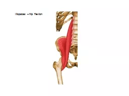
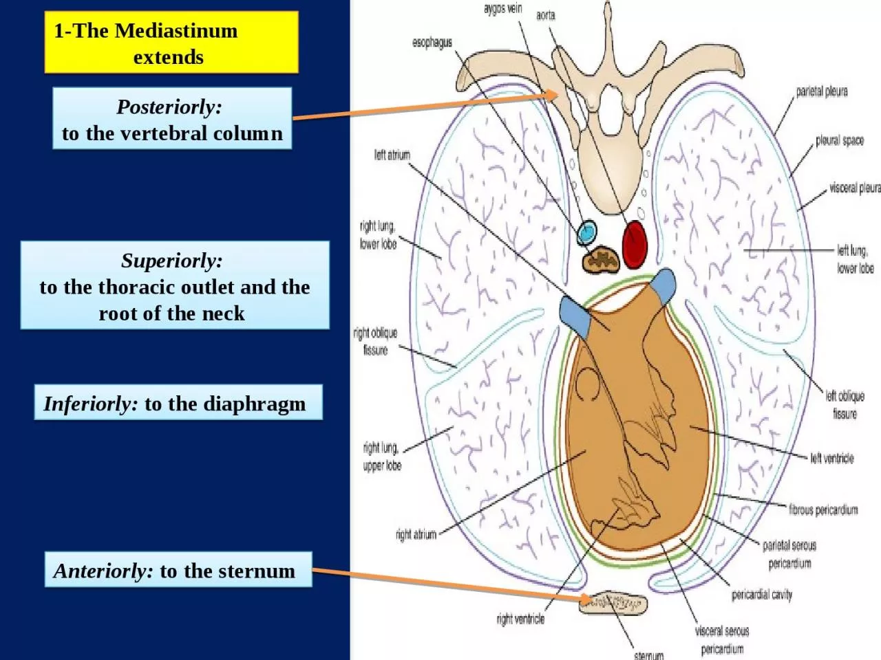
Inferiorly to the diaphragm Superiorly to the thoracic outlet and the root of the neck Anteriorly to the sternum Posteriorly to the vertebral column An imaginary plane passing from ID: 930912
Download Presentation The PPT/PDF document "1-The Mediastinum extends" is the property of its rightful owner. Permission is granted to download and print the materials on this web site for personal, non-commercial use only, and to display it on your personal computer provided you do not modify the materials and that you retain all copyright notices contained in the materials. By downloading content from our website, you accept the terms of this agreement.
Slide1
1-The Mediastinumextends
Inferiorly:
to the diaphragm
Superiorly: to the thoracic outlet and the root of the neck
Anteriorly: to the sternum
Posteriorly: to the vertebral column
Slide2An imaginary plane passing from
the sternal
angle anteriorly to the lower border of the body of the fourth thoracic vertebra posteriorly
divides the mediastinum into:
SUPERIOR AND INFERIOR MEDIASTINA
Which one is larger?
Slide3THE INFERIOR MEDIASTINUM is further subdivided into:
is a space between the
pericardium and the sternum
consists of the pericardium and heart
lies between
THE PERICARDIUM And THE VERTEBRAL COLUMN
3-THE POSTERIOR MEDIASTINUM
2-THE ANTERIOR MEDIASTINUM
1-THE MIDDLE MEDIASTINUM
Slide4Slide5THYMUS
LARGE VEINS?
LARGE ARTERIES? TRACHEA
ESOPHAGUSTHORACIC DUCT SYMPATHETIC TRUNKS
is bounded in front by the manubrium
sterni and behind by the first four thoracic vertebrae
THE SUPERIOR MEDIASTINUM
contains
From anterior to posterior
anterior
posterior
Slide6THE SUPERIOR MEDIASTINUM
THYMUS
LARGE VEINS, right and left brachiocephalic
veins and the upper half of superior vena cava. LARGE
ARTERIES, arch of the aorta with its three large branches.
TRACHEAESOPHAGUSTHORACIC DUCT (g)
phrenic nerves, vagus nerves
and SYMPATHETIC
TRUNKS
Main contents!
Cross-section through the superior mediastinum at the level of vertebra TIII.
Anterior
Posterior
Slide7Structures in the superior mediastinum
Slide8THYMUS
Involved in the early development of the immune system
,
the thymus is a large structure in the child,
begins to atrophy after puberty
In the elderly adult, it is barely identifiable as an organ, consisting mostly of fatty tissue
Slide9Large Veins of the
Thorax
1-Brachiocephalic Veins
A-The right brachiocephalic vein:formed by the union of the right subclavian
and the right internal jugular veins (note: it is shorter and vertical)
the right subclavian
the right internal
jugular veins
Slide10B-The left brachiocephalic vein:
Is formed by the union of
the LEFT subclavian
and the LEFT internal jugular veins ( note: It passes obliquely and it is longer)It joins the right brachiocephalic vein to form
the superior vena cava
Slide11The superior vena cava contains all the venous blood from
the head and neck and both upper limbs
Superior Vena Cava
is formed by the union of the two brachiocephalic veins
It passes downward to end in the right atrium of the heart
The vena azygos joins the superior vena cava just before it enters the pericardium
Slide12The azygos veins consist of:
Azygos
Veins
c-THE SUPERIOR HEMIAZYGOS VEINb-THE INFERIOR HEMIAZYGOS VEIN
a-THE MAIN AZYGOS VEIN
Slide13The inferior vena cava pierces the central tendon of the diaphragm
opposite the eighth thoracic vertebra
and almost immediately enters the lowest part of the right atrium
Inferior Vena Cava
Slide14Aorta
The aorta is the main arterial trunk that delivers oxygenated blood from the left ventricle of the heart to the tissues of the body
. It is divided for purposes of description into the following parts:
Large Arteries of the Thorax
A-ASCENDING AORTA
B-ARCH OF THE AORTA
C-DESCENDING:
THORACIC AORTA (above diaphragm)
ABDOMINAL AORTA
(below diaphragm)
Slide15The ascending aorta lies within the
fibrous pericardium
(what does this mean?) Begins at the base of the left ventricle
Ends at the level of the sternal angle, where it becomes continuous with the arch of the aorta
At its root it possesses three bulges, the sinuses of the aortaBranches
A-Ascending Aorta
The right coronary artery
The left coronary artery
Slide16is a continuation of the ascending aorta
(what does this mean?)Ends at the level of the
sternal angle where it becomes continuous with the descending aorta.
Branchesa-THE BRACHIOCEPHALIC ARTERY arises from the convex surface of the aortic arch
It divides into: 1-THE RIGHT SUBCLAVIAN ARTERY2-RIGHT COMMON CAROTID ARTERY
B-Arch of the Aorta
Slide17Arises from the convex surface of the aortic arch
enters the neck behind the left sternoclavicular
joint.Why we call it subclavian?
arises from the aortic archRuns in a groove in the first rib
b-The left common carotid artery
c-The left subclavian artery
Slide18lies in the posterior mediastinum
begins as a continuation of the arch of the aorta
(opposite the sternal angle). At the level of
the 12th thoracic vertebra, it passes behind the diaphragm (through the aortic opening) in the midline and becomes continuous with the abdominal aorta.
C- Descending Thoracic Aorta
1-Posterior intercostal arteries are given off to the lower nine intercostal spaces
2-Pericardial, esophageal, and bronchial arteries are small branches that are distributed to these organs
.
Branches
READ ONLY
Slide19What do you think about the aorta and mediastina?
Slide20Thoracic Duct
The thoracic duct begins below in the abdomen as a dilated sac,
THE CISTERNA CHYLI It ascends through the aortic opening in the diaphragm
enters the beginning of the left brachiocephalic vein.The thoracic duct thus conveys to the blood all lymph from the lower limbs, pelvic cavity, abdominal cavity, left side of the thorax, and left side of the head, neck, and left arm
Read only
Passes through superior and posterior mediastinum
Slide21Vagus NervesCranial nerve
10
The right vagus nerve
Nerves of the Thorax
The left vagus nerve
Located in the neck Thorax: passes through both
superior and posterior mediastinum
Slide22Phrenic Nerves
The phrenic nerves arise from the anterior rami of the third, fourth, and fifth cervical nerves
It passes in front of the root of the lungs
Its terminal branches pass through the caval opening in the diaphragm to supply the central part of the peritoneum on its underaspect
.Read only
Slide23The thoracic part of the sympathetic trunk
is continuous above with the cervical and below with the lumbar parts of the sympathetic trunk.
Thoracic Part of the Sympathetic Trunk
The first ganglion is often fused with the inferior cervical ganglion to form the
stellate ganglion
Read only
: passes through both superior and posterior mediastinum
Slide24is a tubular structure about 10 in. (25 cm) long
is continuous above with the laryngeal part of the pharynx opposite the sixth cervical vertebra
. It passes through the diaphragm at the level of the 10th thoracic vertebra to join the stomach
Esophagus
The esophagus has three anatomic and physiologic constrictions
where the pharynx joins the upper end
where the aortic arch and the left bronchus cross its anterior surface
where the esophagus passes through the diaphragm into the stomach
These constrictions are
1
2
3
Read only
passes
through both
superior and posterior mediastinum