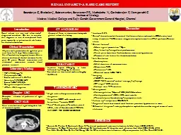
Clinical features Mean age at presentation 65yrs Sexlaterality no significant predominance flank pain hematuria flank tenderness new onset hypertension Presence of risk factors for thromboembolism ID: 933141
Download Presentation The PPT/PDF document "Incidence 0.2% Causes- thromboemboli fro..." is the property of its rightful owner. Permission is granted to download and print the materials on this web site for personal, non-commercial use only, and to display it on your personal computer provided you do not modify the materials and that you retain all copyright notices contained in the materials. By downloading content from our website, you accept the terms of this agreement.
Slide1
Incidence 0.2%Causes- thromboemboli from atrial fibrillation, infective endocarditis, DCM, aortic/renal artery dissection, FMD, trauma, iatrogenic, transplant, vasculitis, APLA syndrome, Cocaine abuseClinical features- Mean age at presentation – 65yrsSex/laterality- no significant predominanceflank pain, hematuria, flank tenderness, new onset hypertensionPresence of risk factors for thromboembolismLab EvaluationLeukocytosisMild elevation of serum creatinineHematuria-50%Proteinuria- 45%Elevated serum LDH -100%Imaging USG-3% sensitiveCECT -80%sensitive- cortical rim sign, flip flop signCT angio- 100% sensitive Isotope scan -97% sensitiveManagement Anticoagulation –Heparin- UFH/LMWH, warfarinAntihypertensives- ACEI, ARBs most suitableThrombolysis/thrombectomy- no prospective trialsFollow up – isotope scan, ECHO, INRPrognosis- most have normal renal function, persistent hypertension in someDifferential diagnosis- acute pyelonephritis, nephrolithiasis, other surgical causes of acute abdomen
TC- 17900/mm^3Polymorphs- 85%Serum creatinine- 1.3-1.7-1.4Urinalysis- 5-8RBC/HPFSerum LDH- 875IU/LCoagulation profile normalAPLA , ANA negative
Soundarya G, Sivabalan J, Subramanian, Saravanan P R, Muthulatha N, Govindarajan P, Ilamparuthi C Institute of UrologyMadras Medical College and Rajiv Gandhi Government General Hospital, Chennai
Laboratory Workup
RENAL INFARCT- A RARE CASE REPORT
Introduction
Doppler USG
CT ANGIOGRAM
Complete absence of perfusion of right kidney
Clinical Presentation
CECT KUB
Discussion
TREATMENT
Injection heparin 80mg/kg iv bolus followed by 15mg/kg/hr infusion
Switched on later to oral warfarin 4mg/day
INR-2.15
Renal infarct is a rare and often under-diagnosed condition. One has to consider this diagnosis in case of unexplained flank pain, especially in patients with risk factors for thromboembolism
45 years male presented with right loin pain and fever for 3 days. No h/o hematuria.
No co-morbidities/surgeries/trauma. He is a chronic smoker and alcohol consumer for the past 30 years. General examination and abdominal examination normal, blood pressure normal.
Non visualisation of right kidney due to lack of contrast uptake
Absence of flow in rt main renal artery with global infarct of rt kidney
High index of suspicion needed for diagnosis
Negative NCCT with unexplained hematuria – consider CECTTriad- flank pain, +/-hematuria, raised LDH, risk of thromboemboli- RENAL INFARCT TO BE CONSIDERED
Korzets Z, Plotkin E, Bernheim J, Zissin R. The clinical spectrum of acute renal infarction. Isr Med Assoc J. 2002;10:781–4.Bolderman R, Oyen R, Verrijcken A, Knockaert D, Vanderschueren S. Idiopathic renal infarction. Am J Med. 2006;119:356. e9-12.Tsai SH, Chu SJ, Chen SJ, Fan YM, Chang WC, Wu CP, et al. Acute renal infarction: a 10-year experience. Int J Clin Pract. 2007;61:62.
TAKE HOME MSG
REFERENCE