/
Milk Production Management and Dairy Development Milk Production Management and Dairy Development

Milk Production Management and Dairy Development - PowerPoint Presentation

Honeybunches
Honeybunches . @Honeybunches
Follow
355 views
Uploaded On 2022-08-03

Milk Production Management and Dairy Development - PPT Presentation

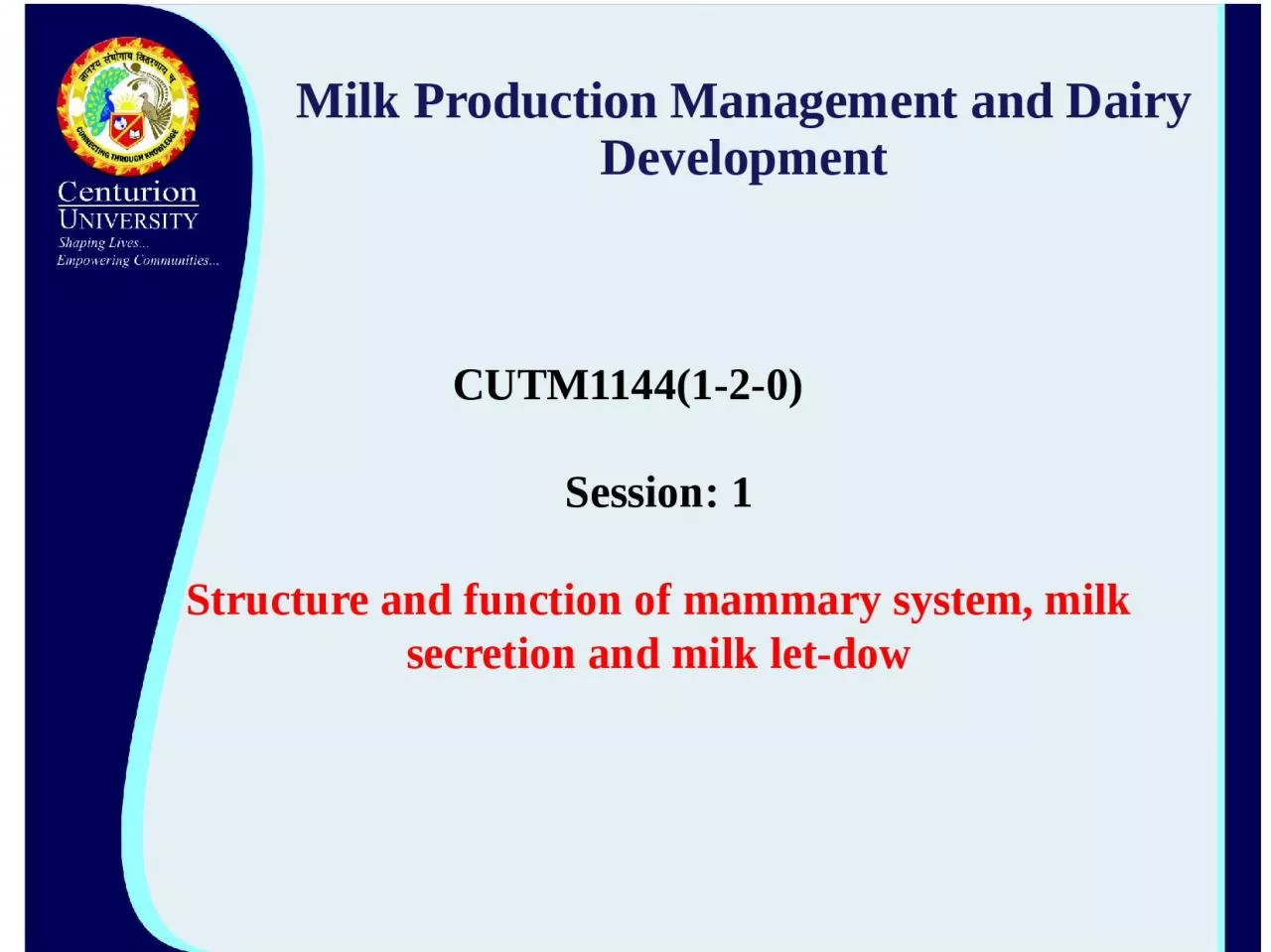
CUTM1144120 Session 1 Structure and function of mammary system milk secretion and milk let dow External Anatomy The udder consists of 4 separate glands Located ID: 933634

milk gland artery mammary gland milk mammary artery tissue udder blood ducts teats external lymph system cistern skin suspensory

Share:

Link:

Embed:

Download Presentation from below link

Download Presentation The PPT/PDF document "Milk Production Management and Dairy Dev..." is the property of its rightful owner. Permission is granted to download and print the materials on this web site for personal, non-commercial use only, and to display it on your personal computer provided you do not modify the materials and that you retain all copyright notices contained in the materials. By downloading content from our website, you accept the terms of this agreement.


Presentation Transcript

Slide1

Milk Production Management and Dairy Development

CUTM1144(1-2-0

)

Session: 1

Structure and function of mammary system, milk secretion and milk let-

dow

Slide2

External

Anatomy

The

udder

consists

of

4

separate

glands

Located

in

the

inguinal

region

of

the

ventral

aspect.

Each

gland

has

one

teat

Each

teat

has

one

opening

The

glands

are

covered

with

hair

Teats

do

not

have

hair.

Slide3

The

right

and

left

halves

are

entirely separate externally indicated by intermammary groove.The rear quarters account for 55-60% of the milk produced and 55-60% of udder weight.

Rear

teats

are

usually

shorter

than the

front

teats.

Slide4

The Teats - (papilla mammae)

Functions

as

the

only exit for the

mammary secretionThe only means for the calf to receive

milk.

Usually,

only

one

teat

drains

one

gland.

No

hair,

sweat

glands

or

sebacious

glands on

the

teats.

Slide5

Supernumerary Teats

About

50%

of

all

cows

have extra teats- supernumerary teats.Some of these extra teats open into a "normal"

gland, but many do not.

Slide6

Slide7

Streak canal (ductus papillaris)

Functions

as

the

only

orifice

of

the

gland

between

the internal

milk

secretory

system

and

the

external

environment.

The

streak

canal

is

the

main barrier

against

infection.

Lined

with

a

skin-like

epidermis.

Closed

by

sphincter

muscles

around

the

streak

canal.

streak

canal

length

increases

with

increasing

lactation

number.

Slide8

The

interior of the gland is made up

of:

Connective

tissue

- fibrous tissue (collagen) and fatty tissue

(adipose cells).Secretory tissue - secretory epithelial cells-produce the

milk.

The

relative

amount

of

connective

vs.

secretory

tissue

varies

from

animal

to

animal,

by

stage

of

mammary

development

Slide9

Gland Cistern - (sinus lactiferus)

Also

called

the

udder

cistern

or milk

cistern

opens

directly

into

the

teat

cistern.

The

cisterns

function

for

milk

storage

(holds

~100-

400

ml).

The

gland

cistern

varies

greatly

in

size

and

shape.

There

are

often

pockets

formed

in

the

cistern

at

the

end

of the

larger

ducts.

The

major

ducts

which

empty

into

the

gland

cistern

sometimes are

called

cisternal

ducts.

Slide10

Secretory

tissue in the udder is

organized

into

lobes-

many

lobules-lobule

contains 150-220 microscopic alveoli.Alveoli - (acini)

Sack-like

structures

where

milk

is

synthesized

and

secreted.

A

single

layer

of

secretory

epithelial

cells

lines

the

lumen.

Contractile myoepithelial

cells

surround

the epithelial

lining.

Slide11

Myoepithelial cells

contract

in

response

to

the

hormone

oxytocin-milk being squeezed out of the alveolar lumen and into the small ducts.Outside

of the myoepithelial cells

the

alveolus

is

surrounded

by

a

connective

tissue

basement

membrane.

Slide12

The capillary

bed on

the

outside

of

the

alveolus

is part of the stromal connective tissue between alveoli.A group of

alveoli can be visualized

as

a

clump

of

grapes,

with

the

stems

acting

as

the

small

ducts

leading

from

the

alveoli.

Slide13

Slide14

Slide15

Lobules -

Clusters

of

150-220

alveoli

are

encapsulated

by a connective tissue sheath and are organized as a lobule (~.7-.8 mm dia.).

Lobes

-

Groups

of

lobules

are

surrounded

by

a

connective

tissue

sheath

and

comprise

a

lobe.

Each

mammary

gland

is

made

of

numerous

lobes.

Slide16

Ducts

-tubules

by

which

milk

drains from

the

alveoli down to the gland cistern.Interlobar or primary ducts drain multiple lobes.

Intralobar

ducts

or

secondary

ducts

are

within

a

lobe

-drain

several

regions

of

the

lobe

.

Intercalary or

tertiary

ducts

-small

ducts

which

exit

from

the

alveolus.

Slide17

Slide18

A

strong

suspensory

system

required-high

producers.

Mammary gland is a

skin gland, and is

therefore

external

to

the

body

cavity.

Slide19

The tissues, which provide some degree

of support for the udder:

Skin

-minor

support..

Superficial

fascia

or

Areolar

subcutaneous

tissue

-attaches

the

skin

to

the

underlying

tissues.

Suspensory

ligaments-main

suspensory

structures.

Suspensory

ligaments

are

three:

Superficial

Lateral

suspensory

ligament

Deep

Lateral

suspensory

ligament

Median

suspensory

ligaments

Slide20

Slide21

Blood supply

to

the

mammary

gland

-

extremely

important for mammary function.All of the milk precursors come from blood.

On

average

400

-

500

units

of

blood

passes

through

the

udder

for

each

unit

of

milk

synthesized

Total

udder

blood

volume

for

lactating

cows

is

about

8%

of

total

body

blood

volume,

non-lactating

cow

-about

7.4%.

There

is

a

2-6

fold

increase

in

blood

flow

in

the

mammary

gland

starting 2-3

days

prepartum

.

Slide22

Arterial System

Blood

leaves

the

heart

and flows towards the rear

of

the

cow

-abdominal

aorta

.When

it

reaches

the

pubic

area

-called

the

common

iliac

arteries.

These

divide

into

the internal

and

external

iliac

arteries.

The

external

iliac

artery

becomes

the

femoral

artery

(supplies

the

leg

muscles)

Slide23

A branch

off

of the

femoral

artery

forms

the

prepubic artery from which branches the posterior abdominal artery and the external pudic (or external pudental) artery.

The external pudic

artery

passes

through

the

inguinal

canal

and

out of

the

body

cavity.

Slide24

The inguinal

canal -orifice

in

the

body

cavity

in

the inguinal region where blood vessels, lymph vessels and nerves enter and leave the body wall to supply the skin in the posterior part of the animal.

As

the

external

pudic

artery

passes

out of

the

body

cavity

it

becomes

the

mammary

artery.

Once

it

enters

the

gland,

the

mammary

artery

then

divides

into

the

anterior

(or

cranial)

and

posterior

(or

caudal)

mammary

arteries,

which

then

it

further

branches

as

it

descends

down

into

the

gland

Slide25

A small amount

of

blood

also

reaches

the

mammary

gland by the perineal artery (from the internal iliac artery), but this only supplies the upper rear portion of gland.

Slide26

Slide27

Venous System

Veins

leave

the

mammary

gland

anti-parallel

to

the arteries.There are three veins on each

side that carry blood

away

from

the

gland:

External

pudic

vein

leaves

the

udder

anti-parallel

to

the

external

pudic

artery

Subcutaneous

abdominal

vein

(milk

vein)

exits

the

gland

at

the

anterior

end

of

the

front

quarters

and

passes

along

the

abdominal

wall-large

vein

visible

under

the

skin

on

the

belly

of the

cow.

Slide28

Slide29

Nervous System

Innervation

of the

udder

is

sparse compared

with

other

tissues.

Sensory

nerves

are

found

in

the

teats

and

skin;

these

are

involved

in

the

afferent

pathway

of

the

milk

ejection

reflex.

There

is

no

parasympathetic

innervation

to

the

gland;

this

is

similar to

other

skin

glands.

There

is

no

innervation

of the

secretory

system:

myoepithelial

cells

are

not

innervated;

they

do

not

contract

in

response

to direct

innervation,

but

rather

they

contract

in

response

to

the

blood-

borne

hormone,

oxytocin

.

Slide30

Slide31

The lymphatic networkThere

is extensive lymph drainage from the

teats.

originates

in

tissue

spaces as

lymphatic

capillaries.

Lymph

capillaries

converge

to

form

larger

vessels.

Lymph

flow

is

undirectional

from

the

udder

through

lymphatic

vessels,

eventually

dumping

lymph

into

the

vena

cava.

Lymph

is

a

clear,

colorless

liquid

with

a

composition

similar to

blood

plasma.

Slide32

Valves in

the

lymphatic

vessels

prevent retrograde

flow.

In the udder, the lymph system

flows through the

supramammary

lymph nodes.

Slide33

Mastitis

Inflammation

of

udder

due

to bacterial or fungal infection.

Physical and chemical composition of the milk will be changed.

In

our

condition

one

of

the

reasons

for

poor

production

from

local

cattle

could

be

due

to

subclinical

mastitis

that

go

on

undetected

in

the field.

Slide34

Mammary gland

abalation:

Surgical

removal

of

mammary

glands

as a remedy to disease process. Carried out only in pets for saving their life.

Slide35

Thank you