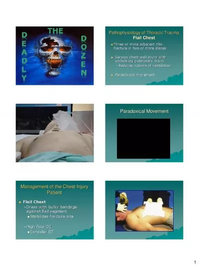
Anatomy Trauma or injury is defined as cellular disruption caused by an exchange with environmental energy that is beyond the bodys resilience Trauma remains the most common ID: 933137
Download Presentation The PPT/PDF document "Majid Pourfahraji chest trauma" is the property of its rightful owner. Permission is granted to download and print the materials on this web site for personal, non-commercial use only, and to display it on your personal computer provided you do not modify the materials and that you retain all copyright notices contained in the materials. By downloading content from our website, you accept the terms of this agreement.
Slide1
Slide2Majid
Pourfahraji
chest trauma
Slide3Anatomy
Slide4Trauma
, or injury, is defined as cellular
disruption
caused by an exchange with environmental energy that is beyond the body's
resilience
.
Trauma
remains the most common cause of death for all individuals between the ages of 1 and 44 years and is the third most common cause of death regardless of age.
trauma
Slide5The
initial
management of seriously injured patients consists of performing
the
primary
survey
(
the "ABCs"—Airway with cervical spine protection, Breathing, and Circulation); the goals of the primary survey are to identify and treat conditions that constitute an immediate threat
to
life.
primary survey
Slide6Blunt
Trauma:
Blunt force to chest.
Penetrating
Trauma
:
Projectile that enters chest causing small or large hole.
Compression Injury: Chest is caught between two objects and chest is compressed.
Main Causes of Chest Trauma
Slide7Chest wall
* Rib fracture
* Flail chest
Airway obstruction
Pneumothorax
* Simple/Closed
* Open Pneumothorax
* Tension Pneumothorax
Hemothorax
Flail Chest and Pulmonary Contusion
Cardiac
Tamponade
Traumatic Aortic RuptureDiaphragmatic Rupture
Trauma to the chest
Slide8Blunt
And
Penetrating
PAIN
Shallow breathing
Atelectasis Shunt: lack of ventilation respiratory and metabolic acidosis
rib
fracture
Slide9Anatomy
Slide10Intercostal
nerve block
Slide11Opening in
lung
tissue that leaks air into chest cavity
Blunt
trauma is main cause
May be
spontaneous : Cough
Usually self correctingS/S
Chest
Pain
Dyspnea
Tachycardia
Tachypnea
Decreased Breath Sounds on Affected Side
Simple
Pneumothorax
Slide12Treatment
for Simple/Closed
ABC’s
with C-spine control
Airway Assistance as needed
If not contraindicated transport in
semi-sitting position Provide
supportive
care
Contact
Hospital
and/or ALS unit as soon as possible
Slide13T
horacocentesis
Chest
Tube or
throcostomy
Treatment
for Simple/Closed
Slide14Chest tube !!
Slide15An open pneumothorax or "
sucking
chest wound" occurs with full-thickness loss of the chest
wall
Causes
the lung to collapse due to
increased pressure
in pleural cavityCan be life threatening and can deteriorate rapidly
Results
in
hypoxia
and
hypercarbia
Complete occlusion of the chest wall defect
without
a
tube
thoracostomy
may convert an open
pneumothorax
to
a tension
pneumothorax
Temporary
management of this injury includes covering the wound with an occlusive dressing that is
taped on
three sides
.
Definitive
treatment requires
closure
of the chest wall defect and
tube thoracostomy remote from the wound.
Open
pneumothorax
Slide16Occlusive Dressing
Slide17Slide18Asherman
Chest
Seal
Slide19Dyspnea
Sudden sharp
pain
Subcutaneous
Emphysema
Decreased lung sounds on affected side Red Bubbles on Exhalation from wound…
S/S of
Open
pneumothorax
Slide20Slide21Slide22Respiratory
distress
Tachypnea
Tachycardia
Poor Color
Anxiety/Restlessness
Accessory Muscle Use *Hypotension* But JVP +
Tracheal deviation
away from the affected side
Lack of or
decreased
breath
sounds on the affected side Subcutaneous emphysema on the affected side Hypotension qualifies the pneumothorax Needle
thoracostomy
with a
14
-gauge
angiocatheter
in the
second
intercostal
space in the
midclavicular
line
Tube
thoracostomy
should be performed immediately
Tension
pneomothorax
Slide23The
normally negative
intrapleural pressure becomes positive, which depresses the ipsilateral
hemidiaphragm
and shifts the
mediastinal structures into the contralateral chest the contralateral lung is compressed and the heart rotates about the superior and inferior vena cava; this decreases venous return and ultimately cardiac output, which results in cardiovascular collapse
Tension
pneomothorax
Slide24Tension
pneomothorax
Slide25Needle
toracostomy
Slide26Needle Decompression
Slide27nEEDLE
tHoracostomy
Slide28Slide29Slide30* Flail
chest occurs when TWO
or more contiguous ribs are fractured in at least
two location* additional
work of breathing and chest wall
pain
caused by the flail segment is sufficient to compromise
ventilation* it is the decreased compliance and increased shunt fraction caused by the associated pulmonary contusion that is typically the source of post injury pulmonary dysfunction* Treatment is intubation and mechanical ventilation (PEEP
mode)
The
patient's initial chest radiograph often underestimates the extent of the pulmonary parenchymal damage
Must chest tube if bleeding
!Flail chest
Slide31Flail chest
Slide32Flail chest
Slide33life-threatening
injury number one
A
massive hemothorax is defined as >1500 mL
of blood or, in the
pediatric
population,
one third of the patient's blood volume in the pleural space tube thoracostomy is the only reliable means to quantify the amount of hemothoraxAfter blunt trauma, a hemothorax usually is due to multiple rib fractures
occasionally bleeding is from lacerated lung
parenchyma
a
massive
hemothorax is an indication for operative interventionIndication of emergency toracotomyhemothorax
Slide34hemothorax
Slide35Hemothorax
Physical Findings
Slide36Rib fracture with
hemothorax
Slide37Rib fracture with
hemothorax
Slide38life-threatening
injury number two
Acutely, <
100 mL of pericardial blood may cause pericardial tamponadeThe classic diagnostic
Beck's triad
—dilated neck veins, muffled heart tones, and a decline in arterial pressure
—often is not observed in the trauma
Increased intrapericardial pressure also impedes myocardial blood flow, which leads to subendocardial ischemiaBest way to diagnose is ultrasound of the pericardium Early in the course of
tamponade
fluid administration
a pericardial drain is placed using ultrasound guidance
Pericardiocentesis
is successful in decompressing
tamponade in approximately 80% of cases : 15 to 20 ccCardiac tamponade
Slide39Cardiac
tamponade
Slide40BECKS
tRIAD
Slide41Pericardial
Tamponade
Physical Findings
Slide42Slide43Pericardiocentesis
Slide44Slide45