/
Analysis of Wear Patterns on Neanderthal Stone Tools Analysis of Wear Patterns on Neanderthal Stone Tools

Analysis of Wear Patterns on Neanderthal Stone Tools - PowerPoint Presentation

Littlespud
Littlespud . @Littlespud
Follow
343 views
Uploaded On 2022-07-28

Analysis of Wear Patterns on Neanderthal Stone Tools - PPT Presentation

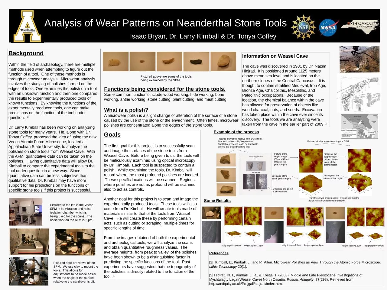
Isaac Bryan Dr Larry Kimball amp Dr Tonya Coffey Goals The first goal for this project is to successfully scan and image the surfaces of the stone tools from Weasel Cave Before being given to us the tools will be meticulously examined using optical microscopy by Dr Kimball Each tool i ID: 930976

kimball tools tool stone tools kimball stone tool polishes cave polish µm span working image functions weasel height microwear

Share:

Link:

Embed:

Download Presentation from below link

Download Presentation The PPT/PDF document "Analysis of Wear Patterns on Neanderthal..." is the property of its rightful owner. Permission is granted to download and print the materials on this web site for personal, non-commercial use only, and to display it on your personal computer provided you do not modify the materials and that you retain all copyright notices contained in the materials. By downloading content from our website, you accept the terms of this agreement.


Presentation Transcript

Slide1

Analysis of Wear Patterns on Neanderthal Stone Tools

Isaac Bryan, Dr. Larry Kimball & Dr

. Tonya Coffey

Goals

The first goal for this project is to successfully scan and image the surfaces of the stone tools from Weasel Cave. Before being given to us, the tools will be meticulously examined using optical microscopy by Dr. Kimball. Each tool is suspected to contain a polish. While examining the tools, Dr. Kimball will record where the most profound polishes are located. These specific locations will be scanned. Regions where polishes are not as profound will be scanned also to act as controls.

Another goal for this project is to scan and image the experimentally produced tools. These tools will also come from Dr. Kimball. He will create tools made of materials similar to that of the tools from Weasel Cave. He will create these by performing certain acts, such as cutting or scraping, multiple times for specific lengths of time.

From the images obtained of both the experimental and archeological tools, we will analyze the scans and obtain quantitative roughness values. The average heights, from peak to valley, of the polishes have been shown to be a distinguishing factor in predicting the specific functions of the tool. Past experiments have suggested that the topography of the polishes is directly related to the function of the tool. [1]

Background

Within the field of archaeology, there are multiple methods used when attempting to figure out the function of a tool. One of these methods is through microwear analysis. Microwear analysis involves the studying of polishes formed on the edges of tools. One examines the polish on a tool with an unknown function and then one compares the results to experimentally produced tools of known functions. By knowing the functions of the experimentally produced tools, one can make predictions on the function of the tool under question. [1]Dr. Larry Kimball has been working on analyzing stone tools for many years. He, along with Dr. Tonya Coffey, proposed the idea of using the new Veeco Atomic Force Microscope, located at Appalachian State University, to analyze the polishes on stone tools from Weasel Cave. With the AFM, quantitative data can be taken on the polishes. Having quantitative data will allow Dr. Kimball to compare the experimental tools to the tool under question in a new way. Since quantitative data can be less subjective than qualitative data, Dr. Kimball may have more support for his predictions on the functions of specific stone tools if this project is successful.

Functions being considered for the stone tools.Some common functions include wood working, hide working, bone working, antler working, stone cutting, plant cutting, and meat cutting.What is a polish?A microwear polish is a slight change or alteration of the surface of a stone caused by the use of the stone or the environment. Often times, microwear polishes are concentrated along the edges of the stone tools.

Pictured to the left is the

Veeco

SPM in its vibration and noise isolation chamber which is being used for the scans. The noise floor on the AFM is 2 pm.

Pictured here are views of the SPM. We use clay to mount the tools. This allows for adjustments to be made easier when the angle of the surface relative to the cantilever is off.

References

[1] Kimball, L., Kimball, J., and P. Allen.

Microwear

Polishes as View Through the Atomic Force Microscope.

Lithic

Technology

20(1).

[2]

Hidjrati

, N. I., Kimball, L. R., &

Koetje

, T. (2003). Middle and Late Pleistocene Investigations of

Myshtulagty

Lagat

(Weasel Cave) North Ossetia, Russia. 

Antiquity

, 77(298), Retrieved from http://antiquity.ac.uk/Projgall/hidjrati/index.html

Example of the process

Information on Weasel Cave

The cave was discovered in 1981 by Dr.

Nazim Hidjrati. It is positioned around 1125 meters above mean sea level and is located on the northern slopes of the Central Caucasus. It is thought to contain stratified Medieval, Iron Age, Bronze Age, Chalcolithic, Mesolithic, and Paleolithic occupations. Because of the location, the chemical balance within the cave has allowed for preservation of objects like wood charcoal, nuts, and seeds. Excavation has taken place within the cave ever since its discovery. The tools we are analyzing were taken from the cave in the earlier part of 2009.[2]

Pictured above are some of the tools being examined by the SPM.

Pictures of what we receive from Dr. Kimball.This tool is around 90,000 years old. Qualitative evidence leads Dr. Kimball to believe it is a wood working tool.

Evidence of a polish is shown here

Picture of the height image (50

µm x

50

µm) made of the polish region

3d image of the same polish region

Picture of the height image (50µm x 50µm) made of the control region

3d image of the same control region

Pictures of what we obtain using the SPM

Some Results

From these two images above, we can see that the polish has a much smoother surface.

h

eight span=2.0

µm

height

span=2.5

µm

h

eight span=3.5

µm

height

span=4.0

µm

h

eight span=1.5

µm

height

span=4.0

µm