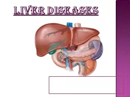
Liver anatomy It is the largest abdominal organ 1500g ribs amp ccs 610 on R 6 amp 7 on L Two lobes Cantles line Two surfaces ID: 930445
Download Presentation The PPT/PDF document "Liver lesion SARAH AWAISHEH, BAU" is the property of its rightful owner. Permission is granted to download and print the materials on this web site for personal, non-commercial use only, and to display it on your personal computer provided you do not modify the materials and that you retain all copyright notices contained in the materials. By downloading content from our website, you accept the terms of this agreement.
Slide1
Liver lesion
SARAH AWAISHEH, BAU
Slide2Liver anatomy
It is the largest abdominal organ
1500g
ribs & cc’s (6-10 on
R
, 6 & 7 on
L
)
Two lobes
Cantle’s line
Two surfaces :
-
Diaphragmatic surface
‘bare area’ of the liver
- Visceral surface
With the exception of the fossa of the gallbladder and porta hepatis, it is covered with peritoneum.
Slide3Liver anatomy
Protected by rib cage
Glisson’s capsule
8 ligaments :
Coronary ligament : anterior and posterior fold
Triangular ligament : right and left
Falciform
ligament :Sickle-shaped
Ligamentum
teres
Ligamentum
venosum
Lesser
omentum
:
hepatogastric
ligament, hepatoduodenal ligament
Slide4Slide5Liver anatomy
Hepatic artery 25%
Dual supply :
Portal vein 75%
72% of the Oxygen
venous drainage by the right, middle and left hepatic veins
physiological functions
Production of :plasma
protiens
and coagulation factors.
Fat soluble vitamins metabolism
storage of :
protiens
(A.A), glucose(glycogen), fat(
cholestrol
) Detoxification
Slide7Portal Hypertension:
sustained elevation of venous portal pressure more than 10 mmHg (15-20 mmHg )
Normal pressure 5-10 mmHg
There are 6 potential routes of portal –systemic collateral blood flow (
ares
of communication):
Slide8Region
Name of clinical condition
Portal circulation
Systemic circulation
Esophageal
Esophageal varices
Esophageal branch of
left gastric vein
Esophageal branches of
azygos vein
Rectal
Rectal varices
Superior rectal vein
Middle rectal veins
and
inferior rectal veins
Paraumbilical
Caput medusae
Paraumbilical veins
Superficial epigastric vein
Retroperitoneal
Splenorenal shunt
[3]
Splenic vein
Renal vein
,
suprarenal vein
,
paravertebral vein
, and
gonadal vein
(no clinical name)
[4]
Right colic vein
,
middle colic vein
,
left colic vein
Retroperitoneal veins of Retzius
Intrahepatic
Hepatic pseudolesions
[5]
Perihepatic veins of Sappey
Superior epigastric vein
Patent ductus venosus
Left branch of portal vein
Inferior vena cava
Slide9Etiology:
1.
Presinusoidal
Extrahepatic : Intrahepatic :
Splenic vein thrombosis Schistosomiasis (Egypt )
Splenomegaly Congenital hepatic fibrosis
Splenic A-V fistula Idiopathic portal fibrosis
Myeloproliferative disorders
scardiosis
Slide10etiology…
2. Sinusoidal
3. Post sinusoidal
Posthepatic
Intrahepatic
Budd Chiari
Cardiac cirrhosis
IVC web
Congestive
Hepatopathy Primary Thrombosis Secondary Compresion
Slide11signs & symptoms
Splenomegaly
esophageal
varice
Caput medusa
Hemorrhoids
spider
angioma
, palmer erythema
Ascitisasteraxis
(hepatic flap)
fetor
hepaticus
Jaundice
confusion and drowsiness
Slide12signs & symptoms…
esophageal varices :
30% of patients with compensated cirrhosis
60% of patients with decompensated cirrhosis
(development of jaundice, ascites, variceal hemorrhage, or hepatic encephalopathy )
1/3 of all patients with varices will experience variceal bleeding
Each episode 20-30% mortality if untreated
70% of patients who survive the initial episode will experience recurrent
haemorrhage
within 2 years
Slide13esophageal varices
Slide14Treatement
of esophageal varices :
Management can be divided into :
the active bleeding episode,
the prevention of
rebleeding
,
the prophylactic measures to prevent the first hemorrhage
Slide15Initial management of active bleeding episode
I.Resuscitation
:
IV line insertion ( 2 large bore cannulas) and IV fluid
Admission to ICU
Obtain blood for grouping and
crossmatching
(blood transfusion without over transfusion just until hemoglobin 9)
Correct coagulopathy: use fresh frozen plasma /Platelets/ coagulation factors Antibiotics
Vasopressin
(octreotide) / IV
II. Urgent endoscopy: both diagnostic and therapeutic (Variceal banding / Injection sclerotherapy)
Slide16If initial attempt failed
Blackmore-
Sengstaken
tube
for temporary blood bleeding control
four ports
1- for gastric aspiration
2- for gastric balloon ( 500 ml)
3- for esophageal balloon (200 ml) 4- for esophageal aspiration prevent aspiration pneumonia
do not leave it in situ for more than 24-36 h (risk of perforation and necrosis)
Slide17▪
Transjagular
intrahepatic
portocaval
shunt
(TIPS): an expandable covered metal shunt
Used when bleeding cannot be stopped after 2 sessions of endoscopic therapy within 5 days.
Advantages: it reduces the portal vein pressure by creating a total shunt and doesn’t have the risk of general anesthesia and surgery.
Disadvantages: increased risk of portosystemic encephalopathy.
Slide18▪ Surgical shunt:
Shunting:
Portocaval
(increase incidence of encephalopathy)
Mesocaval
stent
Distal lienorenal (Warren) (most used
Non shunting: Sigiura ( bleeding uncontrollable)
Liver transplant
Slide19Schistosomiasis
parasitic disease caused by blood flukes
exposure to infested water
Presentation: abdominal pain,
diarrhea,
bloody stool,
hepatomegaly.
Dx: detection of parasite eggs in stool or serum antibodies.
Causes Perisinosoidal portal HT
Tx: Praziquantel Single dose 40-70 mg/kg.
Education
Hygiene
Slide20Liver Abscess
mass filled with pus inside the liver
Classified into :
• Pyogenic liver abscess: which is most often
polymicrobial
, accounts for 80% of hepatic abscess.
• Amoebic liver abscess: due to Entamoeba
histolytica
accounts for 10% of cases.
• Fungal abscess: most often due to Candida species, accounts for less than 10% of cases.
• Iatrogenic abscess: caused by medical interventions
Slide21Amebic abscess
Entamoeba histolytica enter mesenteric venules.
travel to the liver where they typically form one or more abscesses.
The right lobe of the liver is more commonly affected than the left lobe.
amebic abscess have characteristic chocolate appearance.
Slide22Investigation:
CBC,LFT, Direct and indirect serological tests (CF, IHA and ELISA) to detect amoebic protein, stools examination for amebae trophozoites or cysts.
Imaging:
USS and CT: usually large, solitary, thin-walled, poorly defined abscess in the right lobe.
Slide23Management:
Empirical
tx
in areas where the problem is endemic.
metronidazole with chloroquine phosphate usually results in rapid resolution.
Needle aspiration if : -No clinical response within 72 hours
-There was superinfection (treated as pyogenic abscess).
-The abscess is large.