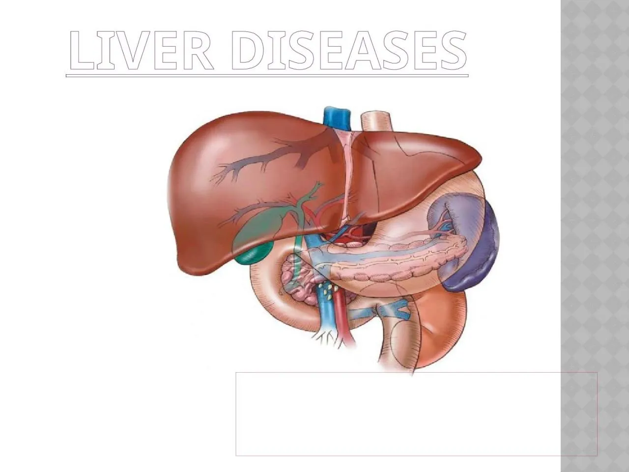
/
LIVER DISEASES   Physiology  of liver LIVER DISEASES   Physiology  of liver

LIVER DISEASES Physiology of liver - PowerPoint Presentation

harper
harper . @harper
Follow
343 views
Uploaded On 2022-05-31

LIVER DISEASES Physiology of liver - PPT Presentation

Liver is the largest organ of the body Sets in the right side of the belly Weighs between 1025 kg Heavier in males than females It is reddish in color amp feel rubbery to touch Have two sectionsright amp left lobe ID: 912557

liver bile amp bilirubin bile liver bilirubin amp blood chronic fat disease gallbladder acute duct cholecystitis acids intestine serum

Share:

Link:

Embed:

Download Presentation from below link

Download Presentation The PPT/PDF document "LIVER DISEASES Physiology of liver" is the property of its rightful owner. Permission is granted to download and print the materials on this web site for personal, non-commercial use only, and to display it on your personal computer provided you do not modify the materials and that you retain all copyright notices contained in the materials. By downloading content from our website, you accept the terms of this agreement.


Presentation Transcript

Slide1

LIVER DISEASES

Slide2

Physiology of liver

Liver is the largest organ of the body.Sets in the right side of the belly.Weighs between 1.0-2.5 kg.Heavier in males than females.It is reddish in color & feel rubbery to touch.

Have two sections-right & left lobe.

The right lobe is further divided into the interior and posterior segments

Slide3

The externally visible falciform ligament divides the left lobe into the medial and

lateral segments.

Liver is supplied with blood from two sources –the hepatic artery & portal vein.

Responsible for 25 %of the basal metabolism & intimately concerned with the metabolism of fats,protiens & CHO.

Slide4

Slide5

structure of liver

Composed of 50,000-100,000 lobules. Each lobule consist of a central vein surrounded by tiny liver cells grouped in the sheet or bundle.Small branch of hepatic vein extends through their centre. .Space b/w these cells contain sinusoids (blood vessels that are irregular in shape, incomplete in size &wider than blood capillaries .)

Slide6

Slide7

Slide8

Slide9

Blood supply

the liver receives blood containing oxygen from the heart. This blood enters the liver through the hepatic artery. The liver also receives blood filled with nutrients, or digested food particles, from the small intestine. This blood enters the liver through the portal vein.

In the liver, the hepatic artery and the portal vein branch into a network of tiny blood vessels that empty into the sinusoids.

The liver cells absorb nutrients and oxygen from the blood as it flows through the sinusoids. At the same time, they secrete sugar, vitamins, minerals, and other substances into the blood.

The sinusoids drain into the central veins, which join to form the hepatic vein. Blood leaves the liver through the hepatic vein.

Each lobule also contains bile capillaries, tiny tubes that carry the bile secreted by the liver cells. The bile capillaries join to form bile ducts, which carry bile out of the liver. Soon after leaving the liver, the bile ducts join together, forming the hepatic duct.

Excess bile flows into the gall bladder, where it is stored for later use.

Slide10

Blood supply

Slide11

Functions of liver

Secretion of bile Storage of glycogen

Metabolism of fats

Deamination of amino acids

Production of the plasma

protiens

.

Storage & transport of vitamins & minerals.

Storage of iron .

Production of clotting factors .

Production of heat .

Detoxification.

Acts as filter

Slide12

Role of branched chain amino acids & aromatic amino acids in liver disease

major hypothesis for the pathogenesis of portal systemic encephalopathy has been termed the altered neurotransmitter Theory. A plasma amino acid imbalance exists

in ESLD in which the branched-chain amino acids( BGAAs) valine, leucine, and isoleucine are decreased, and aromatic amino acids (AAAs) tryptophan, phenylalanine, and tryrosine,plus methionine, glutamine, asparagine, and histidine are increased.

The BCAAs furnish as much as 30% of energy requirements for skeletal muscle, heart, and brain when gluconeogenesis and ketogenesis are depressed .

This causes serum BCAA levels to fall. At the same time, plasma AAA’S and methionine are released into circulation by muscle proteolysis, but the synthesis into protein and liver clearance of AAAs is depressed .

This changes the plasma molar ratio of BCAAs to AAA’S and may contribute to the development of hepatic

encephalopathy.

AAAs may limit the cerebral uptake of BCAA because they compete for carrier-mediated transport at the blood-brain barrier.

..

Slide13

.

Amino Acids Commonly Altered in Liver Disease

Aromatic Amino Acids: serum levels are increased.

Tyrosine

Phenylalanine*

Free tryptophan*

Branched €

hain

Amino Acids: levels are decreased.

Valine*

Leucine"

Isoleucine*

Ammonlogenic

Amino Acids: levels are increased.

Glycine

Serine

Threonine

*

Glutamine

Histidine*

Lysine"

Asparagine

Methionine*

Slide14

Slide15

1.Aspartate aminotransferase

(AST or SGOT)

Located in

cytosol

and mitochondria of

hepatocyte

also in cardiac and skeletal muscle, brain, pancreas, kidney and leukocytes.

An enzyme similar to ALT but less specific for liver disease.

Levels of AST are increased with liver damage or death of hepatocytes.

In some cases

viz

alcoholic hepatitis or shock

liv

the elevation in serum AST level may be, higher than the elevation in serum ALT level.

AST Normal range – 2-40 U/L.

Slide16

2.Alanine aminotransferase

(ALT or SGPT)

It is located in

cytosol

of hepatocytes found in several other body tissues.

It is highest in liver.

ALT level is increased in case of liver cell death, caused due to, Hepatitis, any shock, injury or excess alcohol or drug toxicity.

Normal range – 2-40 U/L

Slide17

3.Serum alkaline phosphatase

An enzyme that is widely distributed in liver, bone, placenta, intestine, kidney, leukocytes.

It is mainly bound to

canalicular

membranes of liver produced in the bile ducts.

This enzyme activity can be increased with bone disorders, pregnancy , normal growth & some malignancies.

40 - 129 U/L

Slide18

4.bilirubinBilirubin is a yellow breakdown product of the normal heme catabolism .It is excreted in bile & urine and elevated levels may indicate certain diseases.

It is responsible for the yellow color of the bruises ,urine & brown color of feces & yellow discoloration of skin.

Slide19

functionsBilirubin is created by the activity of 

biliverdin reductase on biliverdin

, a green tetrapyrrolic bile pigment that is also a product of heme catabolism. Bilirubin, when oxidized, reverts to become biliverdin once again. This cycle, in addition to the demonstration of the potent antioxidant activity of bilirubin, has led to the hypothesis that bilirubin's main physiologic role is as a cellular antioxidant.

Slide20

Types of bilirubin

Unconjugated bilirubin (indirect)Erythrocytes (red blood cells) generated in the 

bone marrow

 are disposed of in the

spleen when they get old or damaged. This releases 

hemoglobin

, which is broken down to 

heme as the globin parts are turned into amino acids. The heme is then turned into unconjugated bilirubin in the reticuloendothelial cells of the spleen. This unconjugated bilirubin is not soluble in water, due to intramolecular hydrogen bonding. It is then bound to 

albumin

 and sent to the 

liver

Conjugated bilirubin(direct)

In the liver it is conjugated with 

glucuronic acid

 by the enzyme 

glucuronyltransferase

, making it soluble in water. Much of it goes into the bile and thus out into the small intestine. However 95% of the secreted bile is reabsorbed by the small intestine. This bile is then resecreted by the liver into the small intestine. This process is known as 

enterohepatic circulation

. About half of the conjugated bilirubin remaining in the large intestine(about 5% of what was originally secreted) is metabolized by colonic bacteria to

urobilinogen

, which can be further metabolized to 

stercobilinogen

, and finally oxidized to 

stercobilin

. Stercobilin gives feces its brown color

Slide21

Blood testsBilirubin is broken down by light. Tubes containing blood or (especially) serum to be used in bilirubin assays should be protected from illumination.

Total bilirubin ("TBIL") measures both BU and BC. Total and direct bilirubin levels can be measured from the blood, but indirect bilirubin is calculated from the total and direct bilirubin.Indirect bilirubin is fat-soluble and direct bilirubin is water-soluble.

Bilirubin (in blood) is in two forms.

Slide22

Blood tests

Abb.

Name(s)

Water-soluble?

Reaction

"BC"

"Conjugated" or

"Direct bilirubin"

Yes (bound

to

glucuronic

acid

)

Reacts quickly when dyes (

diazo

reagent) are added to the blood specimen to

produce

azobilirubin

 "Direct bilirubin"

"BU"

"Unconjugated" or "Indirect bilirubin"

No

Reacts more slowly. Still produces

azobilirubin

. Ethanol makes all bilirubin react promptly then calc: Indirect bilirubin = Total bilirubin - Direct bilirubin

Slide23

Normal Ranges of Bilirubin in Blood -

μ

mol/L

mg/

dL

0.2-1.9,

[

Total bilirubin

5.1–17.0

0.3–1.0

0.1-1.2

Direct bilirubin

1.0–5.1

0-0.3

0.1–0.3

0.1-0.4

Slide24

Hyperbilirubinemia

Hyperbilirubinemia results from a higher than normal level of bilirubin in the blood.

Mild rises in bilirubin may be caused by:

Hemolysis

or increased breakdown of red blood cells

Gilbert's syndrome

- a genetic disorder of bilirubin metabolism that can result in mild jaundice, found in about 5% of the populationRotor syndrome:

non-itching jaundice, with rise of bilirubin in the patient's serum, mainly of the conjugated type.

Moderate rise in bilirubin may be caused by:

Pharmaceutical drugs

(especially antipsychotic, some sex hormones, and a wide range of other drugs)

Sulfonamides are contraindicated in infants less than 2 months old as they increase unconjugated bilirubin leading to kernicterus.

Hepatitis

(levels may be moderate or high)

Chemotherapy

Biliary stricture

(benign or malignant)

Very levels of bilirubin may be caused by:

Neonatal hyperbilirubinaemia,

where the newborn's liver is not able to properly process the bilirubin causing jaundice

Unusually large bile duct obstruction,

e.g. stone in common bile duct, tumour obstructing common bile duct etc.

Severe liver failure with cirrhosis (e.g. primary biliary cirrhosis

Choledocholithiasis

(chronic or acute)

presence of gallstone in the common bile duct

Slide25

5.Gamma-glutamyl transpeptidase

(GGT)

An enzyme associated with

microsomes

and plasma membranes in hepatocytes also present in kidney, pancreas, heart , brain.

An enzyme produced in the bile ducts that like alkaline

phosphatase

, may be elevated in the serum of patients with bile duct disease.

It is increased with liver disease, pulmonary disease, diabetes mellitus and during alcohol ingestion by many drugs.

Slide26

Serum albumin

Main export protein synthesized in the liver and most imp. factor in plasma osmotic pressure maintenance, secreted in the blood.

Major protein circulating in blood stream.

Low serum albumin levels indicate poor liver function.

Serum albumin concentration is usually normal in chronic liver diseases until cirrhosis and liver damage is present.

Albumin – 3.4 - 4.8 g%.

Albumin levels can be low in conditions other than, liver disease like malnutrition.

Albumin is a better index of chronic liver disease.

Increased losses occur with protein losing

enteropathy

,

nephrotic

syndrome burns, GIT bleeding,

exfoliative

dermatitis.

Slide27

Prothrombin time

Most blood coagulation factors are synthesized in the liver.

When liver function is severely abnormal, their synthesis and secretion into blood is decreased.

PT is a type of blood clotting test and is prolonged when blood concentration of some clotting factors made by liver are low.

In chronic liver disease, PT is not usually elevated until cirrhosis is present and liver damage is significant.

In acute liver disease, PT can be prolonged with severe liver damage.

Decreased synthesis of clotting factors increase prothrombin time and risk of bleeding.

Slide28

Serum protein electrophoresis

Major protein in the serum are separated in an electric field and their concentrations are determined.

Four major types of serum proteins measured are albumin, alpha- globulins, beta- globulins and gamma- globulins.

Alpha and gamma globulins are synthesized in liver. Levels increase with chronic liver disease.

In cirrhosis, the albumin may be decreased and the gamma - globulins elevated.

Gamma- globulins can be significantly elevated in some types of autoimmune hepatitis also.

Slide29

Platelet count

Platelets are the smallest of the blood cells, involved in blood clotting.With liver disease the spleen becomes enlarged as blood flow through the liver is impeded.

This can lead to platelets being sequestered in the enlarged spleen.

The platelet count can be abnormal in many conditions other than liver disease.

Slide30

Gall bladder diseases

Slide31

gallbladder

It is a thin walled reservoir situated on the under surface of liver.It can store about 40-50ml of bile. It concentrates bile formed in the liver.Stores it , until needed for digestion.Interference with the flow of bile in gallbladder diseases may cause impaired fat digestion.

Slide32

Functions of gall bladder

Acts as a reservoir for the storage of bile .Mucosal lining of gall bladder reabsorbs the fluid & electrolytes ,thus concentrating the bile constituents.Hormone cholecystokinin secreted by the duodenal walls, cause muscular walls of the gall bladder to contract and expel the bile.

Duodenal peristalsis inhibits the sphincter of oddi & cause it to relax & allow the bile to enter the duodenum.

Slide33

bileBile is the external secretion of the liver .

Produced in the diluted form .Concentrated later by the gall bladder to a viscid greenish fluid .It consists of-Water Bile salts(sodium glycocholate &sodium taurocholate)

Bile acids

Bile pigment( bilirubin).

Cholesterol Mucus

Slide34

Functions

Bile acts to some extent as a surfactant, helping to emulsify the fats in the food.

Since bile increases the absorption of fats, it is an important part of the absorption of the fat-soluble vitamins, such as the vitamins D, E, K and A.

Bile serves also as the route of excretion for bilirubin, a by-product of red blood cells, recycled by the liver.

Bile salts also act as bactericides, destroying many of the microbes that may be present in the food.

Bile salt anions have a hydrophilic side and a hydrophobic side, and therefore tend to aggregate around droplets of fat (triglycerides and phospholipids) to form micelles, with the hydrophobic sides towards the fat and hydrophilic towards the outside.

Slide35

Bile salts

have important Function in assisting the digestive action of the pancreatic enzyme . helps in aiding the absorption of fats & fat soluble vitamins form the small intestine.These salts by lowering the surface tension cause fat droplets to break up or emulsify into small droplets allowing fat digesting enzyme to work more efficiently.

They convert them to fatty acids & glycerol ,the form in which they are absorbed.

Bile salts do not appear in faeces as they are reabsorbed from the small intestine & returned to the liver.

Slide36

Bile acidsThese include40% cholic acid .

40% chemodeoxycholic acid.18% deooxycholic acid .2% lithocholic acid.Total bile acid pool is 2.4 g but it is absorbed & recirculated 6-8 times a day. Thus providing over 15- 20 gm of bile for digestion.

Slide37

Bile pigments

Are derived from the breakdown of the haemoglobin of worn out RBC’s & give the bile its characteristic color.The bile pigments are converted in the bowel to urobilinogen by bacterial action.Some urobilinogen is reabsorbed into the blood & is excreted by the kidneys into the urine.Exposure of urine to the air causes urobilinogen to be oxidized to urobilin.

In the faeces , urobilinogen is altered 7 oxidized to form stercobilin which gives the faeces a dark brown colour.

If there is an obstruction to the excretion of bile , bile pigments accumulate in the blood giving the skin ,a yellow color (jaundice).

Slide38

Bile Storage

The liver is constantly secreting bile, up to 1 liter in a 24 hour period, but most of it is stored in the gallbladder. This hollow organ can only hold 30 to 60 ml of bile and is able to store the large quantities of bile from the liver by concentrating it. The gallbladder is able to achieve this by reabsorption of water, sodium, chloride and other electrolytes through its lining.

The other constituents of bile, like the bile salts, cholesterol, lecithin and bilirubin, stays in the gallbladder.

Slide39

Bile Production

The liver cells (hepatocytes) produce bile which collects and drains into the hepatic duct. From here it can enter the small intestine to act on fats by traveling down the common bile duct, or it can enter the gallbladder through the cystic duct, where it is stored.

The liver manufactures between 600ml to 1 liter of bile in a day. As bile travels down the ducts, the lining of these passages, secrete water, sodium and bicarbonate ions into the bile, thereby diluting it.

These additional substances help to neutralize the stomach acid which enters the duodenum with partially digested food (

chyme

) from the stomach.

Slide40

Secretion & circulation of Bile-`

Slide41

diseases

Cholelithiases

Slide42

Cholelithiasis

The formation of gallstones (calculi) in the absence of infection of the gallbladder is called cholelithiasis. all gallstones form within the gallbladder

Gallstones that pass from the gallbladder into the common bile duct may remain there indefinitely without causing symptoms, or they may pass into the duodenum with or without symptoms.

Choledocholithiasis

develops when stones slip into the bile ducts, producing obstruction, pain, and cramps.

If passage of bile into the duodenum is interrupted,

cholecystitis can develop. In the absence of bile in the intestine, lipid absorption is impaired, and without bile pigments, stools become light in color.

If uncorrected, bile backup can result in jaundice and liver damage (secondary biliary cirrhosis).

Obstruction of the distal common bile duct can lead to

pancreatitis

if the pancreatic duct is blocked.

Slide43

Types of stones

Most gallstones in people are unpigmented cholesterol stones composed primarily of cholesterol ,bilirubin and calcium salts.

Risk factors for cholesterol stone formation include female gender, pregnancy, older age, family history obesity diabetes mellitus, inflammatory bowel disease and drugs (lipid-lowering medications, oral contraceptives and estrogens).

Rapid weight loss (as with jejunoileal and gastric bypass and fasting or severe calorie restriction) is associated with a high incidence of biliary sludge and gallstone formation .

Bacteria may also play a role in gallstone formation. Low-grade chronic infections produce changes in the gallbladder mucosa, which affect its absorptive capabilities. Excess water or bile acid may be absorbed as a result. Cholesterol may then precipitate out and cause gallstone formation .

Slide44

High dietary fat intake over a prolonged period may predispose a person to gallstone formation because of the constant stimulus to produce more cholesterol for bile synthesis required in fat digestion.

Pigmented stones typically consist of bilirubin polymers or calcium salts. They are associated with chronic hemolysis.

Risk factors associated with these stones are age, sickle cell anemia and thalassemia, biliary tract infection, cirrhosis, alcoholism and long-term PN(

parenteral

nutrition).

Slide45

Medical management

treatment of gallstone disease includes cholecystectomy, especially if the stones are numerous, large, or calcified. cholecystectomy may be done as a traditional open laparotomy or as a less invasive laparoscopic procedure. Chemical dissolution with the administration of bile salt with other acids are used but very less.

Patients with gallstones that have migrated into the bile ducts may be candidates for endoscopic retrograde cholangiopancreatography techniques

Slide46

Nutrition therapy

No specific dietary treatment is available to prevent cholelithiasism in susceptible persons. Nutrition-related factors include obesity ,severe fasting and these should be corrected when possible. In cholecystitis, dietary treatment includes a low-fat diet to prevent gallbladder contractions.

After surgical removal of the gallbladder, oral feedings are usually resumed with the return of bowel movement.

after that the diet can be advanced to a regular diet as tolerated.

In the absence of the gallbladder, bile is secreted directly by the liver into the intestine.

Slide47

Cholecystitis

Inflammation of the gallbladder is known as cholecystitis. it may be chronic or acute. It is usually caused by gallstones obstructing the bile ducts , leading to the backup of bile. Bilirubin, the main bile pigment, gives bile its greenish color.

When biliary tract obstruction prevents bile from reaching the intestine, it backs up and returns to the circulation.

Slide48

Acute cholecystitis

Due to infection of gall bladder.It occurs in association with obstruction to the cystic duct or neck of the gall bladder.Gall stones are the cause of obstruction The walls of the gallbladder become inflamed and distended and infection can occur.

During such episodes, the patient experiences upper quadrant abdominal pain accompanied by nausea, vomiting, and flatulence.

Slide49

Nutritional therapy in acute cholecystitis

In an acute attack, oral feedings are discontinued. Parenteral nutrition may be indicated if the patient is malnourished. When feedings are resumed, a low-fat diet is recommended to decrease gallbladder stimulation Patient should be kept in bed & given analgesics.

Large quantities of fluids should be drunk.

For acute cases, an entirely fluid of at least 2 -3

litres

daily given in small feeds at hourly or two hourly intervals is advisable for few days.

Slide50

Chronic cholecystitis

Chronic cholecystitis is long-standing inflammation of the gallbladder. It is caused by repeated mild attacks of acute cholecystitis. This leads to thickening of the walls of the gallbladder. The gallbladder begins to shrink and eventually loses the ability to perform its function: concentrating and storing bile.

Eating foods that are high in fat may aggravate the symptoms of cholecystitis because bile is needed to digest such foods.

Chronic cholecystitis occurs more often in women than in men and the incidence increases after the age of 40.

Risk factors include the presence of gallstones and a history of acute cholecystitis.

Slide51

Nutritional therapy in chronic cholecystitis

Patients with chronic conditions may require a long-term low-fat diet that contains 25% to30% of total kilocalories as fat. Stricter limitation is undesirable because fat in the intestine is important for some stimulation and drainage of the biliary tract. The degree of food intolerance varies widely among persons with gallbladder disorders.

avoid foods that cause flatulence and bloating.

Slide52

Cholestasis

Cholestasis is a condition in which little or no bile is secreted or the flow of bile into the digestive tract is obstructed.This can occur in patients without oral or enteral feeding for a prolonged period and can predispose to a calculous cholecystitis.

Prevention includes stimulation of intestinal and biliary motility.

If this is not possible, drug therapy is used.

Slide53

Diagnostic testsMost common test is oral cholecystography.Presence of stones then can be visualized by roentgenogram.

When stones are not visualized ,intravenous cholangiography is done. It involves administration of an iodine contrast dye Injection of an iodine contrast medium permits visualization of biliary duct.Ultrasonography & computed tomography (ct scan) can also be done.

Slide54

Acute cholangitis

Pathophysiology and medical ManagementInflammation of the bile ducts is known as cholangitis.Patients with acute cholangitis need resuscitation with fluids and broad-spectrum antibiotics. If the patient does not improve with conservative treatment, placement of a percutaneous biliary stent or cholecystectomy may be done.

Slide55

Sclerosing cholangitis

Sclerosing cholangitis is another chronic cholestatic liver disease.Fibrosing inflammation of segments of extrahepatic bile ducts, with or without involvement of intrahepatic ducts.

Progression of the disease leads to complications of portal hypertension, hepatic failure and cholangiocarcinoma.

Primary Sclerosing cholangitis (PSC) is the most common type of Sclerosing cholangitis.

It usually occurs in association with inflammatory bowel disease.

Slide56

Medical Management

Sclerosing cholangitis can result in sepsis and liver failure.Most patients have multiple intrahepatic strictures, which makes surgical intervention difficult. Patients are generally on broad-spectrum antibiotics.

When sepsis is recurrent, patients may require chronic antibiotic therapy.

Slide57

Pancreatic diseases

Slide58

pancreas

The pancreas is an elongated, soft & flattened gland.It lies in the upper abdomen behind the stomach.

It is 12-20 cm long in adults & weighing 75-85 gm.

The head of the pancreas is in the right upper quadrant below the stomach ,adjacent to the duodenum.

Slide59

Main pancreatic duct extends from tail, collecting it’s vital secretions of enzymes.

This portion consists of about 1 million islets of langerhans

, out of which 80% secrete insulin, 15% secrete glucagon, 5% secrete

somatostatin

.

This glandular organ has both an endocrine & exocrine function.

Slide60

functions

The exocrine function is to secrete digestive enzymes in the inactive form . Which only become activated after their release into the pancreatic juices & the small intestine .

the endocrine function is the secretion of insulin & glucagon.

Insulin is imp for the metabolism of CHO, fats & proteins & glucagon is concerned with the breakdown of liver glycogen & with increased glucogenesis.

Slide61

TEST

SIGNIFICANCESECRETIN STIMULATION TESTMEASURES PANCREATIC SECREATION, PARTICULARLY BICARBONATE, IN RESPONSE TO SECRETIN STIMULATION.

GLUCOSE

TOLERANCE TEST

ASSESES

ENDOCRINE FUNCTION OF THE PANCREAS BY MEASURING INSULIN RESPONSE TO A GLUCOSE LEVEL.

72-HOUR

STOOL FAT TEST

ASSESSES

EXOCRINE FUNCTION OF THE PANCREAS BY MEASURING FAT ABSORPTION THAT REFLECTS PANCREATIC LIPASE SECRETION.

TESTS OF PANCREATIC FUNCTION

Slide62

pancreatitis

Pancreatitis is an inflammation of the pancreas and is characterized by oedema, cellular exudate and fat necrosis. Pancreatitis is classified as either acute or chronic.The symptoms of pancreatitis can range from continuous or intermittent pain of varying intensity to severe upper abdominal pain, which may radiate to the back.

Symptoms may worsen with the ingestion of food.

Clinical presentation may also include nausea, vomiting, abdominal distention, and steatorrhea. Severe cases are complicated by hypotension, oliguria, and dyspnea.

Slide63

Chronic Alcoholism

Gall stones

Biliary tract disease

Trauma

Hypertriglyceridemia

hypercalcemia

Certain drugs some viral infections

Pancreatitis

causes

Slide64

PATHOPHYSIOLOGY

Pancreatitis

Diagnosis

Test of pancreatic function

Secretin stimulation test

Glucose tolerance test

72 hrs stool fat test

Clinical findings

Symptoms:

Abnormal pain and distention

Nausea

Vomiting

Steatorrhea

In severe form:

Hypotension

Oliguria

dyspnea

Slide65

P

ANC

R

E

A

T

I

TI

S

NUTRITION MANAGEMENT

Acute:

Withhold oral and

enternal

feeding

If oral nutrition cannot be initiated in 5 to 7 days, start nutrition support

Once oral nutrition is started, provide:

Easily digestible foods

Low fat diet

6 small meals

Adequate protein intake

Increased calories

Chronic:

Provide oral diet as in acute phase

Supplement pancreatic enzymes

Supplement fat soluble vitamins and vitamin B

12

Acute:

Withhold oral feeding

Administer H

2

-receptor antagonism,

somatstatin

Chronic:

Manage intestinal pH with:

Ant acids

H

2

receptor inhibitors

Administer insulin for glucose intolerance

MEDICAL MANAGEMENT

MANAGEMENT

Slide66

Acute pancreatitis

This is a serious disorder which may lead to haemorrhagic necrosis of the pancreas, peritonitis & death.

Biliary tract stone disease & alcohol abuse account for 80% of cases of acute pancreatitis.

Gall stones & biliary sludge

impaires

normal flow of secretion of pancreatic enzymes.

Severe acute pancreatitis results in a hyper metabolic & catabolic state.

Slide67

In these conditions, the effect of

trypsin inhibitor may be overwhelmed.

if unchecked the rapidly activated enzymes literally digest the pancreas in few hours.

This phase is lethal & may lead to chronic pancreatic insufficiency.

Genetic disease like cystic fibrosis may also lead to pancreatic insufficiency.

Slide68

Chronic Pancreatitis

Alcohol & malnutrition are the major causes of chronic pancreatitis.

It is characterized by recurrent attacks of, epigastric pain of long duration that may radiate into the back.

The pain can be precipitated by meals.

The gastric pain is associated with nausea, vomiting or diarrhea.

Rarely, chronic pancreatitis is hereditary.

Principle sign of chronic pancreatitis is malabsorption caused by deficiency of pancreatic enzymes& subsequent steatorrhoea.

Slide69

Objective of therapy

to prevent further damage to the pancreas. decrease the number of attacks of acute inflammation, alleviate pain, decrease steatorrhea, and correct malnutrition. Dietary intake should be as liberal as possible, but modifications may be necessary to minimize symptomsSubstitution of dietary fat with MCT oil may relieve steatorrhea and lead to weight gain.

Antacids, H2-receptor antagonists that reduce gastric acid secretion may be used to

achieve this effect.