/
Histology of Adrenal Gland - Medulla Histology of Adrenal Gland - Medulla

Histology of Adrenal Gland - Medulla - PowerPoint Presentation

SkylineBabe
SkylineBabe . @SkylineBabe
Follow
349 views
Uploaded On 2022-07-28

Histology of Adrenal Gland - Medulla - PPT Presentation

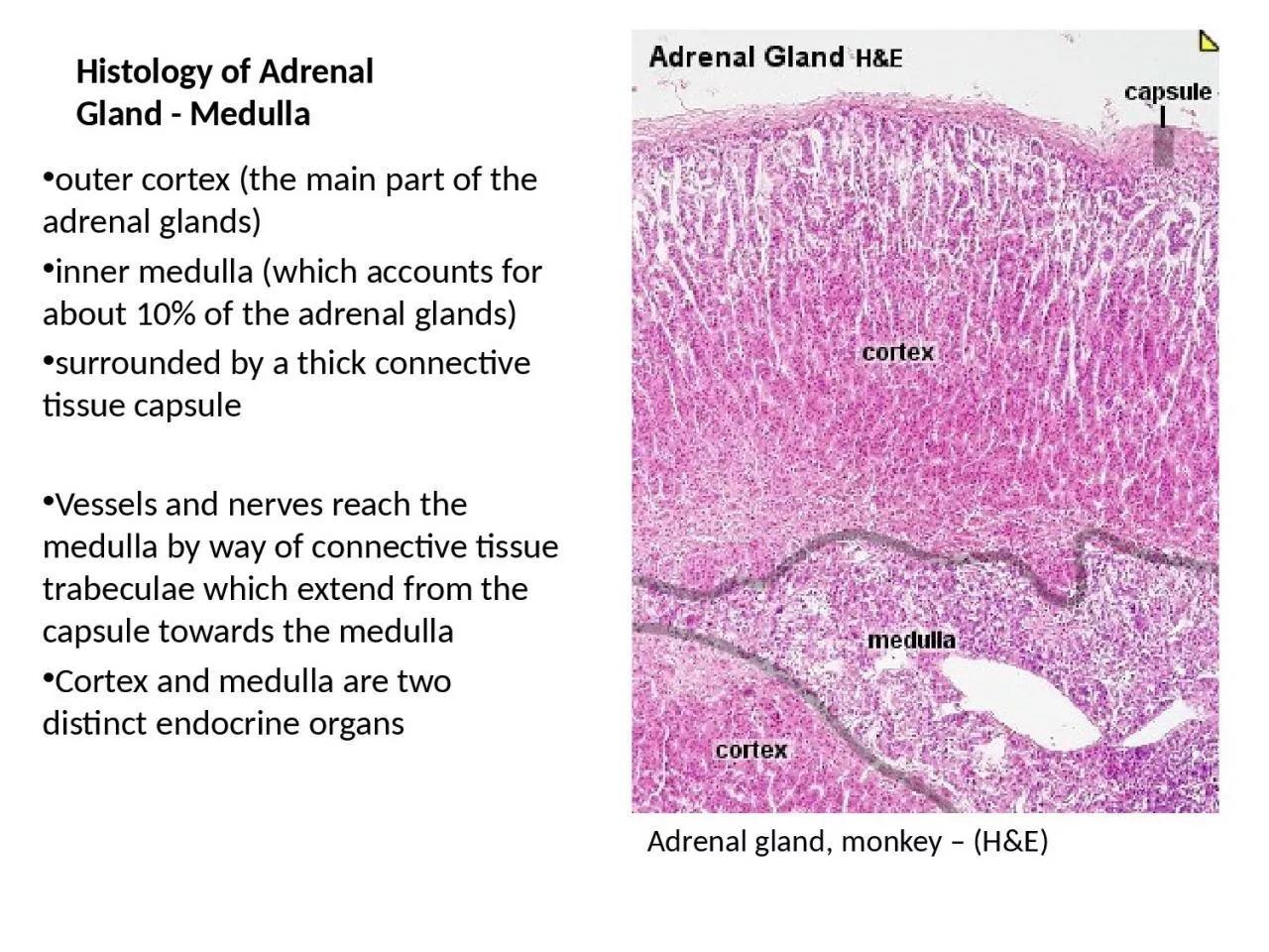
outer cortex the main part of the adrenal glands inner medulla which accounts for about 10 of the adrenal glands surrounded by a thick connective tissue capsule Vessels and nerves reach the medulla by way of connective tissue ID: 931168

adrenal medulla amp cells medulla adrenal cells amp gland arrow pheochromocytes sympathetic nerve groups venous sinusoids tail granules catecholamines

Share:

Link:

Embed:

Download Presentation from below link

Download Presentation The PPT/PDF document "Histology of Adrenal Gland - Medulla" is the property of its rightful owner. Permission is granted to download and print the materials on this web site for personal, non-commercial use only, and to display it on your personal computer provided you do not modify the materials and that you retain all copyright notices contained in the materials. By downloading content from our website, you accept the terms of this agreement.


Presentation Transcript

Slide1

Histology of Adrenal Gland - Medulla

outer cortex (the main part of the adrenal glands)inner medulla (which accounts for about 10% of the adrenal glands)surrounded by a thick connective tissue capsuleVessels and nerves reach the medulla by way of connective tissue trabeculae which extend from the capsule towards the medullaCortex and medulla are two distinct endocrine organs

Adrenal gland, monkey – (H&E)

Slide2

Adrenal gland, medulla (H&E)

boundary between cortex and medulla (arrowheads) is sharp and distinct because of the staining quality of the cells on both sidesnothing separates the cells of the cortex (zona reticularis) from cells of the medulla

Slide3

Adrenal gland, medulla (H&E)

consists of anastomosing cords and trabecuale of groups of cells (arrow) separated by venous sinusoids (arrow with tail)medullary cells a.k.a pheochromocytes or chromaffin cells, synthesize and store catecholamines epinephrine and norepinephrine

- released into the venous sinusoids upon signals by sympathetic neurons whose axons form synapses with pheochromocytes

venous

sinusoids are lined by a fenestrated endothelial layer.

Slide4

Adrenal gland, medulla (H&E)

groups of pheochromocytes are bounded by supporting cells (sustentacular cells) and reticular fiber networkThe sustentacular cells are flat to spindle-shaped and not readily identifiable with routine H&E stainingThe pheochromocytes form part of the neuroendocrine system and can be regarded as postganglionic sympathetic neurons.

Slide5

Adrenal gland, medulla (H&E)

Granules in pheochromocytes (arrow) range in size, with smaller ones containing epinephrine and the larger ones norepinephrineChromogranin proteins package catecholamines in granules.

Slide6

Adrenal gland, medulla (H&E)

medulla contains single or groups of sympathetic ganglions (arrows) and preganglionic sympathetic nerve bundles (arrowheads) coming from splanchnic nervesThe preganglionic nerve axons synapse with pheochromocytes (arrow with tail and squiggly arrow).

Slide7

Adrenal gland, medulla (H&E)

Medulla is shown to contain not only pheochromocytes (arrow with tail) but also ganglion cells (arrows on the right) and nerve bundles (arrowhead on the left).

Slide8

Adrenal gland, medulla (H&E)

The sympathetic ganglions are large plump cells with abundant amphophilic cytoplasm (arrowhead) and a nucleus with a prominent nucleolus (thin arrow).