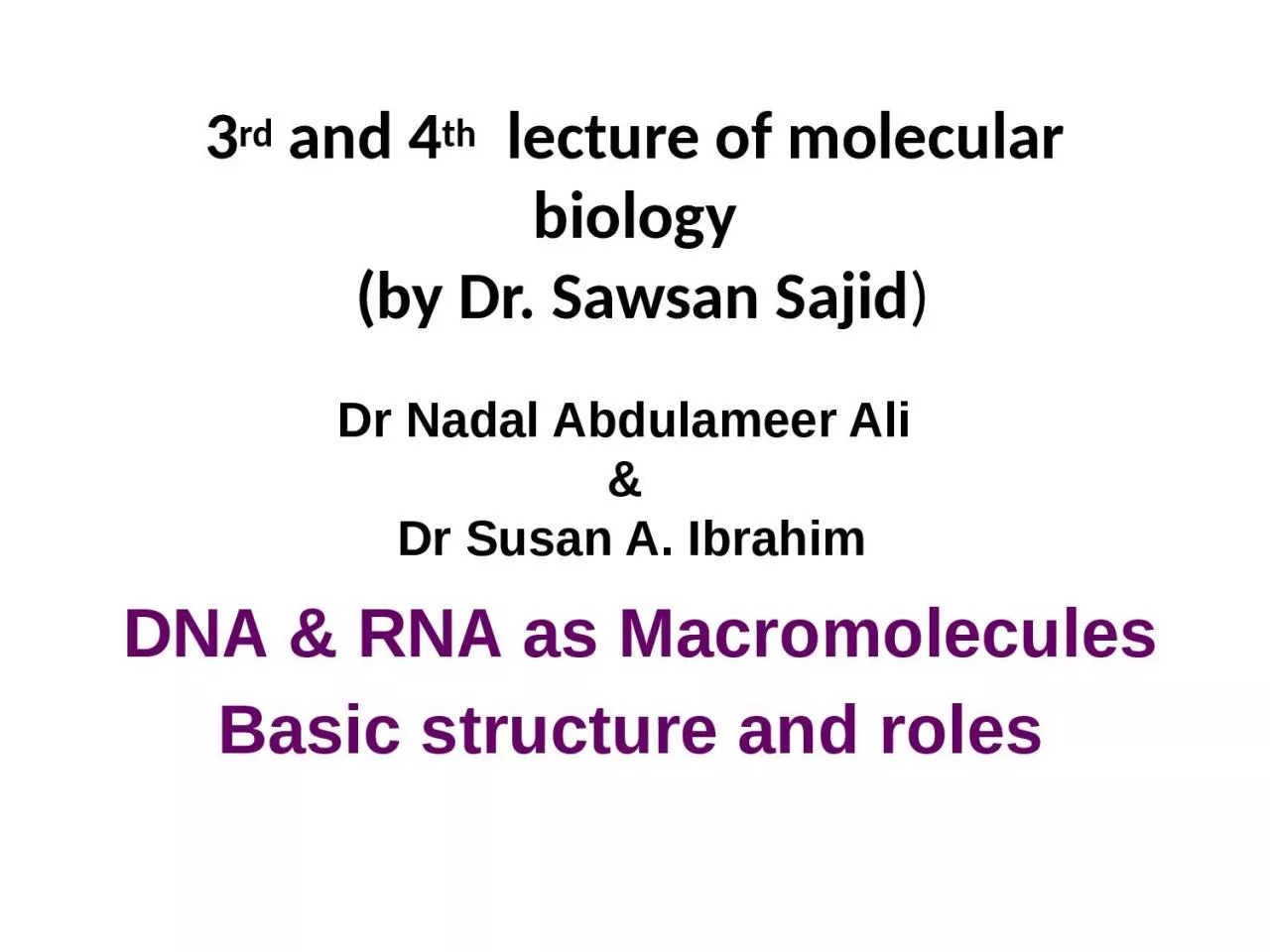
/
3 rd  and 4 th   lecture of molecular biology 3 rd  and 4 th   lecture of molecular biology

3 rd and 4 th lecture of molecular biology - PowerPoint Presentation

SportyChick
SportyChick . @SportyChick
Follow
342 views
Uploaded On 2022-07-28

3 rd and 4 th lecture of molecular biology - PPT Presentation

by Dr Sawsan Sajid DNA amp RNA as Macromolecules Basic structure and roles Dr Nadal Abdulameer Ali amp Dr Susan A Ibrahim Type of Macromolecules الجزيئات العملاقة ID: 930185

base dna acid structure dna base structure acid sugar helix nitrogen nucleotide cell phosphate form bond double rna strand

Share:

Link:

Embed:

Download Presentation from below link

Download Presentation The PPT/PDF document "3 rd and 4 th lecture of molecular bi..." is the property of its rightful owner. Permission is granted to download and print the materials on this web site for personal, non-commercial use only, and to display it on your personal computer provided you do not modify the materials and that you retain all copyright notices contained in the materials. By downloading content from our website, you accept the terms of this agreement.


Presentation Transcript

Slide1

3rd and 4th lecture of molecular biology (by Dr. Sawsan Sajid)

DNA & RNA as MacromoleculesBasic structure and roles

Dr

Nadal

Abdulameer

Ali

&

Dr

Susan A. Ibrahim

Slide2

Type of Macromoleculesالجزيئات العملاقة Nucleic acid :including DNA {Deoxribonucleic

acid} & &RNA { Ribonucleic acid}.Proteins Polysaccharid THE NUCLEIC ACIDS :DNA is the most

importent

molecule in living cells and contains all the information that the cell need to live and to propagate itself .with RNA they maintain the cell through Gene expression. Nearly all of the DNA present in eukaryotic cells can be found in the 

cell nucleus

but it is present in cytoplasm in bacteria.

Slide3

Basic structure of DNAA Nucleic Acid is a polymer consisting from nucleotides (the main structural units in nucleic acid) each nucleotide has the 3 following components:

Cyclic five carbon sugar called ribose (in RNA) and as it devoid hydroxyl group attached to carbon no. 2 DNA structure thus it called deoxyribose,

Nitrogen base the

purine

{adenine A and guanine G}and

pyrimidine

{cytosine C, thymine T and

uracile

U} .the first type consist from 2 rings while second type contain only one ring .

A phosphate group (phosphoric acid) attached to the 5 carbon atom of the sugar by a

phosphoester

linkage .this phosphate group is responsible for the strong negative charge of nucleic acid

Slide4

Structure

of DNA nucleotide

Slide5

5 Nitrogen base :the blue nitrogen atom represent the place where the sugar attached to the nitrogen base

Slide6

N base +sugar→ Nucleoside via N-glycosylic bondN base +sugar+ phosphate group→Nucleotide

Slide7

The bond between base and sugar is N- glycosylic bondthe bond between sugar and phosphate is estar bond the bond between 2 bases is hydrogen bond

Slide8

Carbon no:1 in Sugar attached to nitrogen NO. 9 in case of purine

Slide9

Carbone no:1 in Sugar attached with nitrogen N0.1 in case of pyramidine

Slide10

Nucleotides the basic unit in nucleic acid

Slide11

According to nitrogen base type the nucleotide is named N.B

Nucleoside Nucleotide 1-Adenine Deoxyadenosine

Deoxyadenylic

acid (d AMP)

2-Cytosine

Deoxycytidine

Deoxycytidylic

(d CMP)

3-Guanine

Deoxyguanosine

Deoxyguanylic

(d GMP)

4-Tymine

Deoxythymidine

Deoxythymidylic

(d TMP)

5-

Uracil

Uridine

Uridylic

acid

For the phosphate group:

Usually the nucleotide come with 3 phosphate

group (

α

β

γ

)

when it bind to DNA strand it will lose two groups (pyrophosphate) leafing only one

group

α

attached to the helix . The attachment only from one side (the 3́ OH free end)

Slide12

The attachment always occur from 3́ end ,the bond between 2 adjacent nucleotide is called 3́ 5́

phosphodiester bond

Slide13

Slide14

The primary structure of DNA represent one single strand ,not branched, result from binding polymer of nucleotides between the 3́ OH free end of the upper sugar with the 5́ P end (Pe phosphate) of the following nucleotide

Slide15

The secondary structure of DNA represent by binding two single strand via hydrogen bond A=T and G≡C .the two strand are anti parallel to each (opposite direction )that is to say 3́ end face the 5́ end twisted together .

Slide16

Formation of double helix

Slide17

Nitrogen base in DNA : A T G C without U (Uracile only in RNA) .the back bone is the sugar- phosphate

Slide18

Usually DNA is stored inside the cell as super helix rather than linear to reduce the it size thus DNA is longer than the cell itself

Slide19

Other type of DNA rather than chromosomal DNA

Plasmid DNA :It is a double stranded super helix DNA (extra chromosomal genetic material) usually found in bacterial cytoplasm .plasmid are not confined to bacteria but they have been isolated from yeast, protozoa and plants

Mitochondrial DNA

 (

mtDNA

 or 

mDNA

)

is the 

DNA

 located in 

organelles

 called 

mitochondria

, structures within 

eukaryotic

 cells that convert chemical energy from food into a form that cells can use, 

adenosine

triphosphate

 (ATP).In humans, mitochondrial DNA can be assessed as the smallest 

chromosome

 coding for only 37 genes and containing only about 16,600 base pairs. 

Human mitochondrial DNA

 was the first significant part of the 

human genome

 to be sequenced.

Chloroplast DNA

:

present inside chloroplast responsible for its activity

Slide20

Slide21

 DNA in E. coli bacteria is made up of 4 million base pairs and the whole genome is thus one millimeter long. The single-cell bacterium can copy its genome and divide into two cells once every 20 minutes.The DNA of humans, on the other hand, is composed of approximately 3 billion base pairs, making up a total of almost a meter-long stretch of DNA in every cell in our bodies.In order to fit, the DNA must be packaged in a very compact form. In E. coli the single circular DNA molecule is curled up in a condensed fashion, whereas the human DNA is packaged in 23 distinct chromosome pairs. Here the genetic material is tightly rolled up on structures called histones

.

Slide22

The main difference between DNA and RNA is that the former is double strand while the latter is single strand and Thymine T is replaced by Uracile U

Slide23

The Double helix model as proposed by Watson &Crick 1953

Slide24

In 1953, James Watson and Francis Crick published the structure of DNA for

which they were awarded a Nobel Prize in 1962. They determined that DNA consists of two antiparallel, complementary strands twisted around each other to form a right-handed double helix held in place by interactions between complementary base pairs: adenine (A) with thymine (T) and guanine (G) with cytosine (C). From this structure, it was straightforwardly evident how the genetic information was copied and maintained [2].

Slide25

Watson and Crick used stick-and-ball models to test their ideas on the possible structure of DNA. They depend on structure conformation shape proposed Rosalind Franklin and Maurice Wilkins, who were using X-ray diffraction to understand the physical structure of the DNA molecule.

Slide26

Basic role in DNA double helix B form :1-purines equal to pyrimidine bases within the anti parallel strand l 2-The DNA double helix is stabilized primarily by two forces: hydrogen bonds between nucleotides and 

base-stacking interactions among aromatic nucleobases3-It contain 2 groove, the major groove, is 22 Å wide and the other, the minor groove, is 12 Å wide.

4-the backbone directed outside the helix while N.B oriented inside

Slide27

Slide28

Slide29

Slide30

Compared to B-DNA, the A-DNA form is a wider right-handed spiral, with a shallow, wide minor groove and a narrower, deeper major groove. The A form occurs under non-physiological conditions in partially dehydrated samples of DNA, while in the cell it may be produced in hybrid pairings of DNA and RNA strands, as well as in enzyme-DNA complexes.Segments of DNA where the bases have been chemically modified by methylation may undergo a larger change in conformation and adopt the 

Z form. Here, the strands turn about the helical axis in a left-handed spiral, the opposite of the more common B form. These unusual structures can be recognized by specific Z-DNA binding proteins and may be involved in the regulation of transcription.

Slide31

DNA conformation (A .B.Z SHAPE)

Slide32

Slide33

Compares between 3 conformation DNA shape depending on Xray