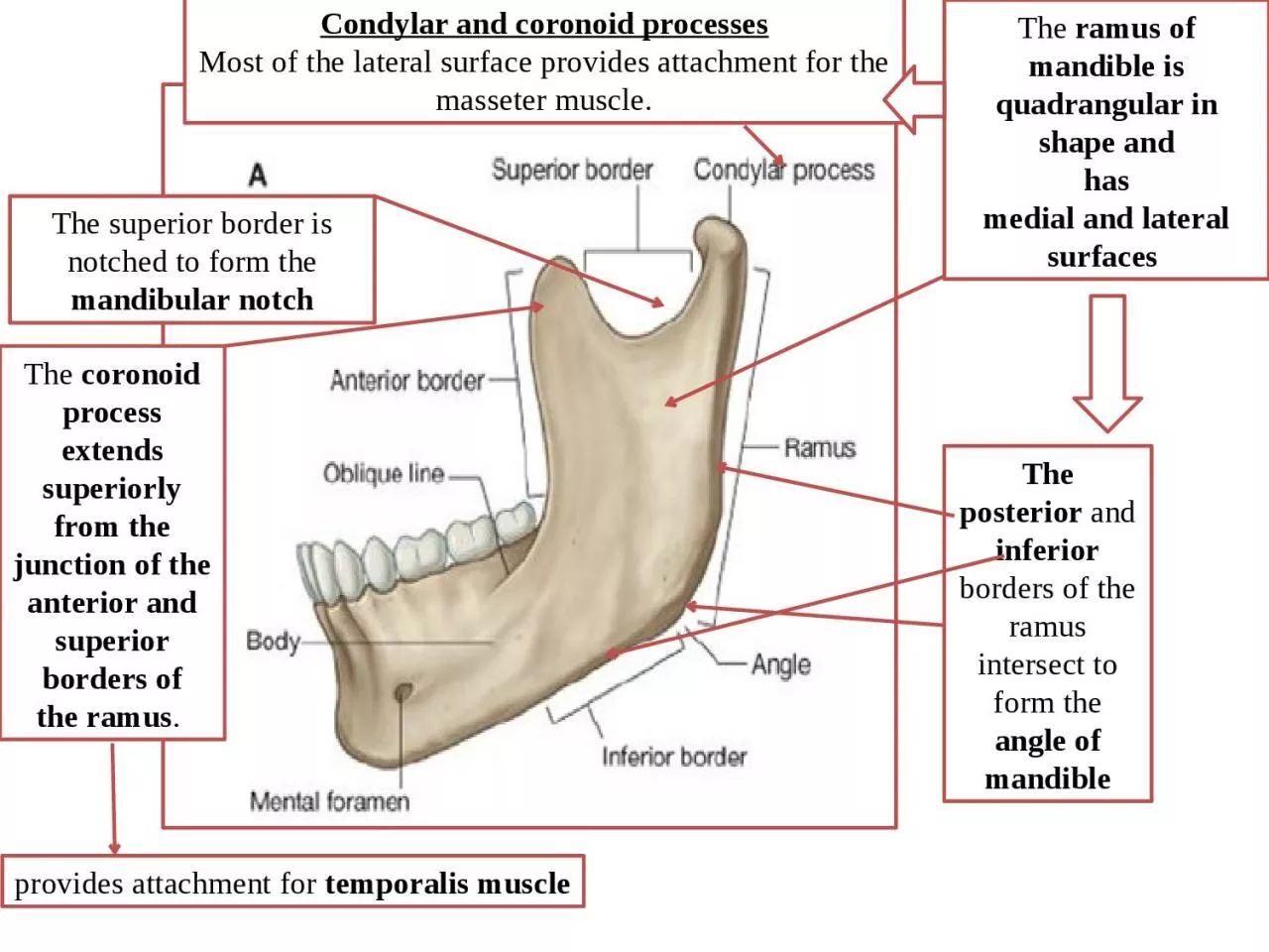
/
The  ramus of mandible is quadrangular in shape and The  ramus of mandible is quadrangular in shape and

The ramus of mandible is quadrangular in shape and - PowerPoint Presentation

SugaryDreams
SugaryDreams . @SugaryDreams
Follow
352 views
Uploaded On 2022-07-28

The ramus of mandible is quadrangular in shape and - PPT Presentation

has medial and lateral surfaces Condylar and coronoid processes Most of the lateral surface provides attachment for the masseter muscle The posterior and inferior borders of the ramus intersect to form the ID: 931038

mandible pterygoid lateral medial pterygoid mandible medial lateral surface muscle mandibular nerve process ramus inferior temporalis temporal innervated foramen

Share:

Link:

Embed:

Download Presentation from below link

Download Presentation The PPT/PDF document "The ramus of mandible is quadrangular i..." is the property of its rightful owner. Permission is granted to download and print the materials on this web site for personal, non-commercial use only, and to display it on your personal computer provided you do not modify the materials and that you retain all copyright notices contained in the materials. By downloading content from our website, you accept the terms of this agreement.


Presentation Transcript

Slide1

The

ramus of mandible is quadrangular in shape and has medial and lateral surfaces

Condylar and coronoid processes

Most of the lateral surface provides attachment for the masseter muscle.

The posterior

and

inferior borders of the ramus intersect to form the angle of mandible

The superior border is notched to form the

mandibular notch

The

coronoid process extends superiorly from the junction of the anterior and superior borders of

the ramus

.

provides attachment for

temporalis muscle

Slide2

1-the

head of mandible, participates in forming thetemporomandibular joint;and

2-the neck of mandible, which bears a shallow depression (the pterygoid fovea) on its anterior

surface for attachment of the lateral pterygoid muscle.

The condylar process is made of:

Slide3

1-Mandibular

foramen, which is the superior opening ofthe

mandibular canal. The inferior alveolar nerve and vessels pass through this foramen.

2-A

triangular elevation (the lingula

) forattachment of the mandibular end of the sphenomandibular

ligamentThe medial surface of the ramus shows

the following features:

4-An

elongate groove (the mylohyoid groove) extends anteroinferiorly

from the mandibular foramen.The nerve to mylohyoid is in this groove

3-Roughened

for attachment of the medial pterygoid muscle

Slide4

Masseter muscle

The masseter muscle is quadrangular in shape Origin

: inferior border and inner surface of the zygomatic arch. insertion: into the lateral surface of the ramus of the mandible and its coronoid process.

The masseter is innervated by the masseteric

nerve from the mandibular

nerve [V3]

Note: the mandible is the only movable bone in the skull !!! Therefore , it receives the insertion of all the muscles of mastication.

Slide5

Temporalis muscle

The temporalis muscle is a large fan-shaped muscle that fills much of the temporal fossa

It originates from the bony surfaces of the temporal fossa superiorly to the inferior temporal line

Tip and medial surface of the coronoid process And anterior border of the ramus of the mandible

Temporalis is a powerful elevator of the mandible, closes the mandible

Temporalis is innervated by deep temporal nerves that originate from the

mandibular nerve

Slide6

Medial pterygoid

The medial pterygoid muscle is quadrangular in shape and has deep and superficial heads

The medial pterygoid mainly elevates the mandible,

closing jawsThe medial pterygoid is innervated by the nerve to medial pterygoid from the mandibular nerve

[V3].

Origin: medial surface of the lateral plate of the

pterygoid process and the pyramidal process of the palatine boneInsertion: medial surface of the ramus of mandible inferior to

mandibular foramen

Slide7

Lateral pterygoid

The lateral pterygoid is a thick triangular muscleThe upper head originates from the roof of the infratemporal fossa (inferior surface of the greater wing of the sphenoid and the infratemporal crest

The lower head is larger and originates from the lateral surface of the lateral plate of the pterygoid process

Insertion: into the neck of mandibleinto the capsule of the Temporomandibular joint

Into the articular disc.

The lateral pterygoid is innervated by the nerve to lateral pterygoid from the mandibular nerve [V

3].