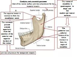
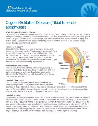
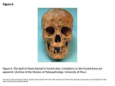
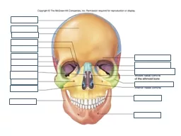
Zygomatic facial foramen 8 Orbital surface of maxilla 9 Temporal fossa 10 Lateral surface of ethmoid 11 Superior orbital fissure 12 Lacrimal bone and groove 13 Optic foramen 14 Ethmoidal ID: 775249
Download Presentation The PPT/PDF document " Above: Frontal View. Legend: 1- Mental ..." is the property of its rightful owner. Permission is granted to download and print the materials on this web site for personal, non-commercial use only, and to display it on your personal computer provided you do not modify the materials and that you retain all copyright notices contained in the materials. By downloading content from our website, you accept the terms of this agreement.
Slide1
Slide2Slide3Slide4Above: Frontal View. Legend: 1- Mental tubercle. 2- Body of mandible. 3- Ramus of mandible. 4- Anterior nasal spine. 5- Canine fossa. 6- Infra-orbital foramen. 7-
Zygomatic
-facial foramen. 8- Orbital surface of maxilla. 9- Temporal fossa. 10- Lateral surface of
ethmoid
. 11- Superior orbital fissure. 12- Lacrimal bone and groove. 13- Optic foramen. 14-
Ethmoidal
foramina. 15- Temporal line. 16- Supra-orbital notch. 17- Glabella. 18- Frontal tuberosity. 19-
Superciliary
arch. 20- Parietal bone. 21-
Naso
-frontal suture. 22- Pterion. 23- Great wing of sphenoid. 24- Orbital surface of great wing of sphenoid. 25- Squamous part of temporal. 26- Left nasal bone. 27-
Zygomatic
bone. 28- Inferior orbital fissure. 27-
Zygomatic
bone. 28- Inferior orbital fissure. 29-
Zygomatic
arch. 30-
Apertura
piriformis
, displaying nasal septum and
infeior
and middle concha. 31- Mastoid process. 32- Incisive fossa. 33- Angle of mandible. 34- Mental foramen. 35-
Symphysis
menti
Slide5Above: Lateral View. Legend: 1- Mental foramen. 2- Body of the mandible. 3- Maxilla. 4- Ramus of the mandible. 5-
Zygomatic
arch. 6-
Styloid
process. 7- External acoustic meatus. 8- Mastoid process. 9-
Asterion
. 10- Superior nuchal line. 11- External occipital protuberance. 12-
Lambdoid
suture. 13- Occipital bone. 14- Lambda. 15-
Obelion
. 16- Parietal bone. 17- Lower temporal line. 18- Upper temporal line. 19- Squamous temporal. 20-
Bregma
. 21- Coronal suture. 22-
Stephanion
. 23- Frontal bone. 24- Pterion. 25- Temporal fossa. 26- Great wing of sphenoid. 27-
Zygomatic
bone. 28-
Zygomatic
-facial foramen. 29- Fossa
sacci
lacrimalis
. 30- Nasal bone. 31- Infra-orbital foramen. 32- Anterior nasal spine
Slide6Above: Basal View of the lower surface of the cranium (mandible is removed). Legend: 1- External Occipital Crest. 2- Superior nuchal line of the occipital bone. 3- Foramen magnum. 4- Occipital condyle. 5- Mastoid notch. 6- Mastoid process. 7- External acoustic meatus. 8-
Styloid
process. 9- Mandibular fossa. 10- Foramen
spinosum
. 11- Angular spine of the sphenoid. 12- Foramen
ovale
. 13- Lateral
pterygoid
lamina. 14-
Hamulus
of medial
pterygoid
lamina. 15-
Vomer
. 16- Posterior nasal spine. 17-
Horzontal
part of palate bone. 18- Palatine process of maxilla. 19- Incisive foramen. 20-
Intermaxillary
suture. 21- Greater palatine foramen. 22-
Zygomatic
process of maxilla. 23- Inferior orbital fissure. 24- Infra-temporal fossa. 25-
Zygomatic
arch. 26- Left
choana
. 27-
Pterygoid
fossa. 28- Scaphoid fossa. 29- Foramen
lacerum
. 30- Opening of osseous part of auditory tube. 31- Carotid canal. 32- Jugular fossa. 33-
Stylo
-mastoid foramen. 34- Jugular process of occipital bone. 35- Groove for occipital artery. 36- Mastoid foramen. 37-
Canalis
condyloideus
. 38- Inferior nuchal line of occipital bone. 39- External occipital protuberance.
Slide7