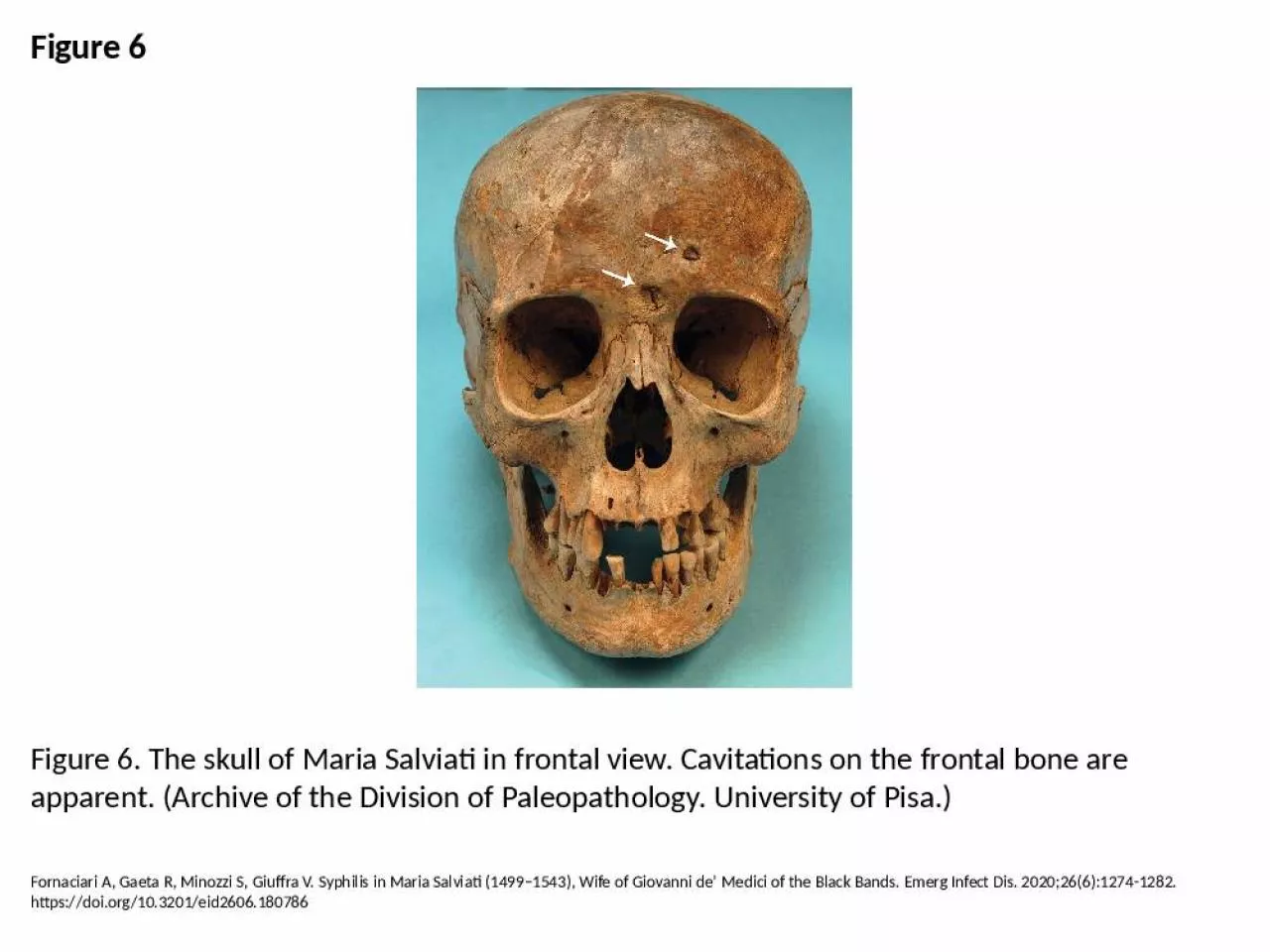
PPT-Figure 6 Figure 6. The skull of Maria Salviati in frontal view. Cavitations on the frontal
Author : tripp682 | Published Date : 2024-10-04
Fornaciari A Gaeta R Minozzi S Giuffra V Syphilis in Maria Salviati 14991543 Wife of Giovanni de Medici of the Black Bands Emerg Infect Dis 202026612741282 httpsdoiorg103201eid2606180786
Presentation Embed Code
Download Presentation
Download Presentation The PPT/PDF document "Figure 6 Figure 6. The skull of Maria Sa..." is the property of its rightful owner. Permission is granted to download and print the materials on this website for personal, non-commercial use only, and to display it on your personal computer provided you do not modify the materials and that you retain all copyright notices contained in the materials. By downloading content from our website, you accept the terms of this agreement.
Figure 6 Figure 6. The skull of Maria Salviati in frontal view. Cavitations on the frontal: Transcript
Fornaciari A Gaeta R Minozzi S Giuffra V Syphilis in Maria Salviati 14991543 Wife of Giovanni de Medici of the Black Bands Emerg Infect Dis 202026612741282 httpsdoiorg103201eid2606180786. Focus on the Skull. Review Anatomical Terms. Anterior/Posterior. Dorsal/Ventral. Medial/Lateral. Superior/Inferior. Axial Skeleton. Includes bones of the skull, vertebral column and thoracic cage. Creates a framework of support and protection for internal organs. Forms the longitudinal axis of the body. Divided into three parts. Skull. Vertebral column. Bony thorax. Figure 5.8a. (a) Anterior view. Phalanges. Metatarsals. Tarsals. Fibula. Tibia. Patella. Femur. 6 single. 11 paired. Cranium. Single. Frontal. Sphenoid. Ethmoid. Occipital. Paired. Parietal. Temporal. Face. Single:. Mandible. Vomer. Paired:. Maxillae. Zygomatic. Lacrimal. Inferior Nasal . Conchae. Longitudinal section. Compact. bone. Spongy. bone. Periosteum. Medullary cavity. Medullary cavity. Osteon. (a) Osteons (Haversian systems) in compact bone and trabeculae in spongy bone. 2. 3. 4. 5. 6. Animalia. Phylum:. Chordata. Class:. Mammalia. Order:. Carnivora. Family:. Felidae. Genus:. Felis. Species:. F. . catus. Cats . are similar in . anatomy . to the other . felines . with strong, flexible bodies, quick reflexes, sharp retractable claws, and teeth adapted to killing small prey. As . Why This Matters. Understanding the anatomy of the skeleton enables you to anticipate problems such as pelvic dimensions that may affect labor and delivery. © 2017 Pearson Education, Inc.. The Skeleton. Infant skull has more bones than the adult skull. Skull bones such as the mandible and frontal bones are unfused . At birth, skull bones are connected by fontanelles. Fontanelles. Unossified remnants of fibrous membranes between fetal skull bones. © 2015 Pearson Education, Inc.. The Skeletal System. Two subdivisions of the skeleton. Axial skeleton. Appendicular skeleton. Parts of the skeletal system. Bones (skeleton). Joints. Cartilages. Ligaments. 8x8 University of Washington PKU Clinic CHDD - Box 357920 Seattle WA 98195 206 685-3015 Toll Free in Washington State 877-685-3015 http//deptswashingtonedu/pkuPelvic bones Ilium Pubis Ischium Temporal bo tissue. Dr. Maria . Zahiri. 3. Dr. Maria Zahiri. . Four . basic tissue types. :. E. pithelial . tissue (surfaces and glands). Connective tissue. . Muscle . tissue (contractile cells). N. ervous . tissue. OVERVIEW OF HUMAN SKULL This appendix contains a brief review of the development of the fetal skull. The goal is to provide a qualitative understanding of the loca Anatomical Features of the Skull 1 2 3 4 5 6 7 8 9 10 11 12 Front View Side ViewAsk A BiologistWeb address: Our brain is one of the most important organs in the human body. It needs special protection...your skull! It’s made up of Figure 1 - Anatomy of the skull in an infant showing the various bones that fuse PathologyCraniosynostosis occurs when one or more of the major sutures close early The cause is unknown It is called
Download Document
Here is the link to download the presentation.
"Figure 6 Figure 6. The skull of Maria Salviati in frontal view. Cavitations on the frontal"The content belongs to its owner. You may download and print it for personal use, without modification, and keep all copyright notices. By downloading, you agree to these terms.
Related Documents