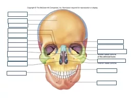
eEdE eEdE157 Nothing To Disclose LifeThreatening Lytic lesion of the Mandible A Lesson Learned Nucharin Supakul MD 1 Juan G Tejada MD 2 1 Ramathibodi Hospital ID: 774600
Download Presentation The PPT/PDF document " Control # 209 Title: Life -Threatenin..." is the property of its rightful owner. Permission is granted to download and print the materials on this web site for personal, non-commercial use only, and to display it on your personal computer provided you do not modify the materials and that you retain all copyright notices contained in the materials. By downloading content from our website, you accept the terms of this agreement.
Slide1
Control #
209
Title:
Life -Threatening Lytic lesion of the Mandible: A Lesson Learned
eEdE
#
eEdE-157
Slide2Nothing To Disclose
Slide3Life-Threatening Lytic lesion of the Mandible “A Lesson Learned”
Nucharin Supakul, MD1Juan G Tejada, MD2
1. Ramathibodi Hospital, Mahidol UniversityBangkok, Thailand2. Indiana University School of Medicine,Eskenazi HealthIndianapolis, Indiana, USA
Slide4Purpose
To review the characteristic imaging findings of mandibular vascular malformations
and
avoid unnecessary
and risky biopsies of the
mandibular
lesions.
To demonstrate interventional treatment options for vascular lesions in the mandible in life-threatening conditions and also in the preoperative setting.
Slide5Introduction
Mandibular Vascular
lesions are divided
in 2
groups
Hemangioma
Failure of
differentiation in the early stages of
embryogenesis
Appears in childhood and
regresses
over
time
Rarely associated with
fatal hemorrhage
Vascular malformation
Disturbance in the late stage of angiogenesis (
truncal
stage) and
results
in persistence of arteriovenous anastomosis
Present at birth and
grows
over
time
Symptoms
depend
on hemodynamic
factors
High flow: AVM, AVF
Fatal hemorrhage
Low flow: Lymphatic, venous, or mixed
Slide6Mandibular AVM
Abnormal direct communication between arteries and veins, bypassing capillary bed
Location:
Usually posterior location within the ramus and posterior mandibular body
Clinical presentation
Gingival bleeding (most common)
Massive bleeding with shock following by the extraction of teeth (most common)
Soft tissue mass (pulsatile/ non pulsatile), bruit, thrill
Painful, alteration of facial morphology
Neurosensory deficit
Slide7Imaging Findings
Cystic radiolucent lesion
Honeycomb (
multilocular
) or soap bubble appearance
Resorption of the adjacent bone/ dental root
teeth floating in the adjacent alveolar osseous erosion
Mimics odontogenic/non-odontogenic lesions
Central giant
cell granuloma
Ossifying fibroma
Traumatic bone cyst
Ameloblastoma
Slide8Imaging Findings
CT and MRI
Evaluates
the extent of the lesion
Bone erosion
Involvement of major vessels
Catheter angiogram
Gold standard in diagnosis and treatment
Super-selective arteriography of the external carotid
evaluates
collaterals and multiple anastomoses of the Internal maxillary artery
Slide9Management
Sclerosing
agents
(sodium
morrhuate,alcohol
, tetracycline etc
.)
ineffective most of the times
Ligation of the external carotid
not
recommended
Numerous
anastomoses (internal carotid, ophthalmic, vertebral, cervical, and contralateral external carotid) and collateral
vessels
Limits
further
angiography and
future
embolization
Direct trans osseous
puncture
of the vascular bed and
embolization
Embolization
(Onyx
,
cyanoacrylate,
polyvinyl alcohol particles,
Gelfoam
, coils
, collagen)
Pre operative embolization in acute phase then surgery within 48 hours to 2 weeks
Usually multiple
stages
of embolization
for
curative results
Slide1019-month-old female with left lower gingiva bleeding
Slide11Skull AP and Towns views were performed. No demonstrable lytic lesion within the mandible is noted. This is an inappropriate study to evaluate a mandibular lesion.
Slide12MRI and MRA of the head and neck were obtained.
A - B:
Coronal T2 (A) and axial T1 fat suppression images show an
expansile
T1
iso
/T2
hyperintense bony lesion within the left-sided mandible involving body, angle and ramus (orange arrows). Involvement of the left canine, left premolar and left 1st molar teeth is noted. Several flow void signals are noted, best seen on T2 images.C - D: Post contrast T1 fat suppression in coronal (C) and axial (D) images show heterogeneous contrast enhancement and increased signal intensity with in the left masseter and let temporalis muscles.E - F: MIP MRA images of the head and neck vessels show enlargement of the left external carotid artery (blue arrows) supplying this mass (pink arrow) with early draining vein to the left external jugular vein (green arrow).
F
E
D
C
B
A
Slide13Conventional angiogram with left external carotid artery catheterization.
A – B:
There is
a
vascular blush of the mass (green arrow) within the left buccal/maxillary region supplied by branches of the left internal maxillary artery (orange arrows) and left facial artery (blue arrows) with AV shunting and venous drainage into the external jugular vein (pink arrows).C: Post PVA embolization via the left internal maxillary artery, superficial temporal, and left facial arteries with nearly complete disappearance of the vascular blush.
A
C
B
Slide147-year-old MALE with right lower gingiva mass with intermittent bleeding for a month. Recent history of active bleeding with shock
Slide15Grossly unremarkable Panoramic radiograph of the mandible
Slide16MRI and MRA of the head and neck without and with contrastA-B: Axial T1 fat suppression (A) and T2 fat suppression (B) images show T1/T2 hyperintense expansile lesion within the body of the right-sided mandible (orange arrows). A few signal voids are noted.C-D: Post contrast axial T1 fat suppression (C) and coronal T1 fat suppression (D) images show heterogeneous contrast enhancement within this mass (orange arrows).D-E: Contrasted MRA images show dilation of the right facial vein (blue arrow) and external carotid artery (pink arrows), related to a feeding artery.F: MIP image shows dilation of the right external carotid artery (pink arrow) with no visualized drain vein.
G
F
E
D
C
A
B
Slide17D
C
B
A
Conventional angiogram with right external carotid artery catheterization
A-B:
Lateral and AP images show abnormal vascular blush with arterial supply from
the right facial (orange arrows)
and
right internal maxillary arteries (blue arrows) and early draining vein to the right external jugular vein (pink arrow).C-D: Post embolization images after gelfoam and NBCA injection show complete occlusion of the mandibular AVM.
Slide1812-year-old male with lower gingiva bleeding
Slide19CT head and neck with contrast
A-B:
Axial and coronal CT without contrast at the level of the mandible show
well-defined lytic lesion within the posterior body of the right mandible associated with tooth root resorption (orange arrows).
C-D: Axial post contrast images show avid enhancement within this lesion (blue arrow) associated with enlargement of the right external carotid artery and right facial artery. Findings are suggestive of AVM.
A
B
C
D
Slide20Conventional angiogram with right external carotid artery catheterization
A-D:
AP and lateral images show abnormal vascular blush with arterial supply from the
right
inferior
alveolar (orange arrows)
and
right facial arteries (blue arrows). Drainage to the right facial vein (pink arrow) is noted. There is a large venous pouch in the right mandibular body (green arrow). E-F: Post embolization images with NBCA demonstrate residual venous pouch (green arrow) and vascular blush lesion. Patient was scheduled for second stage embolization within a month.
F
E
D
B
C
A
Slide21Second stage embolization with NBCA
R
esidual
small AVM
was treated with NBCA. Post embolization
angiogram shows marked decreased flow of the AVM and
increased
venous stagnation.
Slide2215-year-old male with vascular mass found on dental procedure
Slide2315-year-old boy with intra-oral vascular mass identified incidentally during a dental procedure. A: Axial CT image shows a lytic lesion within the posterior body of the right mandible (orange arrow).B: Axial T1 post contrast image shows an enhancing vascular mass in the right mandibular body (blue arrow).C: Doppler US demonstrates an AVM in the right mandibular body draining into a dilated varix (green star).D: DSA lateral image from the right external carotid artery injection shows an AVM supplied by branches of the facial and internal maxillary arteries (pink arrow) with venous drainage predominantly to the right external jugular vein.E – F: Post-embolization lateral images of the right external carotid artery injection show approximately 80% occlusion of the mandibular AVM (yellow arrow = residual AVM).
Slide24Conclusion
Vascular
malformations of the mandible are
extremely rare
potentially
life-threatening
conditions presenting with intractable
hemorrhage after tooth
extractions
or
biopsies.
Occur
predominantly during childhood with a variety
of symptoms
including gingival bleeding, bruit, dental loosening, swelling of the soft tissues of the face,
discoloration of the skin and mucosa and
sometime neurosensory deficits.
Think about vascular AVM in case of gingival bleeding/ lesion in the posterior body of the mandible.
Cross-sectional
imaging especially CT and MRI with contrast are useful imaging modalities
for clarifying the
extent of the lesion, the degree of bone
erosion, and
involvement of major
vessels
(feeding arteries and draining veins).
Radiologist
should be able to recognize the imaging patterns to avoid
risky and unnecessary biopsies
and suggest prompt treatment in case of life-threatening hemorrhage or the need for preoperative
treatment with endovascular or percutaneous embolization
Slide25References
Scholl
, Robert J., et al. "Cysts and Cystic Lesions of the Mandible: Clinical and Radiologic-
Histopathologic
Review 1."
Radiographics
19.5 (1999): 1107-1124.
Singh
V
,
Bhardwaj
PK
. Arteriovenous malformation of mandible: Extracorporeal curettage with immediate replantation technique.
Natl
J
Maxillofac
Surg. 2010 Jan-Jun; 1(1): 45–49.
Kiyosue
,
Hiro
, et al. "Treatment of mandibular arteriovenous malformation by
transvenous
embolization: a case report." Head & neck 21.6 (1999): 574-577.
Noreau
,
Gaétan
, Pierre-É. Landry, and
Dany
Morais
. "Arteriovenous malformation of the mandible: review of literature and case history." Journal-Canadian Dental Association 67.11 (2001): 646-651
.
A
.
Churojana
, R
.
Khumtong
,
D.
Songsaeng
, C
.
Chongkolwatana
,
and S.
Suthipongcha
,.
Life-Threatening
Arteriovenous Malformation of the
Maxillomandibular
Region and Treatment
Outcomes.
Interv
Neuroradiol
. 2012 Mar; 18(1): 49–59.