/
PNS; THE REFLEX ARC AND GENERAL PROPERTIES OF SPINAL REFLEXES PNS; THE REFLEX ARC AND GENERAL PROPERTIES OF SPINAL REFLEXES

PNS; THE REFLEX ARC AND GENERAL PROPERTIES OF SPINAL REFLEXES - PowerPoint Presentation

SunshineSmile
SunshineSmile . @SunshineSmile
Follow
342 views
Uploaded On 2022-08-04

PNS; THE REFLEX ARC AND GENERAL PROPERTIES OF SPINAL REFLEXES - PPT Presentation

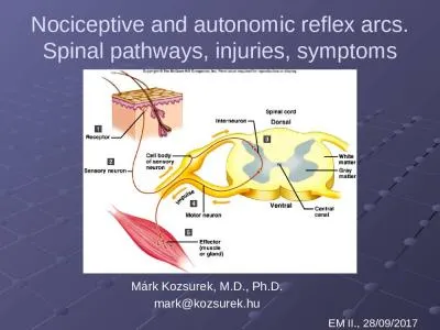
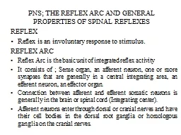
REFLEX Reflex is an involuntary response to stimulus REFLEX ARC Reflex Arc is the basic unit of integrated reflex activity It consists of Sense organ an afferent neuron one or more synapses that are generally in a central integrating area an efferent neuron an effector organ ID: 935006

reflex muscle response spindle muscle reflex spindle response spinal due reflexes synaptic stretch efferent afferents neuron fibers fibres cord

Share:

Link:

Embed:

Download Presentation from below link

Download Presentation The PPT/PDF document "PNS; THE REFLEX ARC AND GENERAL PROPERTI..." is the property of its rightful owner. Permission is granted to download and print the materials on this web site for personal, non-commercial use only, and to display it on your personal computer provided you do not modify the materials and that you retain all copyright notices contained in the materials. By downloading content from our website, you accept the terms of this agreement.


Presentation Transcript

Slide1

PNS; THE REFLEX ARC AND GENERAL PROPERTIES OF SPINAL REFLEXES

REFLEX

Reflex is an involuntary response to stimulus.

REFLEX ARC

Reflex Arc is the basic unit of integrated reflex activity

It consists of ; Sense organ, an afferent neuron, one or more synapses that are generally in a central integrating area, an efferent neuron, an effector organ.

Connection between afferent and efferent somatic neurons is generally in the brain or spinal cord (Integrating center).

Afferent neurons enter through dorsal or cranial nerves and have their cell bodies in the dorsal root ganglia or homologous ganglia on the cranial nerves.

Slide2

Efferent fibers leave via the ventral roots or corresponding motor cranial nerves

BELL-MAGENDIE LAW

In the spinal cord - dorsal roots are sensory

- ventral roots are motor.

Slide3

STRETCH REFLEX (MYOTATIC REFLEX)

Monosynaptic reflex

Stretch of skeletal muscle with intact nerve fiber

Response : contraction (stretch reflex)

Sense organ: Muscle spindle

Impulses originating in the spindle are conducted in the CNS by fast sensory fibers that pass directly to the motor neurons supplying the same muscle.

Neurotransmitter - glutamate

Slide4

CLINICAL EXAMPLES

Knee Jerk – Tapping of the patella tendon elicits knee jerk.

Tap on the tendon stretches the quadriceps

femoris

.

Manual stretch of the muscle produces same response

Tapping on the triceps

brachii

causes extensor response at the elbow

Tapping on the Achilles tendon causes an ankle jerk

Tapping on the side of the face causes a stretch reflex in the masseter

Slide5

MUSCLE SPINDLES

The receptors which detect stretch in muscle are known as muscle spindles.

Each muscle spindle consists of 3 to 12 small muscle fibres called

intrafusal

muscle fibres.

Each

intrafusal

fibre is between 1- 5mm in length and it is striated.

The fibres of the main muscle in which the muscle spindle lies are known as

extrafusal

fibres.

There are two types of

intrafusal

muscle fibres:

Nuclear bag fibres: commonly 2 per spindle; they are longer and thicker than the other and have a group of nuclei clustered around the centre of the fibre.

Nuclear chain fibres: which are shorter and thinner and have fewer nuclei arranged in a chain along the centre of the fibre; there are commonly 4 to 5

per spindle.

Slide6

STRUCTURE OF MUSCLE SPINDLES

REPRESENTATION OF THE MAIN COMPONENTS OF MAMMALIAN MUSCLE SPINDLE

Slide7

Afferents from spindle:

Ia

– from the nuclear bag fibers (

annulospiral

endings) – responds to change in length (static) and rate of change in length (dynamic)

II – from the nuclear chain fibers (flower spray endings) – static response only.

Afferents synapse directly on the anterior horn cell or

motoneurone which innervates the muscle group in which the spindle is situated. Spindle afferents stimulate the motorneurone resulting in muscle contraction.

Slide8

GAMMA (

γ

) –EFFERENT

Motor nerve supply to the contractile ends of the

musle

spindle.

Originate from smaller

motoneurone

, the γ-motoneurones in the anterior horn of the cord.Stimulation cause contraction of the spindle resulting in stretch of the nuclear bag

leading to increase afferent impulses from the muscle spindle

causing an increase in muscle tone

Slide9

γ

–efferent activity is under the control of the extra-pyramidal system particularly the

Reticulo

-spinal and

Vestibulo

-spinal tracts.

Increased

γ

–efferent activity is responsible for the increased tone seen in decerebrate animals (decerebrate rigidity).Clinical uses of Myotatic reflexHyperreflexia occurs in anxiety

May be a sign of damage to descending inhibitory pathways,

commomly

associated with upper motor neuron lesion.

Slide10

Absent reflexes

may be due to emotional depression

Damage to afferents (

tabes

dorsalis

) or to

α

-motor neuron (poliomyelitis) or damage to both afferent and efferent nerve fibers as in peripheral neuropathies lead to absent tendon reflexes and poor muscle tone.Abnormal tendon reflexes may take the form of a pendular knee jerk as in cerebellar dysfunction or a prolongation of the contraction and relaxation time as in hypothyroidism.

Slide11

GENERAL FUNCTIONS OF THE MUSCLE SPINDLE

Monitors and regulates muscle length, especially when the muscle is stretched

Contributes towards

propioception

.

Some collaterals from spindle afferents make connections with pathways to the cerebellum (

spinocerebellar

tracts) and cerebral sensory cortex (dorsal column

lemniscal pathways) and thus contribute to proprioception.

Slide12

INVERSE STRETCH REFLEX (Autogenic Inhibition).

When muscle is overstretched:

T

he

reflex contraction suddenly gives way and the muscle

relaxes.

Receptor

for this reflex is the

Golgi tendon organA high threshold receptor and responds to increase in stretchAfferents are Group Ib fibers which synapse on inhibitory interneurone This is the cause of the “clasp-knife or lengthening reaction seen in decerebrate

preparations.

Slide13

FLEXOR/WITHDRAWAL REFLX

Spinal reflex in response to a

norciceptive

/noxious stimulus

There is reflex flexion of the limb resulting in its withdrawal from the noxious stimulus.

CROSS EXTENSOR REFLEX

When nociceptive stimulus is strong:

Affected limb undergoes flexor/withdrawal

Irradiation of impulse to contraleteral limb induces extension Reflex helps maintain upright posture while the other leg is withdrawn.

Slide14

EXPERIMENTAL PREPARATIONS FOR STUDYING CORD REFLEXES.

SPINAL ANIMAL

Spinal cord is transected in the neck so that most of the cord still remains intact: Spinal animal results

In toad, cut the head behind the tympanic membrane

or single pith to destroy the brain

Slide15

Results in syndrome of Spinal Shock

Complete loss of reflex activity (days in cats, months in humans)

When reflexes return they are no longer under the influence of brain stem and do not follow normal patterns

eg

. A light touch on the foot can cause activation of all flexor muscles of body.

Slide16

Excitability

of

motoneurons

ultimately becomes greater than normal, causing particular groups of muscles to contract continuously

.

2

. DECEREBRATE

ANIMAL

Brain is transected above the pontine reticular formation and results in DECEREBRATE RIGIDITY (Spasticity).In frog, cut the head behind the eyesInhibitory inputs from cortex and cerebellum to reticular formation are removed

α

&

γ

- antigravity

motoneurons

are subjected to intense excitation

.

Excessive firing of these

motoneurons

causes the antigravity muscles (leg extensors and arm flexors) to contract

continuosly

.

Firing of

γ

motoneurons

activates the gamma loop, causing increased discharge of the 1a afferent fibers.

Slide17

3.

DECORTICATE

/ THALAMIC

PREPARATION

Due to removal of Cerebral

hemispheres

Thalamus is intact

Can regulate body temperature (because hypothalamus is intact).

Movement occurs but poorly. Initiated mostly by extrapyramidal pathways. Sensations are crudely appreciated, especially pain and temperature. Decorticate rigidity – moderate rigidity due to loss of frontal inhibitory areas.

Slide18

Exhibits sham rage – Destruction of cortical association areas enhance primitive emotions (which are usually suppressed).

Animal exhibits primitive emotions when noxious stimulus is applied (struggles and fights).

Slide19

REFLEXES OF POSTURE AND LOCOMOTION

Positve

Supportive Reaction,

Pressure on the footpad of a

decerebrate

animal causes

Extension of the limb against the pressure

Reflex can support animal in a rigid position – the positive supportive reaction.Cord Righting reflexesWhen a spinal cat is laid on its side It will make uncoordinated movements that indicate that It is trying to raise itself to standing position Illustrates that complicated reflexes are associated with posture.

Slide20

PROPERTIES

OF SPINAL REFLEXES

1. INHIBITION

Pre-synaptic: Inhibitory neuron acts on pre-synaptic fiber and reduces the magnitude of action potential, causing release of less transmitter

Post-synaptic: Inhibitory neuron connects with post-synaptic membrane, releases inhibitory transmitter which causes hyperpolarization.

Inhibitory neuron

Presynaptic

(a) and Post-synaptic (b) Inhibition

(a)

(b)

Slide21

2. SUMMATION

Spatial: Sub threshold stimuli in 2 or more afferents add to cause excitation of post-synaptic membrane

.

(

a

)

(b)(b) Temporal: Sub threshold stimuli in the same afferent coming in close succession (time and temporal summation) leading to summation and excitation of post-synaptic membrane.

Spatial (a) and Temporal (b) Summation

Slide22

3. FACILITATION

Fringe of a

Overlap of subliminal fringes

Fringe of b

2 neurons stimulated separately:

Independent responses are obtained

An area of sub-threshold excitation, the subliminal fringe, is created

b

a

Slide23

If the 2 neurons are stimulated simultaneously,

2 subliminal fringes will overlap in the middle area

Additional neurons in that area will reach threshold due to spatial summation

So response will exceed the sum of individual responses.

Slide24

4. OCCLUSION

Occlusion in the CNS. (Sharing of neurons)

Occlusion occurs when 2 afferents are stimulated simultaneously and the response is less than the sum of the individual responses.

Due to sharing of the same efferent

neurone

by 2 or more afferents.

Slide25

5. AFTER- DISCHARGE

Persistence of the response after the stimulus is over

Due to

reverberatory

circuits

On stimulation, the primary path elicits an initial response but due to

reverberatory

circuits 2,3&4, which are longer, the excitation continues and the response persists.

2

3

4

1

After-discharge

Effector

Slide26

6. POST-TETENIC FACILLITATION

Increased

response of post-synaptic membrane following brief but rapid repetitive (tetanic) series of impulses. This is due to excessive release of calcium ions during the period of stimulation.

7

. FATIGUE

Due

to excessive and continuous stimulation at a rapid rate. This causes the transmitter substance to be exhausted.