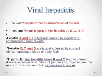
S Abdullah CABM FICMSGampH Viral hepatitis Viral hepatitis must be considered in any patient presenting with hepatitis on LFTs high ALT amp AST Viral hepatitis is usually caused by 2 ID: 933907
Download Presentation The PPT/PDF document "Viral Hepatitis Dr. Abdulwahhab" is the property of its rightful owner. Permission is granted to download and print the materials on this web site for personal, non-commercial use only, and to display it on your personal computer provided you do not modify the materials and that you retain all copyright notices contained in the materials. By downloading content from our website, you accept the terms of this agreement.
Slide1
Viral Hepatitis
Dr.
Abdulwahhab
S. Abdullah
CABM, FICMS-G&H
Slide2Viral hepatitis
Viral hepatitis must be considered in any patient presenting with hepatitis on LFTs (high ALT & AST).
Viral hepatitis is usually caused by 2
enterally
-transmitted and 2 parenterally-transmitted hepatitis viruses.
The enterically-transmitted viruses (hepatitis A and hepatitis E) cause acute self-limited hepatitis.
The parenterally-transmitted viruses (hepatitis B and C) can cause acute or chronic hepatitis.
Slide3Causes of viral hepatitis
Common: Hepatitis A
Hepatitis B (± D)
Hepatitis C
Hepatitis E
Less common: Cytomegalovirus
Epstein–Barr virus
Rare: Herpes simplex
Yellow fever
Slide4Clinical features of acute viral hepatitis
Acute viral hepatitis can be: Asymptomatic
Anicteric
Icteric (jaundice)
A typical episode includes:
Prodromal period (few days to 2 weeks):
malaise, nausea, anorexia.
Icteric period (1-4 weeks):
jaundice heralded by darkening of urine often with improvement of prodromal symptoms.
Recovery period (few weeks to several months):
rapid in children, slow in adults
Slide5Clinical features of acute viral hepatitis
Signs may include Jaundice, tender hepatomegaly, splenomegaly, and cervical lymphadenopathy.
Possible complications include:
Acute liver failure (especially HEV in pregnancy)
Chronic liver disease & cirrhosis (HBV & HCV)
Prolonged cholestasis (HAV & HEV)
Relapses (especially HAV)
Aplastic anemia
Slide6Lab tests in acute viral hepatitis
Hepatitic
pattern of LFTs ( ALT and AST several hundreds to several thousands U/L and ALP less than 2-3 times the upper limit of normal).
PT (INR) indicates the severity of hepatitis, and PT >25 sec usually indicates acute liver failure.
CBC may only show relative lymphocytosis.
Serological tests (
IgM
Ab
) confirm the diagnosis.
Slide7Management of acute viral hepatitis
Outpatient supportive therapy is appropriate for most patients. Antivirals are not used.
Hepatotoxic drugs (e.g.
paracetamol
), sedatives, and alcohol should be avoided.
No special diet is recommended.
Elective surgery must be postponed after recovery.
Persistent nausea/vomiting, any mental confusion, and prolonged PT warrants hospitalization.
Liver transplantation may be indicated in patients with the rare complication of fulminant hepatitis.
Slide8Hepatitis A
Hepatitis A virus (HAV) is the commonest cause of acute viral hepatitis.
HAV is an RNA
picornavirus
(
enterovirus
).
HAV is highly infectious, transmitted
feco
-orally with an average incubation period of 2-4 weeks.
Infection is more common in overcrowded areas with poor hygiene and sanitation.
HAV is present in stool for 2 weeks before and 2 weeks after onset of symptoms with maximal infectivity just before the onset of jaundice.
Slide9Slide10Electron microscopy of hepatitis A
virions
in feces. These are shown as 27 nm spheres. ( × 250 000.)
Slide11Hepatitis A
The presence and severity of symptoms depend on the patient’s age:
About 70% of adults develop symptoms, including jaundice. In contrast, only 10-30% of children <6 years old develop symptoms, which usually are non-specific and flu-like without jaundice.
Mortality (fulminant hepatitis) occur in 0.1% of patients aged <14
yr
and in 2% of those >40 yr.
Slide12Investigations in hepatitis A
Liver biochemistry:
Prodromal stage:
Serum bilirubin is normal (
bilirubinuria
may be found).
Serum ALT & AST is elevated.
Icteric stage:
Serum bilirubin is elevated and ALP is usually <3x ULN.
ALT reach a peak 1–2 days after onset of jaundice.
After jaundice subsides, ALT & AST may remain elevated for some weeks and occasionally for up to 6 months.
Slide13Investigations in hepatitis A
Hematological tests:
Leucopenia with a relative lymphocytosis.
Very rarely
Coombs’
-positive hemolysis or aplastic anemia.
PT is prolonged in severe cases.
ESR is raised.
Viral markers:
IgM
anti-HAV is diagnostic of acute hepatitis A.
IgG
anti-HAV denotes past exposure and immunity.
Slide14Course and prognosis of hepatitis A
Prognosis is excellent in most patients.
Mortality in young adults is 0.1% but it increases with age.
Recovery may be complicated by relapse of the hepatitis in 5-15% of cases followed by resolution.
Adult patients may have a prolonged cholestatic phase with elevated ALP for up to several months.
Patients with underlying chronic liver disease and the elderly with comorbidities may have a serious or life-threatening disease course.
Slide15Prevention of hepatitis A
In the community, improving social conditions of overcrowding, poor hygiene and sanitation is vital.
Active immunization with inactivated vaccine, especially during outbreaks, and for people at risk of severe disease, such as the elderly and patients with chronic hepatitis B or C is recommended.
Passive immunization (post-exposure prophylaxis) of close contacts within 2 weeks using immune serum globulin can prevent secondary spread of HAV.
Slide16Hepatitis E
Hepatitis E is caused by an RNA virus that is endemic in India and the Middle East.
Transmission, clinical features, diagnosis and management of HEV are similar to that of HAV.
HEV vaccine has been developed recently.
HEV differs from HAV in two aspects:
High mortality (20%) reported in pregnancy.
Chronic infection reported in some immune-compromised patients.
Slide17Take a deep breath
It calms the mind
Slide18What is “chronic hepatitis”?
Chronic hepatitis is a liver disorder in which hepatic
necroinflammatory
activity continues for at least 6 months.
Hepatitis B and C are the most common causes of chronic hepatitis worldwide.
Patients with chronic viral hepatitis are usually asymptomatic until they develop cirrhosis.
Slide19Hepatitis B
What’s the size of the problem?
Globally, hepatitis B is a major cause of cirrhosis (chronic liver disease), fulminant hepatitis (acute liver failure), and hepatocellular cancer, with over 1 million deaths annually.
Prevalence of chronic hepatitis B varies geographically (0.5-2% in Western Europe and USA and 10-20% in parts of Africa, the Middle East and the Far East).
Slide20Slide21Structure of HBV (Dane particle)
DNA genome
e antigen
(HBeAg)
core antigen (
HBcAg
)
surface antigen
(HBsAg)
Slide22Slide23How is HBV transmitted?
Perinatal (mother to child at birth)
Close contact among young children
Sexual
Contaminated needles and tools (iv drug use, tattooing, acupuncture, razors, etc..)
Infected unscreened blood products (rare)
Slide24How is HBV transmitted?
The main route of transmission varies by geographical region:
In high-prevalence areas:
vertical (perinatal)
horizontal (close contact among toddlers)
In low-prevalence areas:
sexual
iv drug abuse
Slide25Does HBV cause acute or chronic infection?
Upon exposure to HBV, the risk of progression from acute to chronic infection is inversely related to the patient’s
age
at time of infection:
Slide26Risk of chronic HBV infection
Rate of chronic
infection
Age at time of infection
Slide27Risk of chronic HBV infection
Rate of chronic
infection
Age at time of infection
90-95%
at birth
Slide28Risk of chronic HBV infection
Rate of chronic
infection
Age at time of infection
90-95%
at birth
20-50%
children < 5 years-old
Slide29Risk of chronic HBV infection
Rate of chronic
infection
Age at time of infection
90-95%
at birth
20-50%
children < 5 years-old
1-10%
Older
children and adults
Slide30What are the possible clinical presentations of hepatitis B?
Silent chronic infection is common in children while symptomatic acute infection often occur in adults.
Acute hepatitis B: Jaundice, malaise, and RUQ pain may develop 1-4 months after infection.
Acute (fulminant) liver failure is rare (up to 1% of adults).
Chronic hepatitis B
: defined by persistent HBsAg in serum for >6 months. Patients are usually asymptomatic until cirrhosis develops.
Slide31What are the complications and prognosis of chronic hepatitis B?
Cirrhosis and decompensation (chronic liver failure), such as ascites, variceal bleeding, encephalopathy, etc…
Hepatocellular carcinoma (usually in cirrhotics)
Other: Membranous glomerulonephritis
Polyarteritis nodosa (vasculitis)
Slide32How to diagnose hepatitis B?
Surface antigen (HBsAg)
Surface antibody (anti-HBs)
IgM core antibody (IgM anti-
HBc
)
IgG core antibody (IgG anti-
HBc
)
e antigen (HBeAg)
e antibody (anti-
HBe
)
HBV DNA (viral load)
>20,000 IU/L
<20,000 IU/L
Current infection (6/12?)
Immunity (anti-
HBc
?)
Recent infection (flare?)
Remote infection (HBsAg?)
High replication (always)
?Low replication (DNA?)
High replication (HBeAg±)
Low replication (
HBeAg
-)
Slide33What is the natural history of chronic hepatitis B?
Chronic HBV infection is a dynamic process that can be divided into 4 phases. These are not necessarily sequential and not all patients will go through all of them.
HBV is not directly cytopathic but a prolonged ineffective immune response in patients with chronic hepatitis B can mediate liver damage.
Slide34tolerance
clearance
inactive carrier
reactivation
mutantHBV
DNA
ALT
time (years)
HBeAg-negative
HBeAg-positive
chronic
infection
No inflammation or fibrosis
Significant inflammation mild fibrosis
Mild or no inflammation
Variable fibrosis
Significant inflammation & fibrosis/Cirrhosis
Slide35How to manage a patient with acute hepatitis B?
Supportive care and monitoring for acute liver failure (<1% ). Antivirals are usually not used.
Full recovery expected in 90-95% of immune-competent adults and remaining 5-10% develop chronic infection that usually persists for life.
Resolution occurs within 6/12 and is shown by:
HBsAg (-), HBeAg (- ), anti-HBs (+)
Progression to chronicity is shown on follow up by:
HBsAg (+) for >6/12
or
HBeAg (+) for >3/12
Slide36How to manage a patient with chronic hepatitis B?
The goal is to prevent cirrhosis and cancer (HCC) by clearing HBsAg but this is
not
yet achievable.
Current aims: HBeAg seroconversion (if HBeAg+)
low or undetectable HBV DNA
normal serum ALT
Treatment is indicated for patients with:
-high viral load
and
-active hepatitis (↑ALT
or
significant inflammation & fibrosis on
liver biopsy
)
Slide37Which drugs are used for chronic hepatitis B?
Monotherapy with 1 of 2 types of drugs is used:
nucleos
(t)ide analogues (single daily oral tab):
Best for HBeAg- patients and those with cirrhosis
Entecavir
or
Tenofovir
: 1
st
choice but expensive
Lamivudine
: cheaper but resistance is a problem
Adefovir,Telbivudine
: expensive & not so effective
pegylated interferon-
α
(weekly
s.c.
injection):
Best for pre-cirrhotics with HBeAg+ and
↑ALT.
Side effects is the main problem.
Slide38How to prevent hepatitis B?
Avoiding risky behaviors
Active immunization (vaccine) for:
all newborns
all “at risk” persons
patients with chronic liver disease
Post-exposure prophylaxis with active-passive immunization (vaccine & immune globulin) in e.g.:
needle-stick injury from HBsAg+ patient
neonate of HBsAg+ mother
sexual partner of HBsAg+ patient
Slide39What about hepatitis D?
HDV is an RNA-defective virus that has no independent existence (requires HBV to replicate) and has the same sources and modes of spread.
Can be acquired as
coinfection
or superinfection and can be acute or chronic.
Cirrhosis is more frequent & rapid with chronic hepatitis B+D than with chronic hepatitis B alone.
Diagnosis is by serum anti-HDV (IgM & IgG).
Prevention of hepatitis B prevents hepatitis D.
Slide40What about Hepatitis C?
HCV is an RNA
flavivirus
that has a worldwide distribution & is a major cause of cirrhosis & HCC.
Initial infection is usually clinically silent & leads to chronic infection in 80% of cases who will stay mostly asymptomatic until cirrhosis develops.
Exposure to infected blood & blood products is the most important mode of transmission.
Sexual & perinatal transmission is uncommon.
Males, alcoholics, and HIV-coinfected patients has more aggressive disease course.
Slide41Estimated HCV prevalence by region. (Source:
Perz
J
et al
., unpublished data. Centers for Disease Control,
2002
.)
Slide42How to diagnose HCV infection?
Screening is by serum anti-HCV antibody and confirmation is by serum HCV RNA (PCR).
Anti-HCV Ab persist even after HCV clearance, whether spontaneous or post-treatment.
HCV genotype has no effect on natural course of disease but affect therapeutic response.
LFTs may be normal or show fluctuating ALT.
ALT & AST in hepatitis C are poor predictors of liver damage and cirrhosis may be present with normal ALT & AST.
Slide43How to treat chronic hepatitis C?
The aim is to achieve a sustained viral response evidenced by undetectable HCV RNA 12 or 24 weeks after completion of therapy (SVR12 or SVR24).
Traditional HCV therapy ( weekly
s.c.
peg IFN-
α
injections with daily oral ribavirin) has now been replaced by more effective and convenient oral regimens using direct acting antiviral drugs (DAAs).
No active or passive immunization against HCV is available yet.
Slide44What about liver transplantation for hepatitis B and C?
In chronic viral hepatitis, liver transplantation is only indicated for decompensated end-stage cirrhosis and certain patients with early HCC.
HCV almost always recurs in the allograft but current IFN-free regimens can effectively eradicate the virus in pre- and post-transplant patients.
Recurrent HBV post-transplant is markedly reduced by pre-transplant antiviral drugs and post-transplant drugs and immunoglobulin.
Slide45Thank you