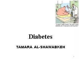
/
Diabetes TAMARA AL-SHAWABKEH Diabetes TAMARA AL-SHAWABKEH

Diabetes TAMARA AL-SHAWABKEH - PowerPoint Presentation

Tornadomaster
Tornadomaster . @Tornadomaster
Follow
342 views
Uploaded On 2022-08-04

Diabetes TAMARA AL-SHAWABKEH - PPT Presentation

1 INTRODUCTION Diabetes mellitus  DM is a group of metabolic disorders characterized by Hyperglycemia Abnormalities in carbohydrate fat and protein metabolism 1 Classification a ID: 935761

glucose insulin patients diabetic insulin glucose diabetic patients type treatment therapy neuropathy diabetes control risk hyperglycemia increased common glycemic

Share:

Link:

Embed:

Download Presentation from below link

Download Presentation The PPT/PDF document "Diabetes TAMARA AL-SHAWABKEH" is the property of its rightful owner. Permission is granted to download and print the materials on this web site for personal, non-commercial use only, and to display it on your personal computer provided you do not modify the materials and that you retain all copyright notices contained in the materials. By downloading content from our website, you accept the terms of this agreement.


Presentation Transcript

Slide1

Diabetes

TAMARA AL-SHAWABKEH

1

Slide2

INTRODUCTION

Diabetes mellitus (DM) is a group of metabolic disorders characterized by Hyperglycemia

Abnormalities in carbohydrate, fat, and protein metabolism.

1. Classification

a

. Type I

IDDM--

approximately 5% of all diabetic patients

b. Type II NIDDM—90% or more of all diabetic patients.

c. Impaired glucose tolerance.

Slide3

Type 1 DM (5%–10% of cases)(juvenile onset ) results from

autoimmune destruction of pancreatic β-cells, leading to absolute deficiency of insulin. This is characterized by a severe deficiency of insulin •Associated with HAA-B8/B15 , HLA-DR3/435-50% IN IDENTICAL twinsMale=female

It usually presents in children and

adolescents

.

The

onset is typically in youth (before age 20), but can occur at any age.

Not

related to obesity.

Usually have a lean body build.

Amylin

(a hormone

cosecreted

from pancreatic β-cells with insulin) suppresses

inappropriate

glucagon secretion,

slows

gastric emptying, and

causes

central satiety;

Amylin

is also deficient in type 1 DM due to β-cell destruction.

Slide4

Type 2 DM (90% of cases) (NIDDM) 

• Insulin levels are usually normal to high but may diminish over many years of having diabetes. • Insulin resistance (due to obesity) plays a major role. •

It often goes undiagnosed for many years.100% in identical twins

Slide5

Pathogenesis of type II diabetes.

a. Risk factors • Obesity (greatest risk factor) • Genetics • Age (insulin production decreases with age)

b. Obesity (plays a major role) • Obesity is associated with increased plasma levels of free fatty acids, which make muscles more insulin resistant, reducing glucose uptake. Therefore, obesity exacerbates insulin resistance. • In the liver, free fatty acids increase the production of glucose.

c

. Lack of compensation in type II diabetic patients • In normal individuals, the pancreas secretes more insulin in response to free fatty acids, thus neutralizing the excess glucose. • In type II diabetic patients, free fatty acids fail to stimulate pancreatic insulin secretion. Therefore, compensation does not occur and hyperglycemia develops; β-cells become desensitized to glucose, leading to decreased insulin secretion.

Slide6

Slide7

Gestational diabetes mellitus (GDM) is defined as glucose intolerance which is first recognized during

pregnancy. Hormone changes during pregnancy result in increased insulin resistance, and GDM may ensue when the mother cannot adequately compensate with increased insulin secretion to maintain

normoglycemia. In most, glucose intolerance first appears near the

beginning of the third trimester.

If

DM is diagnosed prior to pregnancy, this is not GDM, but rather pregnancy with

preexisting

DM.

Detection

is important, as therapy will reduce perinatal morbidity and mortality.

Slide8

CLINICAL PRESENTATION

TYPE 1 DIABETES MELLITUSDue to insulin deficiency 1) increased lipolysis >>> weight loss ,

ketogenesis and metabolic acidosis 2) hyperglycemia

>> osmotic diuresis >> polyuria and dehydration

3) dehydration can

leads to renal impairment ,

hyperosmolarity

( leads to polydipsia ,

polyphagia

) and

hyperkalemia

( high aldosterone due to RAAS activation )

Lathergy

Individuals are often thin and are prone to develop diabetic ketoacidosis if insulin is withheld or under conditions of severe stress.

TYPE 2 DIABETES MELLITUS

Patients are

often asymptomatic

and may be diagnosed secondary to unrelated blood testing.

Lethargy, polyuria,

nocturia

, and polydipsia

can be present.

Significant

weight loss is less common;

most patients are overweight or obese

.( central obesity ) , small amount of

insuline

can supress lipolysis

Slide9

DIAGNOSIS

Criteria for diagnosis of DM include any one of the following:A1C of 6.5% or more ( reflect glycemic control over 3 month )

Fasting (no caloric intake for at least 8 hours) plasma glucose of 126 mg/

dL

or more

Two-hour

plasma glucose of 200 mg/

dL

or

more during

an oral glucose tolerance test (OGTT) using a glucose load containing the equivalent of 75 g anhydrous glucose dissolved in water

Random plasma glucose concentration of 200 mg/

dL

or

more

with classic symptoms of

hyperglycemia

or

hyperglycemic

crisis.

In

the absence of unequivocal

hyperglycemia

, criteria 1 through 3 should be confirmed by repeat testing

.

Urine analysis ( not reliable ) increased due to a decreased urinary threshold pregnancy , young age )

Ketones in serum or urine (dx and follow up of DKA)

Slide10

Diagnosis

1-Symptom of DM + RBS >=200mg/dl 2- at least two separate occasion FBS>=126RBS>=2002 H POST PRANDIAL >=200HbA1C >=6.5

3- prediabetes 1- Impaired fasting glucose (IFG) is FPG 100 to 125 mg/dL

.

2- Impaired

glucose tolerance (IGT) is diagnosed when the 2-hour

postload

sample of OGTT is 140 to 199 mg per

dL

.

Slide11

Dawn Phenomenon and Somogyi Effect

• Both cause morning hyperglycemia. • The dawn phenomenon is probably due to an increase in the nocturnal secretion of growth hormone. This phenomenon is independent of the Somogyi effect.

• The Somogyi effect is a rebound response to nocturnal hypoglycemia—that is, counterregulatory

systems are activated in response to hypoglycemia, leading to morning hyperglycemia

.

• If morning hyperglycemia is present, check the glucose level at 3:00 am. If the glucose level is elevated, the patient has the dawn phenomenon and his or her evening insulin should be increased to provide additional coverage in the overnight hours. If the glucose level is low, the patient has the

Somogyi

effect and his or her evening insulin should be decreased to avoid nocturnal hypoglycemia.

Slide12

TREATMENT

Slide13

Desired Outcome

The primary goals of DM management are to Reduce the risk for microvascular and macrovascular disease complications,

Ameliorate symptoms,

R

educe

mortality,

I

mprove

quality of life

Slide14

Treatment selection should be based on multiple

factorsDuration of diabetesComorbiditiesThe amount of glucose lowering required

to achieve the goalProblem with post-prandial or fasting blood glucose

Adverse

effect

profile,

Contraindications

,

hypoglycemia

potential, and

tolerability

Motivation, resources, and potential

difficulties with adherence

Age

Non-

glycemic

effects

Slide15

Slide16

Slide17

Non-Pharmacological

Slide18

Slide19

Slide20

Pharmacological Therapy

Although nonpharmacological therapy is the cornerstone of treatment for all patients with DM, insulin is required for type 1 DM and nonpharmacological therapy alone is rarely sufficient for type 2 DM

In type 1 DM, the insulin regimen should be tailored to the patient’s lifestyle. This almost always involves a basal-bolus treatment

strategy based

on SMBG readings

In type 2 DM patients need an

early use of

antihyperglycemic

medications in conjunction with diet and

exercise

Slide21

Drug Treatments of Choice

Type 1 DM must be treated with insulin, though adjunct medications may improve glycemic control

Most patients with type 2 DM are initially treated with metformin due its long track record of use in clinical practice,

efficacy, weight

neutrality, low risk of

hypoglycemia

, and low cost. Common alternatives to metformin if intolerance

( GI side effect

diarrhea

, vomiting , nausea ) or contraindicated ( renal failure,

cr

> 135 , Age > 80 )

include

sulfonylureas(GLYBURIDE ) ,

DPP-4

inhibitor ( dipeptidyl peptidase )(LINAGLIPTIN ) ,

GLP-1

(glucagon like peptide ) receptor agonist (

liraglutide

),

or SGLT2

inhibitor

If the HbA1c is more than 1% to 1.5

%

above goal, early

dual therapy

may be

warranted

A patient with

type 2 DM

experiencing symptomatic hyperglycemia at the time of diagnosis

should initially be treated with insulin therapy and transitioned to oral therapy or a GLP-1 receptor agonist once good glycemic control has been

achievedType 2 DM patients who are mildly symptomatic (

ie, without significant weight loss), may be started on early dual therapy

Slide22

Other oral hypoglycemics include

: • Sulfonylureas • Thiazolidinediones (glitazones) •

Alpha glucosidase inhibitors (acarbose, miglitol)

Incretins

Pramlintide

Repaglinide

/

nateglinide

Slide23

2- Sulfonylureas(

Gliclazide ,glipizide,chlorpromide ) Can be used as add on therapy in metformin fail to achieve A1C target Stimulate the release of insulin from the pancreas

Can cause weight gain , best for non-obese daibetic

Can cause hypoglycemia

3- Alpha-

glucosidase inhibitors, delay carbohydrate absorption by inhibiting

disaccharidases

.

Acarbose

.

Side effect (

abd

pain ,

flatulance

,diarrhea )

4- GLP-1

Glucagon like peptide

) increase

insuline

secretion and

inhibt

glucagon secretion , can used as third agent with ( met and

sulf

)

Cause

wt

loss< used if BMI>=35kg/m2

Slide24

•5- SGLT2 inhibitors, The Sodium and Glucose Transporter 2 inhibitors, dapagliflozin

, empagliflozin .They carry 25% reduction in glucose reabsorption , Empaglifozin carries 35% reduction in CV mortality and hospital admissions.6- DPP-4 Inhibitos

Slide25

3. Insulin a. Method of administration.

• Self-administered by SC injection in abdomen, buttocks, arm, leg. • Given intravenously for emergency ketoacidosis. b. Regimens

• Most type I diabetic patients require 0.5 to 1.0 unit/kg per day to achieve acceptable glycemic control.

• Start with a conservative dose and adjust the regimen according to the patient’s glucose levels.

Many different regimens exist, and every patient has unique needs (see Table 4-7)

Slide26

c. Intensive insulin therapy • Long-acting insulin is given once daily in the evening. Regular insulin is given 30 to 45 minutes before each meal, and should be adjusted according to

preprandial home glucose measurements. • These more aggressive therapies have been shown to significantly decrease the incidence of diabetes complications such as retinopathy and microalbuminuria when compared to prior regimens.

All attempts should be made to get patients on more aggressive treatment protocols. • With intensive insulin therapy, the risk for hypoglycemia is a serious concern

.

• Alternatively, a continuous SC infusion of insulin can be given via an insulin pump

.

Preprandial

boluses are given in addition to the basal infusion

.

d. If the patient is unable/unwilling to carry out an intensive insulin program

:

• Give 70/30 units before breakfast and before the evening meal for basal coverage.

Give a short-acting insulin (regular) for prandial control if necessary.

Adjust doses according to fasting and 4 pm glucose determinations.

Slide27

e. Inpatient management of diabetic patients (sliding scale). •

An insulin sliding scale (SSI) of regular insulin doses given according to bedside finger-stick glucose determinations is helpful in controlling blood glucose levels in the hospital setting. • In general, SSI should be used in addition to a regimen of intermediate-acting insulin. If given alone, hyperglycemia usually results. • Monitor blood glucose four times per day: before meals and at bedtime.

• If the home insulin dose is unclear, or if the patient anticipates greater requirements of insulin due to an illness, use the following approach to adjust appropriate insulin doses

Slide28

• Take the total number of units of regular insulin that the patient required in 1 day (while on the sliding scale).

• Add two-thirds of this to the prebreakfast dose and one-third before dinner. It should be given as 70/30 (i.e., 70% NPH/30% regular). f. Modifying insulin doses (see also Clinical Pearl 4-11) • Physical activity—depending on the intensity of the activity, decrease insulin dosage 1 to 2 units per 20 to 30 minutes of activity. • During illness, administer all of the routine insulin. Many episodes of DKA occur during episodes of illness

. • Stress and changes in diet require dosing adjustments

.

• Patients undergoing surgery should get one-third to one-half of the usual daily insulin requirement that day, with frequent monitoring and adjustments as necessary

Slide29

4. Surgical treatment—Surgical weight loss therapy (i.e., gastric bypass) is an effective treatment for some patients, including adolescents. Additionally, islet cell transplantation offers definitive treatment for selected qualified patients.

Slide30

Antihyperglycemic Therapy Recommendations in Type 2 Diabetes. General Recommendations

Slide31

Chronic Complications of Diabetes Mellitus

A. Macrovascular complications 1. The main problem is accelerated atherosclerosis

, which puts patients at increased risk of stroke, MI, and CHF (Figure 4-11). The accelerated atherosclerosis in diabetics is the reason the target

BP is lower

in diabetics (130/80) than in general population (140/90), and the reason the

target LDL

is lower in diabetics is less than 100 mg/

dL

. The cause of accelerated atherosclerosis is not known, although glycation of lipoproteins and increased platelet adhesiveness/aggregation are thought to be two potential causes. In addition

,

the process of fibrinolysis may be impaired in diabetic patients.

2

. The manifestations of atherosclerosis include the following:

a

. Coronary artery disease (CAD).

Risk of CAD is two to four times greater in diabetic than in nondiabetic persons

.

• Most common cause of death in diabetic patients

.

• Silent myocardial infarctions are common.

b

. Peripheral vascular disease—in up to 60% of diabetic patients.

c

. Cerebrovascular disease (strokes).

Slide32

B. Microvascular complications—risk can be markedly reduced by achieving tight glucose control

1. Diabetic nephropathy—most important cause of end-stage renal disease (ESRD) a. Pathologic types • Nodular glomerular sclerosis (Kimmelstiel–Wilson syndrome)—hyaline deposition in one area of glomerulus—pathognomonic for DM

• Diffuse glomerular sclerosis—hyaline deposition is global—also occurs in HTN

Isolated glomerular basement membrane

thickening

b.

Microalbuminuria/proteinuria

• If microalbuminuria is present, strict glycemic control is critical (has been shown to limit progression from microalbuminuria to clinical proteinuria)

Slide33

Without effective treatment, the albuminuria gradually worsens—HTN usually develops during the transition between microalbuminuria

and progressive proteinuria. Persistent HTN and proteinuria cause a decrease in glomerular filtration rate (GFR), leading to renal insufficiency and eventually ESRD• HTN increases the risk of progression of diabetic nephropathy to ESRD. Control BP aggressively. • Initiate ACE inhibitors or ARB immediately. These agents are proven to decrease the rate of progression of nephropathy.

• Microalbuminuria is the screening test!

Slide34

• It usually takes 1 to 5 years for microalbuminuria to advance to full-blown proteinuria. However, with proper treatment (i.e., using ACE inhibitors to control BP) this can be prolonged.

c. Once diabetic nephropathy has progressed to the stage of proteinuria or early renal failure, glycemic control does not significantly influence its course. ACE inhibitors and dietary restriction of protein are recommended.

Slide35

2. Diabetic retinopathy

a. Prevalence is approximately 75% after 20 years of diabetes. Annual screening of all diabetic patients by an ophthalmologist is recommended. b. Background (nonproliferative) retinopathy accounts for the majority of cases.

• Funduscopic examination shows hemorrhages, exudates, microaneurysms, and venous dilatation

.

• These patients are usually asymptomatic unless retinal edema or ischemia involves the central macula

.

• Edema of the macula is the leading cause of visual loss in diabetic patients.

HTN and fluid retention exacerbate this condition.

c

. Proliferative retinopathy

Key characteristics are new vessel formation (neovascularization) and scarring

.

• Two serious complications are

vitreal

hemorrhage and retinal detachment

.

• Can lead to blindness.

Laser

photocoagulation is the treatment.

Slide36

3. Diabetic neuropathy (Figure 4-12)

a. Peripheral neuropathy (distal symmetric neuropathy) • Usually affects sensory nerves in a “stocking/glove pattern”—Usually begins in feet, later involves hands (longest nerves affected first). Numbness and paresthesias are common.

• Loss of sensation leads to the following: ulcer formation (patients do not shift their weight) with subsequent ischemia of pressure point areas; Charcot joints.

Painful diabetic neuropathy—hypersensitivity to light touch; severe “burning” pain (especially at night) that can be difficult to tolerate. Treatment is with gabapentin, tricyclic antidepressants, or

pregabalin

.

b. CN complications—secondary to nerve infarction

.

• Most often involves CN III, but may also involve CN VI and IV.

Diabetic third nerve palsy: eye pain, diplopia, ptosis, inability to adduct the eye; pupils are spared

Slide37

c. Mononeuropathies—secondary to nerve infarction

. • Median nerve neuropathy, ulnar neuropathy, common peroneal neuropathy. • Diabetic lumbosacral plexopathy—severe, deep pain in the thigh; atrophy and weakness in thigh and hip muscles; recovery takes weeks to months.

• Diabetic truncal neuropathy—pain in distribution of one of the intercostal nerves. d. Autonomic neuropathy •

Impotence in men (most common presentation)

Neurogenic bladder—retention,

incontinence

• Gastroparesis—chronic nausea and vomiting, early satiety • Constipation and diarrhea (alternating)

Postural hypotension

4

. Diabetic foot

a

. Caused by a combination of artery disease (ischemia) and nerve disease (neuropathy)—can lead to ulcers/infections and may require amputation

.

Slide38

b. With neuropathy, the patient does not feel pain, so repetitive injuries go unnoticed and ultimately lead to nonhealing

. c. In addition, neuropathy may mask symptoms of PVD (claudication/rest pain). Also, calcific medial arterial disease is common and can cause erroneously high BP readings in lower extremities. 5

. Increased susceptibility to infection a. This results from impaired WBC function, reduced blood supply, and neuropathy

.

Wound healing is impaired in diabetic patients, and this can be problematic postoperatively

.

b. Diabetic patients are at increased risk for the following infections: cellulitis, candidiasis, pneumonia, osteomyelitis, and

polymicrobial

foot ulcers

.

c. Infections of ischemic foot ulcers may lead to osteomyelitis and may require amputation.

Slide39

Specific treatment of chronic diabetic complications 1

. Macrovascular disease—treatment involves reduction of risk factors (e.g., BP reduction, lipid-lowering agents, smoking cessation, exercise), a daily aspirin (if not contraindicated), and strict glycemic control. 2. Nephropathy—ACE inhibitors, benefits of which include: a. Slow progression of microalbuminuria to proteinuria. b. Slow decline of GFR.

3. Retinopathy—Treatment involves referral to an ophthalmologist and possible photocoagulation. 4. Neuropathy—Treatment is complex. Pharmacologic agents that may be helpful include NSAIDs, tricyclic antidepressants, and gabapentin. For gastroparesis, a

promotility

agent such as metoclopramide can be helpful, in addition to exercise and a low-fat diet

.

5. Diabetic foot—The best treatment is prevention: regular foot care, regular podiatrist visits. Amputation is a last resort

Slide40

Thank You

40