/
Motility of the bacteria Motility of the bacteria

Motility of the bacteria - PowerPoint Presentation

alexa-scheidler
alexa-scheidler . @alexa-scheidler
Follow
534 views
Uploaded On 2017-05-20

Motility of the bacteria - PPT Presentation

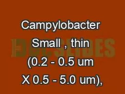
Biology and Biotechnology department A large number of bacteria are motile Most possess one or more flagella on their surface that allow them to swim The pattern of flagellation ID: 550353

flagella bacteria motile slide bacteria flagella slide motile agar line growth motility basal tumble body organism cover microscope surface

Share:

Link:

Embed:

Download Presentation from below link

Download Presentation The PPT/PDF document "Motility of the bacteria" is the property of its rightful owner. Permission is granted to download and print the materials on this web site for personal, non-commercial use only, and to display it on your personal computer provided you do not modify the materials and that you retain all copyright notices contained in the materials. By downloading content from our website, you accept the terms of this agreement.


Presentation Transcript

Slide1

Motility of the bacteria

Biology and Biotechnology departmentSlide2

A large number of bacteria are motile. Most possess one or more

flagella on their surface

that allow

them to swim

.

The

pattern of

flagellation

is an

important feature in

identification

of motile bacteria

.

The

figure illustrates the commonly observed arrangements of flagella. Slide3
Slide4

Polar flagella occur at one or both ends of the bacterium (Vibrio

cholerae

and some species of

Pseudomonas

).

They may be

single

or in

tufts

.

Peritrichous

flagella are

distributed around the surface of the organism

(many

Proteus species

).

Most motile bacteria

move in a straight line for a brief time, then turn and randomly change directions before swimming again.

Slide5

The straight line movement is called a run and the turn is called a tumble.

Runs and tumbles are controlled by the

clockwise

or

counterclockwise rotation of the basal body of the flagellum

, the motor that is

anchored in the cell membrane

.

Some bacteria

do not tumble

, but rather

reverse direction

when they reverse the rotation of the basal body.

Slide6

Chemotactic bacteria contain receptors in the cell membrane that bind to certain chemicals and cause the basal body to direct either a run or tumble

(or

forward

and

reverse directions

).

Many flagellated bacteria can

move toward useful chemicals

and

away from harmful ones

.

This

ability to control movement in response to chemical stimuli is termed

chemotaxis

. Slide7

When the chemical stimulus is an attractant, such as a rich nutrient source, the basal body is made to rotate so that the bacteria swim in straight lines toward the signal for long periods of time

.

If the stimulus is a

repellant

, such as a

poison

, the basal body

reverses direction

and causes the bacterium to

tumble more

often (or reverse direction). Slide8

Flagellar Stain :- Flagella are too thin to be seen by the

ordinary light microscope

.

Flagella should be

amplified

(enlarged). Use a

stain

that is specifically deposited on Flagella thus

increasing diameter

.

Some

flagellar

stains employ

rosaniline

dyes

and a

mordant

, applied to a

bacterial suspension fixed

in

formalin

and spread across a glass slide.

Slide9

The formalin links to, or “fixes,” the flagellar and other surface protein of the cells.

The

dye

and

mordant

then

precipitate around these

“fixed” surfaces,

enlarging their diameters

, and

making

flagella visible when viewed under the microscope

. Slide10

Another method, a ferric-tannate mordant and a silver nitrate solution are applied to a bacterial suspension.

The

resulting dark precipitate that forms on the bacteria

and their flagella allows them to be easily visualized under the microscope.

This

silver-plating technique

is also used to stain the

very slender spirochetes.

Note

: The techniques are somewhat sensitive.Slide11
Slide12

Hanging Drop Technique:- This method is commonly used to view living organisms for the rapid determination of motility.

The

hanging drop is prepared by

suspending

a fluid sample from

a

coverslip over a depression well in a specially designed microscope slide. Slide13

Wet mounts can be used for the same purpose, however, wet mounts tend to dehydrate rapidly. Hanging drops, on the other hand, are sealed within the depression and retain their liquid for longer periods of time

.

In both methods

,

the living specimen is unstained

.

For

best results,

reduce the amount of light passing

through the specimen. Slide14

Procedure:- 1. Place a drop of the bacterial culture (optimally from a young broth culture

) in the middle of a cover slip.

2. Place a thin line of

petroleum jelly

around the edge of the cover slide.

3. Turn the depression slide upside-down (depressed area facing down) and gently touch the cover slide. The jelly holds the cover slip to the slide and also keeps the suspension from drying out

.

Slide15

Positive control: Proteus vulgaris. Negative controle: Staph. Epidermidis.

4. Now flip the entire microscope slide/cover slip combination over. It should look like the diagram below. Slide16
Slide17

NOTES: You should be able to differentiate true motility from Brownian motility 2. Brownian movement is usually caused by the activity of water molecules. (characterized by back and forth movement)

3. True motility (the bacterial cells runs and tumble). Slide18

Motility Agar :- Motile bacteria require liquid to move

.

Thus bacteria can propel themselves in

broth

or

across the surface of a

wet agar plate

.

They

will not however move

when embedded in

1.5% agar

, the minimum concentration

found

in most agar media

.

Semisolid

agar

has a

reduced agar concentration

(

0.4 %

) that allows flagellated bacteria to migrate from the site of inoculation

.

Slide19

Semisolid media are prepared in tubes and are inoculated through most of their length by stabbing with a needle

.

Thus after

48 hours of incubation

, growth of a motile organism will be observed as a turbid region extending from the stab.

No motile bacteria will

only grow along the stab line

.

Positive

control:

Proteus vulgaris

. Negative

controle

:

Staph.

EpidermidisSlide20

Procedure Using aseptic techniques, inoculate the tube by stabbing with the needle to approximately

three-quarters

of its depth. Be careful to bring the needle into the center of the medium and not to touch the side of the tube.

2. Incubate at

room temperature

for

48 hours

.

3. Examine for growth

.Slide21

Interpretation : Pattern of growth of a motile organism. The entire medium is turbid with the growth of the organism, which has moved away from the stab line.

(B) Pattern of growth of a

nonmotile

organism. Only the stab line is turbid with growth.

Note

: Semi solid media with

tetrazolium

chloride (color indicator) Slide22