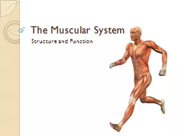
The Muscular System Muscles are responsible for all types of body movement Three basic muscle types are found in the body Skeletal muscle Cardiac muscle Smooth muscle Characteristics of Muscles ID: 920329
Download Presentation The PPT/PDF document "The Muscular System Structure and Functi..." is the property of its rightful owner. Permission is granted to download and print the materials on this web site for personal, non-commercial use only, and to display it on your personal computer provided you do not modify the materials and that you retain all copyright notices contained in the materials. By downloading content from our website, you accept the terms of this agreement.
Slide1
The Muscular System
Structure and Function
Slide2The Muscular System
Muscles are responsible for all types of body movementThree basic muscle types are found in the body
Skeletal muscle
Cardiac muscle
Smooth muscle
Slide3Characteristics of Muscles
Skeletal and smooth muscle cells are elongated (muscle cells are called muscle fibers)Contraction of muscles is due to the movement of microfilaments
All muscles share some terminology
Prefixes
myo
and
mys
refer to “muscle”
Prefix
sarco
refers to “flesh”
Slide4Comparison of Skeletal, Cardiac, and Smooth Muscles
Table 6.1 (1 of 2)
Slide5Comparison of Skeletal, Cardiac, and Smooth Muscles
Slide6Skeletal Muscle Characteristics
Most are attached by tendons to bonesCells are multinucleate
Striated — have
visible banding
Voluntary —
subject to conscious control
Slide7Connective Tissue Wrappings of Skeletal Muscle
Cells are surrounded and bundled by connective tissue
Endomysium
— encloses
a single muscle fiber
Perimysium
— wraps
around a fascicle (bundle) of muscle fibers
Epimysium
— covers
the entire skeletal muscle
Facicle
— bundle of muscle fibers
wraped
by
perimysium
Slide8Slide9Skeletal Muscle Attachments
Epimysium blends into a connective tissue attachmentTendons
— cord-like
structures
Mostly collagen fibers
Often cross a joint due to toughness and small size
Aponeuroses
— sheet-like
structures
Attach muscles indirectly to bones, cartilages, or connective tissue coverings
Slide10Skeletal Muscle Attachments
Sites of muscle attachmentBones
Cartilages
Connective tissue coverings
Slide11Smooth Muscle Characteristics
Lacks striationsSpindle-shaped cellsSingle nucleus
Involuntary—no conscious control
Found mainly in the walls of hollow organs
Slide12Cardiac Muscle Characteristics
StriationsUsually has a single nucleus
Branching cells
Joined to another muscle cell at an intercalated disc
Involuntary
Found only in the heart
Slide13Skeletal Muscle Functions
Produce movementMaintain postureStabilize jointsGenerate heat
Slide14Microscopic Anatomy of Skeletal Muscle
Sarcolemma
— specialized
plasma membrane
Myofibrils
— long
organelles inside muscle cell
Sarcoplasmic
reticulum
— specialized
smooth endoplasmic
reticulum
Used to store calcium
Slide15Microscopic Anatomy of Skeletal Muscle
Figure 6.3a
Slide16Microscopic Anatomy of Skeletal Muscle
Myofibrils are aligned to give distinct bands
I band =
light band
Contains only thin
filaments (
actin
)
A band =
dark band
Contains the entire length of the thick
filaments (myosin)
Slide17Microscopic Anatomy of Skeletal Muscle
Sarcomere
— contractile
unit of a muscle fiber
Organization of the
sarcomere
Myofilaments
Thick filaments = myosin filaments
Thin filaments =
actin
filaments
Slide18Microscopic Anatomy of Skeletal Muscle
Thick
filaments
= myosin filaments
Composed of the protein myosin
Has
ATPase
enzymes
Myosin filaments have heads (extensions, or cross bridges)
Myosin and
actin
overlap somewhat
Thin filaments
=
actin filaments
Composed of the protein
actin
Anchored to the Z disc
Slide19Microscopic Anatomy of Skeletal Muscle
Figure 6.3c
Slide20Microscopic Anatomy of Skeletal Muscle
At rest, there is a bare zone that lacks actin filaments called the H zone
Sarcoplasmic
reticulum (SR)
Stores and releases calcium
Surrounds the myofibril
Slide21Stimulation and Contraction of Single Skeletal Muscle Cells
Excitability (also called responsiveness or irritability
) — ability
to receive and respond to a stimulus
Contractility
— ability
to shorten when an adequate stimulus is received
Extensibility
— ability
of muscle cells to be stretched
Elasticity
— ability
to recoil and resume resting length after stretching
Slide22The Nerve Stimulus and Action Potential
Skeletal muscles must be stimulated by a motor neuron (nerve cell) to contractMotor
unit
— one
motor neuron and all the skeletal muscle cells stimulated by that neuron
Slide23The Nerve Stimulus and Action Potential
Neuromuscular junctionAssociation site of axon terminal of the motor neuron and muscle
Synaptic cleft
Gap between nerve and muscle
Nerve and muscle do not make contact
Area between nerve and muscle is filled with interstitial fluid
Slide24The Nerve Stimulus and Action Potential
Figure 6.5a
Slide25Transmission of Nerve Impulse to Muscle
Neurotransmitter — chemical released by nerve upon arrival of nerve impulse
The neurotransmitter for skeletal muscle is
acetylcholine
(
ACh
)
Acetylcholine attaches to receptors on the
sarcolemma
Sarcolemma
becomes permeable to sodium (Na
+) and potassium (K
+
)
Slide26Transmission of Nerve Impulse to Muscle
Figure 6.5c
Slide27Transmission of Nerve Impulse to Muscle
Sodium rushes into the cell generating an electrical current called an
action potential
Once started, muscle contraction cannot be stopped
Slide28Action potential (2:18)
Slide29The Sliding Filament Theory of Muscle Contraction
Activation by nerve causes myosin heads (cross bridges) to attach to binding sites on the thin filament
Myosin heads then bind to the next site of the thin filament and pull them toward the center of the sarcomere
This continued action causes a sliding of the myosin along the actin
The result is that the muscle is shortened (contracted)
Slide30The Sliding Filament Theory
of Muscle Contraction
Figure 6.7a–b
Slide31Sliding Filament Theory (1:14)
Slide32Sliding Filament Theory (0:57)
Slide33Contraction of a Skeletal Muscle
Steps of contraction:Motor neuron axon releases acetylcholine.
Acetylcholine diffuses across synaptic cleft.
Sarcolemma
is stimulated and the impulse travels over the muscle fiber surface and down transverse tubules to the
sarcoplasmic
reticulum (SR).
Calcium ions diffuse out of the SR into the
sarcoplasm
and bind to
troponin
.
Tropomyosin
moves and exposes sites on
actin
filaments.
Actin
and myosin form linkages.
Actin
filaments are pulled inward by myosin cross-bridges (sliding filament theory).
Muscle fibers shortens as contraction occurs.
Slide34Contraction of a Skeletal Muscle
Steps of Relaxation:Acetylcholine is decomposed and the muscle is no longer stimulated.
Calcium ions are actively transported back into the
sarcoplasmic
reticulum.
Linkages between
actin
and myosin break.
Myosin cross-bridges
recock
.
Troponin
and
tropomyosin
inhibit the interaction between myosin and
actin
.
Slide35Contraction of Skeletal Muscle
Muscle fiber contraction is “all or none”Within a skeletal muscle, not all fibers may be stimulated during the same intervalDifferent combinations of muscle fiber contractions may give differing responses
Graded
responses
— different
degrees of skeletal muscle shortening
Slide36Contraction of Skeletal Muscle
Graded responses can be produced by changingThe frequency
of muscle stimulation
The
number
of muscle cells being stimulated at one time
Slide37Types of Graded Responses
Twitch
Single, brief contraction
Not a normal muscle function
Tetanus
(summing of contractions)
One contraction is immediately followed by another
The muscle does not completely
return to a resting state
The effects are added
Slide38Types of Graded Responses
Unfused
(incomplete) tetanus
Some relaxation occurs between contractions
The results are
summed
Fused (complete) tetanus
No evidence of relaxation before the following contractions
The result is a sustained muscle contraction
Slide39Muscle Response to Strong Stimuli
Muscle force depends upon the number of fibers stimulatedMore fibers contracting results in greater muscle tension
Muscles can continue to contract unless they run out of energy