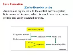
/
Role of Urea Urea is  a waste product of many living organisms, and is the major organic Role of Urea Urea is  a waste product of many living organisms, and is the major organic

Role of Urea Urea is a waste product of many living organisms, and is the major organic - PowerPoint Presentation

amber
amber . @amber
Follow
374 views
Uploaded On 2022-06-11

Role of Urea Urea is a waste product of many living organisms, and is the major organic - PPT Presentation

Urea contributes to the establishment of the osmotic gradient in the medullary pyramids and the ability to form a concentrated urine in the collecting ducts Urea transport is mediated by urea transporters presumably by facilitated diffusion There are at least four ID: 916460

water acidosis failure urine acidosis water urine failure kidney symptoms occurs osmotic urea current blood counter renal medullary kidneys

Share:

Link:

Embed:

Download Presentation from below link

Download Presentation The PPT/PDF document "Role of Urea Urea is a waste product of..." is the property of its rightful owner. Permission is granted to download and print the materials on this web site for personal, non-commercial use only, and to display it on your personal computer provided you do not modify the materials and that you retain all copyright notices contained in the materials. By downloading content from our website, you accept the terms of this agreement.


Presentation Transcript

Slide1

Role of Urea

Urea is

a waste product of many living organisms, and is the major organic component of human urine. This is because it is at the end of chain of reactions which break down the amino acids that make up proteins

.

Urea contributes to the establishment of the osmotic gradient in the

medullary

pyramids and the ability to form a concentrated urine in the collecting ducts. Urea transport is mediated by urea transporters, presumably by facilitated diffusion. There are at least four

isoforms

of the transport protein UT-A in the kidneys (UTA1 to UT-A4) . The amount of urea in the

medullary

interstitium

and in the urine varies with the amount of urea filtered, and this in turn varies with the dietary intake of protein. Therefore, a high-protein diet increases the ability of the kidneys to concentrate the urine

Slide2

Water

Diuresis

The feedback mechanism controlling vasopressin secretion and the way vasopressin secretion is

stimulated by a rise and inhibited by a drop in the effective osmotic pressure of the plasma

. The water

diuresis

produced by drinking large amounts of hypotonic fluid begins about 15 minutes after ingestion of a water load and reaches its maximum in about 40 minutes. The act of drinking produces a small decrease in vasopressin secretion before the water is absorbed, but most of the inhibition is produced by the decrease in plasma

osmolality

after the water is absorbed.

Slide3

Osmotic

diuresis

:

is increased urination caused by the presence of certain substances in the small tubes of the kidneys. The excretion occurs when substances such as glucose enter the kidney tubules and cannot be reabsorbed (due to a pathological state or the normal nature of the substance). The substances cause an increase in the osmotic pressure within the tubule, causing retention of water within the lumen, and thus reduces the reabsorption of water, increasing urine output (i.e.

diuresis

). The same effect can be seen in therapeutics such as

mannitol

, which is used to increase urine output and decrease extracellular fluid volume.

Slide4

Substances in the circulation can also increase the amount of circulating fluid by increasing the

osmolarity

of the blood. This has the effect of pulling water from the interstitial space, making more water available in the blood and causing the kidney to compensate by removing it as urine. In hypotension, often colloids are used intravenously to increase circulating volume in themselves, but as they exert a certain amount of osmotic pressure, water is therefore also moved, further increasing circulating volume. As blood pressure increases, the kidney removes the excess fluid as urine.

Osmotic diuresis results in dehydration from polyuria and the classic polydipsia (excessive thirst) associated with DM.

Slide5

What are the symptoms of an osmotic

diuresis

?

Osmotic

diuresis

does not cause symptoms unless dehydration occurs. Symptoms of dehydration include faintness, dizziness, rapid pulse, and dry mouth.

URINE ACIDIFICATION

Source of acids

Carbonic acids and Non carbonic acids

Source of H+

:

Diet

CO2

produced as end of metabolism

Secretion of H by renal tubules

Slide6

Majority of urine acidification occurs in DCT & PCT .Three main buffers remove free H

1. Bicarbonate system.

2.Phosphate system.

3. Ammonia system

Buffer systems in kidney bicarbonate system secreted H in PCT reacts with

HCO3 to form H2CO3 and help in HCO3 reabsorption

1.No net H secretion occurs.

2. therefore pH remains unchanged

3. H secreted in excess is buffered by two other systems

Slide7

phosphate systems

H secreted is buffered with HPO4

H is secreted in exchange for Na and N

1. 70%-75% of filtered HPO4 is reabsorbed

2. 25% available for buffering a 2 HPO4 in to Na H2PO4

3. Na H2PO4 excreted in urine

Ammonia system

SITE ( DCT and PCT)

Secreted and synthesized

not by filtrate, Formation of ammonia Mainly form glutamine

NH3 diffuses form tubular cells into lumen where it combines with H to form NH4 is lipid insoluble, therefore remains in lumen

Slide8

Uremia or

uraemia

:

is a term used to loosely describe the illness accompanying kidney failure (also called renal failure), in particular the nitrogenous waste products associated with the failure of this organ. This is not to be confused with

uricemia

, or

hyperuricemia

, a build up of uric acid in the blood.

In kidney failure, urea and other waste products, which are normally excreted into the urine, are retained in the blood. Early symptoms include anorexia and lethargy, and late symptoms can include decreased mental acuity and coma. Other symptoms include fatigue, nausea, vomiting, cold, bone pain, itch, shortness of breath.

Slide9

Because uremia mostly is a consequence of kidney failure, its signs and symptoms often occur concomitantly with other signs and symptoms of kidney failure, such as hypertension due to volume overload,

hypocalcemic

tetany

, and anemia due to erythropoietin deficiency.

Many individuals who have uremia need to be hospitalized. Doctors treat the condition with dialysis, a medical procedure in which a machine filters and purifies the blood. Medical practitioners might also make recommendations regarding dietary changes or prescribe medication to control the symptoms.

Slide10

Patients who have uremia occasionally develop acute tubular necrosis, a condition in which the tissues in the kidneys become severely damaged. These patients might eventually develop acute kidney failure, a condition where the kidneys suddenly stop working. Other patients with uremia might have convulsions, heart failure or coma. Untreated uremia can be fatal.

Slide11

Acidosis

is a condition in which there is too much acid in the body fluids. It is the opposite of alkalosis (a condition in which there is too much base in the body fluids).

Causes

The kidneys and lungs maintain the balance (proper pH level) . Acidosis occurs when acid builds up or when bicarbonate (a base) is lost. Acidosis is classified as either

respiratory acidosis

or

metabolic acidosis.

Respiratory

acidosis:

is a condition that occurs when the lungs cannot remove all of the carbon dioxide the body produces. This causes body fluids, especially the blood, to become too acidic.

Metabolic acidosis :

develops when too much acid is produced or the kidneys cannot remove enough acid from the body. There are several types of metabolic acidosis:

Slide12

Diabetic acidosis

(also called diabetic ketoacidosis and DKA) develops when substances called ketone bodies (which are acidic) build up during uncontrolled diabetes.

Lactic acidosis

is a buildup of lactic acid. This can be caused by:

Alcohol ,Cancer, Liver failure, Low blood sugar (hypoglycemia) ,Medications , Prolonged lack of oxygen from shock, heart failure, or severe anemia.

Other causes of metabolic acidosis include:

Kidney disease (distal renal tubular acidosis and proximal renal tubular acidosis) and Severe dehydration.

Slide13

What is Counter current mechanism?

Counter current mechanism is the process by which renal medullary interstitial fluid becomes hyper-osmotic.

Components of Counter Current Mechanism:

Counter current multiplier (Loop of Henle acts here)

Counter current exchanger (vasa recta acts here)

Slide14

Descending limb is not permeable to electrolytes. So it is of no importance in establishing hyper

osmolarity

in

medullary

interstitium

.

Thin and thick segments of ascending limb transports electrolytes (mainly Na+ and

Cl

-) to the

medullary

interstitium

by the process of simple diffusion and 1 Na+1K+-2Cl- co transporter system respectively. This process occurs repeatedly and more and more solutes (not water) are trapped in

medullary

interstitium

which results in

hyperosmotic

renal

medullary

interstitium

(1200-1400

mosm

/L)

Slide15

Counter current exchanger:

Vasa

recta maintain

hyperosmolarity

of

medullary

interstitium

as counter current exchanger.

Vasa

recta forms a U shaped bend parallel to loop of

Henle

.

Osmolarity

in renal medulla is graded (

Osmolarity

increases in downward direction as descending limb of ā€œUā€ gradually passes through

hyperosmotic

medulla,

solute enter into it from medulla

. But ascending limb passes progressively through decreased

osmolarity

of medulla (as

osmolarity

lowers in upward direction). Hence

solutes return back to renal medulla

(which entered in descending limb of earlier)

So counter current exchanger functions by-

Tendency of electrolytes to be

recirculated

Tendency of water to be removed by

vasa

recta

Slide16

Micturition

: is the process where the urinary bladder empties after being filled.

-Micturition reflex: mediated through the pelvic nerves (S2-S3) which contain sensory fibers for the detection of bladder wall stretching and the motor fibers (parasympathetic) ending on bladder wall ganglion cells that send post ganglionic fibers to the detrusor muscles of the bladder.

The urinary bladder begins to fill up to 100-150 ml and issuing a first desire to urinate due to stretch receptors in the wall of the urinary bladder causing a reflex contraction. After some time, the contraction disappears.

Slide17

When the volume reaches 400-500 ml, the reflex contraction increases & if the pressure builds more than the external sphincter (voluntary control), opening of the bladder neck occurs and leads to another reflex through the

pudendal

nerve (S1-S2) to inhibit the external sphincter. If this inhibition overcomes the voluntary control, urination occurs

Slide18