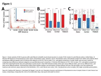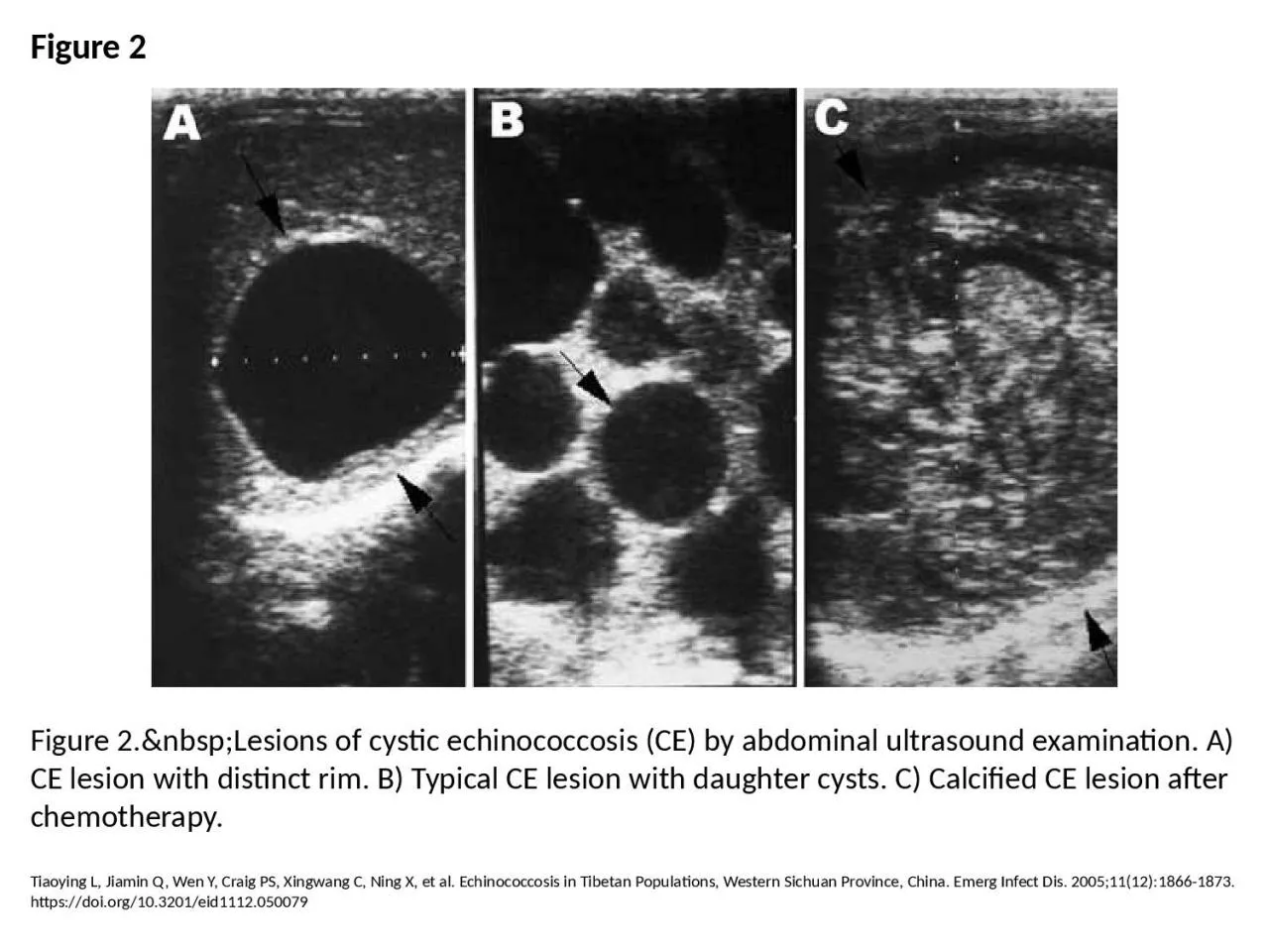
PPT-Figure 2 Figure 2. Lesions of cystic echinococcosis (CE) by abdominal ultrasound
Author : amey | Published Date : 2023-05-31
Tiaoying L Jiamin Q Wen Y Craig PS Xingwang C Ning X et al Echinococcosis in Tibetan Populations Western Sichuan Province China Emerg Infect Dis 2005111218661873
Presentation Embed Code
Download Presentation
Download Presentation The PPT/PDF document "Figure 2 Figure 2. Lesions of c..." is the property of its rightful owner. Permission is granted to download and print the materials on this website for personal, non-commercial use only, and to display it on your personal computer provided you do not modify the materials and that you retain all copyright notices contained in the materials. By downloading content from our website, you accept the terms of this agreement.
Figure 2 Figure 2. Lesions of cystic echinococcosis (CE) by abdominal ultrasound: Transcript
Download Rules Of Document
"Figure 2 Figure 2. Lesions of cystic echinococcosis (CE) by abdominal ultrasound"The content belongs to its owner. You may download and print it for personal use, without modification, and keep all copyright notices. By downloading, you agree to these terms.
Related Documents