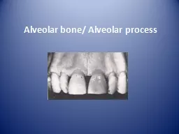
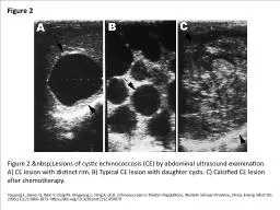
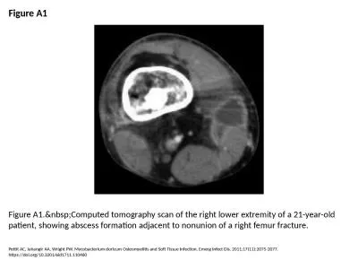
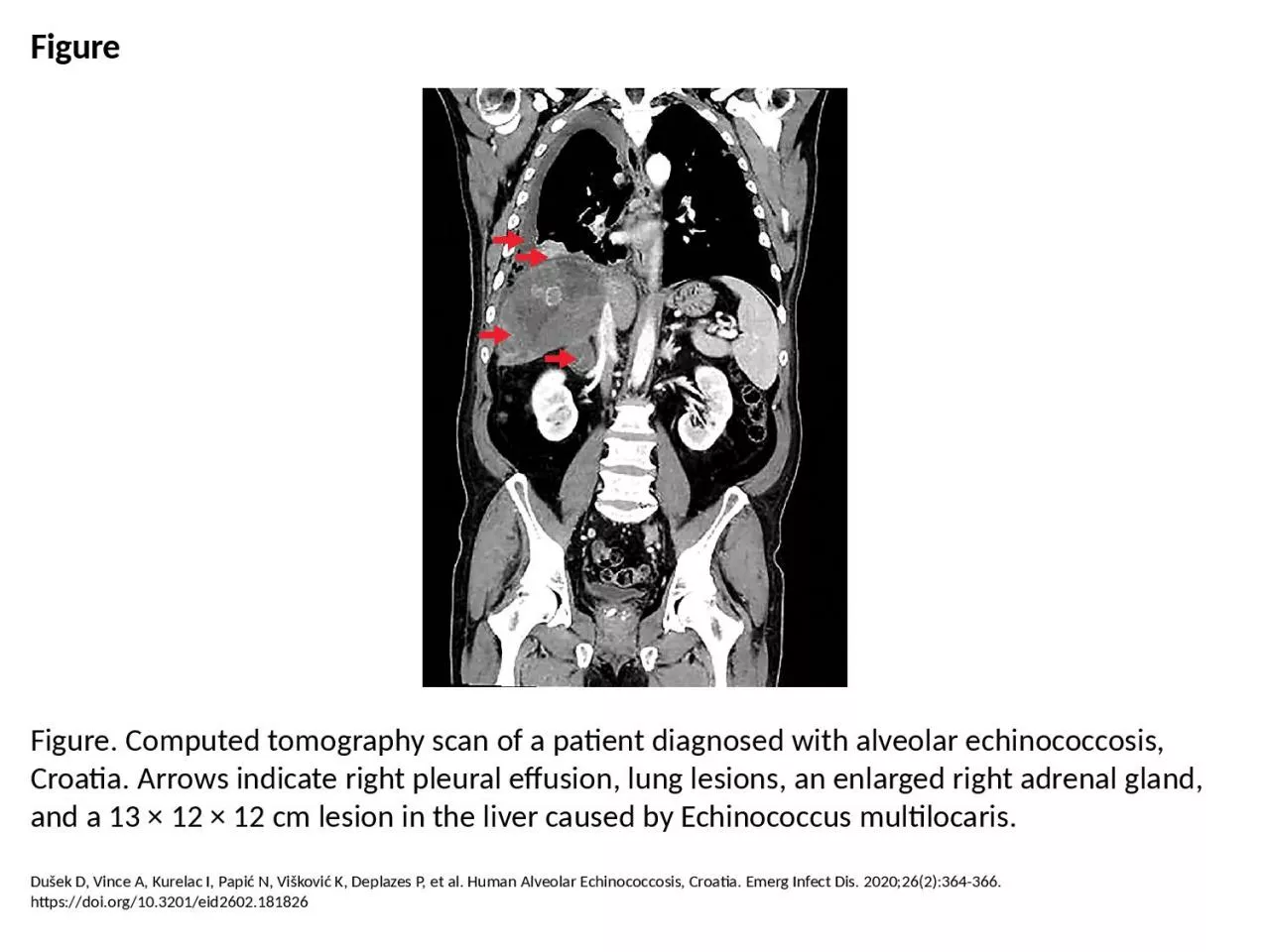
PPT-Figure Figure. Computed tomography scan of a patient diagnosed with alveolar echinococcosis,
Author : freya | Published Date : 2023-11-22
Dušek D Vince A Kurelac I Papić N Višković K Deplazes P et al Human Alveolar Echinococcosis Croatia Emerg Infect Dis 2020262364366 httpsdoiorg103201eid2602181826
Presentation Embed Code
Download Presentation
Download Presentation The PPT/PDF document "Figure Figure. Computed tomography scan ..." is the property of its rightful owner. Permission is granted to download and print the materials on this website for personal, non-commercial use only, and to display it on your personal computer provided you do not modify the materials and that you retain all copyright notices contained in the materials. By downloading content from our website, you accept the terms of this agreement.
Figure Figure. Computed tomography scan of a patient diagnosed with alveolar echinococcosis,: Transcript
Dušek D Vince A Kurelac I Papić N Višković K Deplazes P et al Human Alveolar Echinococcosis Croatia Emerg Infect Dis 2020262364366 httpsdoiorg103201eid2602181826. However perhaps we should broaden our horizons and look back through the centuries to obtain a more complete view of the development of human thought and aspirations which led to the invention of a device without which it would be dif64257cult to im Earls MD FSCCT GB John Mancini MD Koen Nieman MD PhD Gianluca Pontone MD Gilbert L Raff MD FSCCT CoChair University of British Columbia Vancouver Canada University of Texas Southwestern Medical Center Dallas Texas University of Erlangen Erlangen Is the portion of maxilla and mandible that forms and supports the tooth socket (alveoli).. It forms when tooth erupts to provide the osseous attachment to the forming PDL; It disappears gradually after tooth is lost.. echinococcosis: . number and rate per 100 000 population, EU/EEA, 2010–2014. Source: Country reports. Legend: Y = yes, N = no, C = case based, · = no report, ASR: age-standardised rate. Suggested citation: European Centre for Disease Prevention and Control. Annual epidemiological report 2015. . Is the portion of maxilla and mandible that forms and supports the tooth socket (alveoli).. It forms when tooth erupts to provide the osseous attachment to the forming PDL; It disappears gradually after tooth is lost.. This information is provided by Cleveland Clinic and is not intended to replace the medical advice of your doctor or health care provider. Please consult your health care provider for advice about a s Limitations of Radiography. 3D body rendered in 2D. Structures superimposed on image. Structure of interest viewed through underlying / overlying structures. Multiple views used to adequately visualize structure.. DaviD. . sebua. . Elene. . Lortkipanidze. Introduction. History of Radiotherapy. The history of radiotherapy starts from the discovery of X-rays in 1895 by Wilhelm Conrad Rontgen. It was not intended for treatment rather than for imaging.. Physics department. Calculation The Risk of Computed Tomography. Prepared by: Rayan F .Hassan. Fatima A .Mahmud. Supervised by:Msc.Rozhan. Outline. Objective. Introduction. Radiation dose from CT scans. Geometry and historical development. Basic principles. Mathematical principles of CT were first developed in 1917 by Radon. Proved that an image of an unknown object could be produced if one had an infinite number of projections through the object. Tiaoying L, Jiamin Q, Wen Y, Craig PS, Xingwang C, Ning X, et al. Echinococcosis in Tibetan Populations, Western Sichuan Province, China. Emerg Infect Dis. 2005;11(12):1866-1873. https://doi.org/10.3201/eid1112.050079. Pettit AC, Jahangir AA, Wright PW. Mycobacterium doricum Osteomyelitis and Soft Tissue Infection. Emerg Infect Dis. 2011;17(11):2075-2077. https://doi.org/10.3201/eid1711.110460. . EGU 2020. Ilaria Barone. Emanuel. . Kästle. Claudio . Strobbia. Giorgio . Cassiani. realtimeseismic. Università degli Studi di Padova. 2D SURFACE WAVE . tomograpHY. USING ACTIVE SEISMIC . DATA. Tortoli E, Mariottini A, Pierotti P, Simonetti TM, Rossolini G. Mycobacterium yongonense in Pulmonary Disease, Italy. Emerg Infect Dis. 2013;19(11):1902-1904. https://doi.org/10.3201/eid1911.130911.
Download Document
Here is the link to download the presentation.
"Figure Figure. Computed tomography scan of a patient diagnosed with alveolar echinococcosis,"The content belongs to its owner. You may download and print it for personal use, without modification, and keep all copyright notices. By downloading, you agree to these terms.
Related Documents