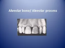
Is the portion of maxilla and mandible that forms and supports the tooth socket alveoli It forms when tooth erupts to provide the osseous attachment to the forming PDL It disappears gradually after tooth is lost ID: 207337
Download Presentation The PPT/PDF document "Alveolar bone/ Alveolar process" is the property of its rightful owner. Permission is granted to download and print the materials on this web site for personal, non-commercial use only, and to display it on your personal computer provided you do not modify the materials and that you retain all copyright notices contained in the materials. By downloading content from our website, you accept the terms of this agreement.
Slide1
Alveolar bone/ Alveolar processSlide2
Is the portion of maxilla and mandible that forms and supports the tooth socket (alveoli).
It forms when tooth erupts to provide the osseous attachment to the forming PDL; It disappears gradually after tooth is lost.Slide3
Development
The alveolar bone begins to first form by an intramembranous ossification with in the
ectomesenchyme
surrounding the developing tooth.
This first formed bone is called as woven bone is less organized and is replaced with more organized lamellar one.Slide4
When a deciduous tooth is shed, its alveolar bone is
resorbed
. The
succedaneous
permanent tooth moves in to place, developing its own alveolar bone from its own dental follicle.
Mandibular
basal bone begins mineralization at the exit of mental nerve from mental foramen.
Maxillary basal bone begins at the exit of
infraorbital
nerve from
infraorbital
foramenSlide5
Parts
Alveolar bone proper
Supporting alveolar bone
- Cortical plates
- Spongy boneSlide6
Composition
Inorganic Organic
(65%) (35%)
Cellular components-
Osteoblast
Osteocytes
Bone lining cells
Osteoclast
Osteoprogenitor Cells Slide7
Periosteum
The tissue covering the outer surface of bone is called
periosteum
.
Consists of two layers-
- inner layer
- outer layer Slide8
Endosteum
Tissue lining the internal bone cavities is called as
endosteum
.
Made up of single layer of
osteoblast
and small amount of connective tissue.Slide9
Fenestration
Isolated areas in which the root is denuded of bone and the root surface is covered only by
periosteum
and overlying gingiva.
In these areas the marginal bone is intactSlide10
Dehiscence
When the denuded areas extend through the marginal bone, the defect is called as dehiscence.Slide11
1. The inactive osteoblasts
are-
Osteocytes
Osteoclasts
Osteoprogenitor
cells
Bone lining cellsSlide12
2. The term lamina dura
refers to the radiographic image of
Periodontal ligament space
Cortical plates
Cancellous
bone
Alveolar crestSlide13
3.Following is true about osteoclast
except
Mononucleated
cells
having branching processes
Derived from circulating blood cells
monocytes
found in
Howship’s
lacunaeSlide14
4. Main type of collagen in bone and ligament are respectively
Type I and type II
(b)Type II and type I
(c) Type II in both
(d) Type I in bothSlide15
5. The enzyme closely associated with the new bone formation is
Acid
phosphatase
(b) Alkaline
phosphatase
(c)
Succinic
dehydrogenase
(d) Both A and BSlide16
THANK YOU