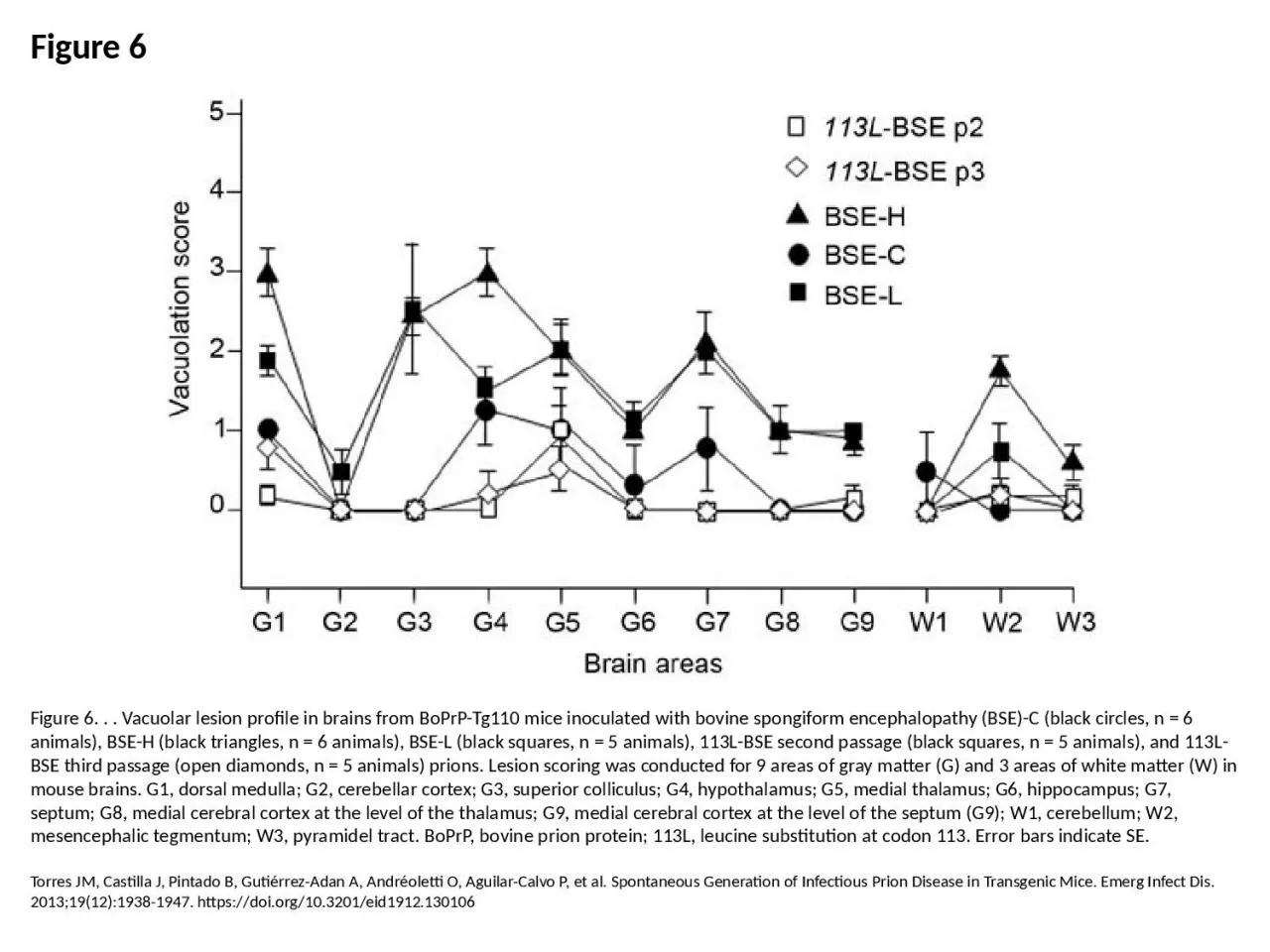
PPT-Figure 6 Figure 6. . . Vacuolar lesion profile in brains from BoPrP-Tg110 mice inoculated
Author : anderson | Published Date : 2023-07-27
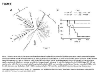
Torres JM Castilla J Pintado B GutiérrezAdan A Andréoletti O AguilarCalvo P et al Spontaneous Generation of Infectious Prion Disease in Transgenic Mice Emerg Infect
Presentation Embed Code
Download Presentation
Download Presentation The PPT/PDF document "Figure 6 Figure 6. . . Vacuolar lesion p..." is the property of its rightful owner. Permission is granted to download and print the materials on this website for personal, non-commercial use only, and to display it on your personal computer provided you do not modify the materials and that you retain all copyright notices contained in the materials. By downloading content from our website, you accept the terms of this agreement.
Figure 6 Figure 6. . . Vacuolar lesion profile in brains from BoPrP-Tg110 mice inoculated: Transcript
Download Rules Of Document
"Figure 6 Figure 6. . . Vacuolar lesion profile in brains from BoPrP-Tg110 mice inoculated"The content belongs to its owner. You may download and print it for personal use, without modification, and keep all copyright notices. By downloading, you agree to these terms.
Related Documents