/
Motor  neuronal   tracts Motor  neuronal   tracts

Motor neuronal tracts - PowerPoint Presentation

anderson
anderson . @anderson
Follow
355 views
Uploaded On 2022-06-14

Motor neuronal tracts - PPT Presentation

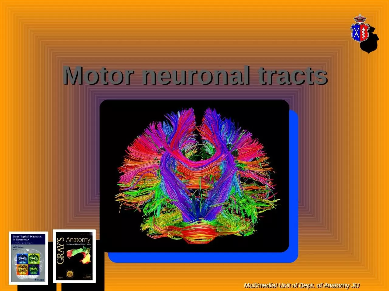
Multimedial Unit of Dept of Anatomy JU A nerve tract is a bundle of nerve fibers axons connecting nuclei of the central nervous system in the peripheral nervous system this is known as a nerve fascicle ID: 917612

tracts motor cortex neurons motor tracts neurons cortex pyramidal tract nerve system fibers spinal cord upper cerebral called connect

Share:

Link:

Embed:

Download Presentation from below link

Download Presentation The PPT/PDF document "Motor neuronal tracts" is the property of its rightful owner. Permission is granted to download and print the materials on this web site for personal, non-commercial use only, and to display it on your personal computer provided you do not modify the materials and that you retain all copyright notices contained in the materials. By downloading content from our website, you accept the terms of this agreement.


Presentation Transcript

Slide1

Motor

neuronal tracts

Multimedial

Unit of Dept. of Anatomy JU

Slide2

A nerve tract is a bundle of nerve fibers (axons) connecting nuclei of the central nervous system

(in the peripheral nervous system this is known as a nerve fascicle)

Slide3

The main nerve tracts in the central nervous system are

of three types:association fiberscommissural fibersprojection fibersCaution: a tract may be referred to as commissure, fasciculus, or

decussation.

Slide4

Association fibers

The tracts that connect cortical areas within the same hemisphere are called association tracts. Long association fibers connect different lobes of a hemisphere to each other whereas short association fibers connect different gyri within a single lobe. Among their roles, association tracts link perceptual and memory centers of the brain.

Slide5

Commissural fibers

Commissural tracts connect corresponding cortical areas in the two hemispheres. They cross from one cerebral hemisphere to the other through bridges called commissures. The great majority of commissural tracts pass through the corpus callosum. A few tracts pass through the much smaller anterior and posterior commissures. Commissural tracts enable the left and right sides of the cerebrum to communicate with each other.

Slide6

Projection

fibersProjection tracts connect the cerebral cortex with the corpus striatum, diencephalon, brainstem and the spinal cord. The corticospinal tract for example, carries motor signals from the cerebrum to the spinal cord. Other projection tracts carry signals upward to the cerebral cortex. Superior to the brainstem, such tracts form a broad, dense sheet called the internal capsule between the thalamus and basal nuclei, then radiate in a diverging, fanlike array to specific areas of the cortex.

Slide7

Projection fibers are of two main types:

afferent (sensory)efferent (motor)

Slide8

Motor tracts are divisible into:

pyramidalextrapyramidal

Slide9

Pyramidal neurons (pyramidal cells) are a type

of multipolar neuron found in areas of the brain including the cerebral cortex, the hippocampus, and the amygdala. Pyramidal neurons are the primary excitation units of the mammalian prefrontal cortex and the corticospinal tract. Pyramidal neurons are also one of two cell types where the characteristic sign, Negri bodies, are found in post-mortem rabies infection. Pyramidal neurons were first discovered and studied by Santiago Ramón y Cajal. Since then, studies on pyramidal neurons have focused on topics ranging from neuroplasticity to cognition

Slide10

Upper motor neurons are motor neurons that originate either in the motor region of the cerebral cortex or in the brain stem and carry motor information down to the lower motor neurons. The main

effector or motor neurons (efferent neurons) for voluntary movement lie within layer V of the primary motor cortex, and are a type of giant pyramidal cell called Betz cells. The cell bodies of these neurons are the largest in the brain, approaching nearly 0.1mm in diameter.

Slide11

The primary motor cortex or precentral gyrus, is one of the most important areas in the frontal lobe. The precentral gyrus is the most posterior gyrus of the frontal lobe and it lies anterior to the central sulcus. The pyramidal cells of the precentral gyrus are also called upper motor neurons. The fibers of the upper motor neurons project out of the precentral gyrus ending in the brainstem, where they will decussate (intersect) within the lower medulla oblongata to form the lateral corticospinal tract on each side of the spinal cord. The fibers that do not decussate will pass through the medulla and continue on to form the anterior corticospinal tracts. The upper motor neuron descends in the spinal cord to the level of the appropriate spinal nerve root. At this point, the upper motor neuron synapses with the lower motor neuron, each of whose axons innervate a fiber

of skeletal muscle.

Slide12

These neurons connect the brain to the appropriate level in the spinal cord, from which point nerve signals continue to the muscles by means of the lower motor neurons.

The neurotransmitter glutamate transmits the nerve impulses from upper to lower motor neurons, where it is detected by glutamatergic receptors.

Slide13

Pyramidal tracts are mainly consisted of:

corticospinalcorticobulbar

Slide14

Corticospinal and corticobulbar tracts.

Slide15

Primary motor area/precentral gyrus (area 4),

premotor cortex (area 6), and prefrontal eye field (area 8).

Slide16

Course of the pyramidal tract, upper portion: the corona

radiata and internal capsule

Slide17

Microarchitecture

of the motor cortex (Golgi stain).

Slide18

Course of the pyramidal tract

Slide19

In anatomy, the

extrapyramidal system is a biological neural network that is part of the motor system causing involuntary actions. The system is called "extrapyramidal" to distinguish it from the tracts of the motor cortex that reach their targets by traveling through the "pyramids" of the medulla. The pyramidal pathways (corticospinal and some corticobulbar tracts) may directly innervate motor neurons of the spinal cord or brainstem (anterior (ventral) horn cells or certain cranial nerve nuclei), whereas the extrapyramidal system centers on the modulation and regulation (indirect control) of anterior (ventral) horn cells.

Slide20

Extrapyramidal

tracts are chiefly found in the reticular formation of the pons and medulla, and target lower motor neurons in the spinal cord that are involved in reflexes, locomotion, complex movements, and postural control. These tracts are in turn modulated by various parts of the central nervous system, including the nigrostriatal pathway, the basal ganglia, the cerebellum, the vestibular nuclei, and different sensory areas of the cerebral cortex. All of these regulatory components can be considered part of the extrapyramidal system, in that they modulate motor activity without directly innervating motor neurons.

Slide21

The

vestibulospinal tract is comprised of a lateral and medial pathway. The function of these tracts is to maintain equilibratory reflexes from the input of the vestibular apparatus. They will reach the axial muscles, i.e. intercostal and back muscles, as well as the extensors of the limbs.The reticulospinal tract is responsible primarily for locomotion and postural control.The tectospinal tract (also known as colliculospinal tract) is a nerve pathway that coordinates head and eye movements

Slide22

Brain structures involved in motor function and

the descending tracts that originate in them

Slide23

Synapses of the descending motor tracts onto anterior horn neurons

.

Slide24

Thank you very much