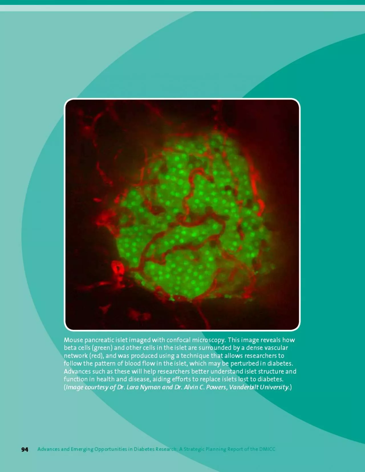
/
Mouse pancreatic islet imaged with confocal microscopy This image rev Mouse pancreatic islet imaged with confocal microscopy This image rev

Mouse pancreatic islet imaged with confocal microscopy This image rev - PDF document

anderson
anderson . @anderson
Follow
342 views
Uploaded On 2022-08-16

Mouse pancreatic islet imaged with confocal microscopy This image rev - PPT Presentation

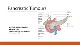
THE BETA CELL Adult Mouse Cells Reprogrammed To Become InsulinProducing Cells Demonstration that Beta Cell Mass Is Dynamically Regulated Insulin As Beta Cell Growth Factor and Regulator of Islet Fun ID: 937634

cell x0003 diabetes beta x0003 cell beta diabetes cells islet insulin type function human islets wkh research imaging x000f

Share:

Link:

Embed:

Download Presentation from below link

Download Pdf The PPT/PDF document "Mouse pancreatic islet imaged with confo..." is the property of its rightful owner. Permission is granted to download and print the materials on this web site for personal, non-commercial use only, and to display it on your personal computer provided you do not modify the materials and that you retain all copyright notices contained in the materials. By downloading content from our website, you accept the terms of this agreement.


Presentation Transcript

Mouse pancreatic islet imaged with confocal microscopy. This image reveals how beta cells (green) and other cells in the islet are surrounded by a dense vascular follow the pattern of blood flow in the islet, which may be perturbed in diabetes. Advances such as these will help researchers better understand islet structure and function in health and disease, aiding efforts to replace islets lost to diabetes. Advances and Emerging Opportunities in Diabetes Research: A Strategic Planning Report of the DMICC THE BETA CELL Adult Mouse Cells Reprogrammed To Become Insulin-Producing Cells Demonstration that Beta Cell Mass Is Dynamically Regulated Insulin As Beta Cell Growth Factor and Regulator of Islet Function Endoplasmic Reticulum Stress Signaling in Pancreatic Beta Cells Identification of Gene Mutations in Rare Forms of Diabetes Provides New Insights Multi-Pronged Approach to Preservation of Beta Cell Function in Human Diabetes Infrastructure Creation To Improve Human Islet Isolation and Promote Basic Islet Research contents: 95  INTRODUCTION Beta cells, which are found in the pancreas within tiny cell clusters called islets, are the body’s sole source of the essential hormone insulin. Diabetes is characterized by the body’s inability to produce and/or respond appropriately to insulin, and results in the inability of the body to absorb and use glucose as cellular fuel. These defects result in persistent elevation of blood glucose levels and other metabolic abnormalities, which, in turn, lead to the development of disease complications. The most common forms of diabetes are type diabetes, in which the immune system launches misguided attack, destroying the beta cells of the pancreas, and type diabetes, in which the body becomes resistant to insulin signaling, with subsequent impaired insulin production. While the causes of beta cell loss or failure differ, all major forms of diabetes share common bond in the pancreatic beta cell. This chapter highlights basic and clinical science focused on the beta cell that will allow the development of strategies to prevent, treat, and cure diabetes. More broadly, an understanding of integrated islet physiology at the cellular and biochemical level could allow researchers to develop means of increasing insulin secretion and enhancing beta cell mass. Determining the molecular mechanisms underlying beta cell dysfunction and failure could identify new targets for pharmacological intervention in diabetes. Efforts to optimize islet transplantation sites and improve islet survival are needed to overcome some of the obstacles that impede the widespread implementation of islet transplantation as treatment for diabetes. Advances in basic stem cell biology and regenerative medicine will need to be harnessed if the promise of new cellular therapies for type and type diabetes is to be realized. The development of novel methods to accurately assess the post-transplant islet mass will be necessary not only to monitor long-term changes at the transplant site, but also to assess the effectiveness of future therapeutic strategies directed at replacing and regenerating beta cells. Generation of Insulin-Producing Cells from Human Embryonic Stem Cells: Taking cues from developmental biology, it has been possible to obtain insulin-producing cells using a step-wis

e protocol to direct the differentiation of human embryonic stem (ES) cells*. The process was designed to mimic how the pancreas forms during fetal development by directing cells through VWDJHVUHVHPEOLQJWKLVSURFHVV LHGHÀQLWLYHHQGRGHUPgut-tube endoderm, pancreatic endoderm, and endocrine precursors). Some of the human ES cell-derived insulin-producing cells have insulin content approaching that of adult beta cells. Similar to fetal beta cells, these cells release C-peptide in response to multiple secretory stimuli; however—unlike the adult beta cells they need to *The NIH supports research using human embryonic stem cells within the NIH Guidelines for Human Stem Cell Research. 96 Advances and Emerging Opportunities in Diabetes Research: A Strategic Planning Report of the DMICC      replace—they are not very responsive to glucose. Although these cells do not yet display regulated insulin secretion, QRULVWKHSURFHVVWRSURGXFHWKHPKLJKO\HIÀFLHQWWKLVmajor achievement provides proof-of-principle that it is possible to recapitulate, in vitro, the steps leading to WKHSURGXFWLRQRILQVXOLQSURGXFLQJFHOOV³DVLJQLÀFDQWleap forward toward the goal of developing beta cell replacement therapies to cure type 1 or severe type 2 diabetes. Induced Pluripotent Stem Cells: In addition to ES cells, there is now evidence that induced pluripotent stem (iPS) cells can be coaxed to differentiate into insulin-producing cells. Although such cells share the main shortcomings of ES cell-derived insulin-producing cells, their derivation raises the possibility of obtaining immuno­FRPSDWLEOHRUHYHQSDWLHQWVSHFLÀFLQVXOLQSURGXFLQJFHOOVAdult Mouse Cells Reprogrammed To Become Insulin-Producing Cells: Research performed in diabetic mice has shown that introducing expression of MXVWWKUHHJHQHVLVVXIÀFLHQWWRUHSURJUDPQRQLQVXOLQproducing adult pancreatic cells (and potentially other cell types) into beta-cell-like insulin-producing cells. The reprogrammed cells lowered blood glucose in diabetic animals—important progress towards harnessing regenerative medicine to treat diabetes. The reprogrammed cells were shown to be stable and effective for the life of the mice. A common technique, called viral gene transfer, was used to introduce the three genes into mouse cells. Future research that could lead to possible applications of this technology to people with diabetes includes achieving similar results without using a virus to carry the genes, testing the approach in oth

er cell types, DQGUHSOLFDWLQJWKHÀQGLQJVLQKXPDQFHOOV:KLOHPXFKwork remains to be done before this becomes a safe and effective therapy, the principle of adult cell reprogramming is a major step forward and could serve as a model for other applications of regenerative medicine. Stem cells can differentiate into a number of different cell types, a characteristic that may allow researchers to develop therapies for diabetes to replace destroyed, damaged, or malfunctioning beta cells and islets. (Image credit: Donald Bliss, Medical Arts and Photography Branch, NIH.) Demonstration that Beta Cell Mass Is Dynamically Regulated: Surveys of human cadavers have shown that pancreata from people with type 1 diabetes exhibit a profound loss of beta cells and have low insulin levels, while the loss of beta cells in people with type 2 diabetes is less severe. However, in type 1 diabetes, beta cell depletion is often not absolute, and scattered insulin-immunoreactive cells may often be observed even after many years of disease. Studies in people with type 2 diabetes have also shown an intriguing correlation between duration of diabetes and decline of beta cell mass. Animal studies have shown that the progression from insulin resistance to type 2 diabetes is associated with an initial dynamic increase in beta cell mass to ��   accommodate the metabolic demand for insulin, followed by progressive loss of beta cell mass. Similarly, animal studies of how changes in beta cell mass are regulated during pregnancy to meet increased insulin demands have rendered new insights that could help explain gestational GLDEHWHV2QHRIWKHGLIÀFXOWLHVLQVWXG\LQJEHWDFHOOregeneration has been the lack of robust, synchronized animal system that would allow the controlled destruction of beta cells and study of subsequent cell proliferation in the adult pancreas. Several new transgenic mouse models have been developed that permit the study of the dynamics of beta cell regeneration from diabetic state. Lineage tracing analyses have indicated that enhanced proliferation of surviving beta cells, or perhaps other pancreatic lineages under extreme conditions of tissue damage, contributes to beta cell regeneration. These advances have enabled investigators to measure changes in beta cell proliferation and survival as function of disease progression, leading to the hypothesis that diabetes is primarily the result of impaired beta cell mass, and that the seemingly irreversible course of the disease DQGLWVJURZLQJUHIUDFWRULQHVVWRLQWHUYHQWLRQVUHÁHFWGHÀFLWLQWKHQXPEHURIIXQFWLRQLQJEHWDFHOOVAdvances in Whole Islet Biology: Beta cells do not function in isolation. They are embedded in the pancreatic islets, which include several different endocrine

cell types, blood vessels, and nerve endings. While it has been known for decades that hormone products of different endocrine cells have profound effects on neighboring islet cells (paracrine effects), the extent of regulation among islet FHOOW\SHVWKHVSHFLÀFUROHVRIQHXURWUDQVPLWWHUVDQGWKHmolecular underpinnings of these interactions have only started to come to light in the last decade. Finally, a new cell type has been discovered in the pancreatic islet—the ghrelin-producing cell, which is thought to exert both autocrine (self) and paracrine effects. The ghrelin receptor (GHsr) is present not only on ghrelin-producing cells, but also on beta cells and the glucagon-producing alpha cells. A better understanding of these complex intra-islet paracrine and autocrine regulatory roles could lead to novel therapeutic strategies to improve regulated insulin secretion and/or beta cell mass in type 2 diabetes. Insulin As Beta Cell Growth Factor and Regulator of Islet Function: While the paracrine effects of other endocrine cell types on the beta cell are fairly well-documented, the autocrine effect of insulin on beta cell function remains a matter of debate. Historically, it has been suggested that insulin exerts a negative effect on beta cells, but recent data provide evidence for a positive role of insulin on beta cell function and survival. Moreover, insulin signaling, once thought to be exclusively important in peripheral target cells, has now been shown to be critical to maintain beta cell mass, and for compensatory islet growth in insulin-resistant states. This new understanding of the role of insulin will help investigators in efforts to replenish and maintain beta cells in the islet. Extracellular Matrix and Cell-Cell Interaction: In humans and other vertebrates, the endocrine pancreas has developed into a complex network of cells. Signals that diffuse through the intercellular space of the islets interplay with signaling cascades that depend on membrane proteins and are concentrated at points of cell contact. These mechanisms mediate both indirect (e.g., neurotransmitter-, hormone-, ion-, nucleotide-mediated) and direct (e.g., cell adhesion molecule-, integrin-, receptor-, and junction-mediated) islet cell-to-cell communication. These cell-to-cell interactions combine to ensure key functions and properties of the pancreatic islet, such as synchronized insulin secretion in response to glucose, or maintenance of islet size and architecture. In UHFHQWDGYDQFHVLQYHVWLJDWRUVKDYHLGHQWLÀHGLQGLYLGXDOcell surface proteins involved in these integrated responses. These observations raise the exciting prospect OO98 Advances and Emerging Opportunities in Diabetes Research: A Strategic Planning Report of the DMICC ��    &#x

0003;   that proper expression of these proteins may help foster the development of novel cell sources that would retain, at least partially, the ability to respond properly to acute glucose stimulation—a function which depends on proper cell-to-cell communication. Practical applications that can be explored using this knowledge include improved islet transplantation protocols, novel strategies for islet repair or remodeling, pancreatic tissue engineering using stem or progenitor cells, and the design of three-dimensional VFDIIROGVIRUWKHGHYHORSPHQWRIDELRDUWLÀFLDOendocrine pancreas. Vascular Endothelium and Islet Function: The pancreatic islets are one of the most vascularized tissues in the body, a characteristic that could inform efforts to restore islet function. Recent studies have demonstrated that islet endothelial cells that line the internal walls of capillaries feeding the islet—the microendothelium—are not only involved in the delivery of oxygen and nutrients to endocrine cells, but also induce insulin gene expression during islet development, affect adult beta cell function, promote beta cell proliferation, and produce a number of factors promoting blood vessel dilation/constriction and new growth, including vascular endothelial growth factor A (VEGF-A) and human growth factor (HGF). These islet endothelial cells also play a critical role in the early phase of type 1 diabetes by increasing the expression of surface leukocyte-homing receptors, thereby enabling immune cells to enter the endocrine tissue and cause EHWDFHOOGHVWUXFWLRQ7KHVHÀQGLQJVDUHLPSRUWDQWQRWonly for understanding islet biology, but also for islet transplantation efforts, as the ability of transplanted islets to revascularize with host vascular elements may be of great importance for islet graft function and survival. Moreover, it has recently been shown that residual intra­islet endothelial cells in islets processed for transplant may also participate in revascularization of pancreatic islets subsequent to transplantation. Preservation of intra-islet endothelial cell mass may improve long-term graft function by masking foreign antigens. Together, these advances suggest an important role of the islet microendothelium in normal islet physiology and its possible involvement in type 1 and 2 diabetes pathogenesis, as well as in islet revascularization in transplantation settings. Endocrine/Exocrine Cell-Cell Communication: The pancreas hosts two major tissue types, endocrine tissue (the islets) that secretes insulin, glucagon, and other hormones, and exocrine tissue that secretes digestive enzymes. The endocrine and exocrine tissues of the pancreas have traditionally been considered to be two separate entities, structurally and functionally. However, recent observations suggest that the pancreas is a functionally integrated organ in which endocrine and exocrine glands are structurally interconnected, with a continuous matrix supporting the microcirculation, and ductal function allowing for a well-orchestrated functional response. In rodent models and in humans with type 2 diabetes, a widening of the islet/exocrine interface has been observed as a result of a loss of the interstitial matr

ix and is DVVRFLDWHGZLWKLQÁDPPDWRU\FHOOLQÀOWUDWLRQDQGÀEURVLVin the islet/exocrine interface. This damage is thought to lead to a loss of cellular paracrine communication that may disrupt signals between the endocrine and exocrine pancreas and gut (i.e., inducing a dysfunctional insulin-acinar-ductal-incretin gut hormone axis), potentially H[SODLQLQJWKHSDQFUHDWLFLQVXIÀFLHQF\DQGGHFUHDVHLQlevels of glucagon-like peptide-1 (GLP-1)—a gut hormone WKDWLQÁXHQFHVLVOHWIXQFWLRQDQGEHWDFHOOPDVV³NQRZQto exist in at least some individuals with pre-diabetes and overt type 2 diabetes. Cellular Nutrient Sensing and the Beta Cell: QXPEHURIVFLHQWLÀFDGYDQFHVKDYHFRQWULEXWHGWRWKHunderstanding of the complex signaling pathways that are essential to beta cell function. Transgenic mouse models of type diabetes combined with detailed biochemical analyses have revealed that the cellular response to nutrients is mediated by the concerted action of variety of key signaling pathways. These critical pathways include the adenosine monophosphate (AMP)-activated protein kinase (AMPK) pathway, which responds to changes in energy charge; the mammalian target of rapamycin (mTOR) pathway, which responds primarily to amino acids; the hexosamine pathway, which is responsible for the synthesis of O-linked N-acetylglucosamine, and which responds to amino acids, glucose, and products of fat metabolism; and pathways controlled by the sirtuins, class of deacetylases that is dependent on nicotinamide adenine dinucleotide (NAD). In turn, these essential pathways interact with, and serve to modulate, homeostatic mechanisms such as insulin signaling, and the transforming growth factor-beta (TGF-beta) and mitogen-activated protein kinase (MAP kinase) signaling cascades. The principal job of the beta cell is to orchestrate the endocrine response to nutrients through these complex, interconnected signaling pathways. Deregulation of these pathways could lead to the beta cell pathology associated with diabetes. Endoplasmic Reticulum Stress Signaling in Pancreatic Beta Cells: The endoplasmic reticulum (ER) is cellular compartment specialized for folding DQGPRGLÀFDWLRQRIQDVFHQWSURWHLQV'LVUXSWLRQRI(5homeostasis activates the unfolded protein response (UPR). In pancreatic beta cells, glucose-regulated insulin production requires an intact UPR. When the UPR in the beta cell is dysfunctional, ER stress ensues, and beta cells undergo cell death, or apoptosis. Genetic and biochemical evidence in humans and mice support critical role for the UPR in preserving ER homeostasis and in preventing beta cell failure, mechanisms that are fundamental in the etiology of diabetes. Identification of Gene Mutations in Rare Forms of Diabetes Provides New Insights into the Beta Cell: The rare syndrome of permanent neonatal diabetes mellitus (PNDM) has provided an opportunity to identify genes that regulate beta cell development and function. It was found that, in subset of affected children, there are missense mutations in the insulin gene that in turn lead to the production of misfolded proinsulin molecules. These mutant proteins are thought to affect beta cell function by in

terfering with the processing of proinsulin to generate insulin and causing ER stress. While the relevance of these observations to more common forms of the disease is at this point unclear, other studies in experimental animals and transformed cell lines have shown that ER stress, the UPR, autophagy, apoptosis, premature senescence, and germane cellular biological abnormalities play important roles in beta cell failure in general—suggesting that the mechanistic foundation for these phenomena should be investigated in greater detail in vivo Still other, more common forms of neonatal diabetes have been attributed to mutations in genes encoding the two protein subunits of potassium ion channel that regulates insulin secretion; the mutations prevent the normal release of insulin from pancreatic beta cells (see the “Genetic Basis of Type Diabetes, Type Diabetes, Obesity, and Their Complications” chapter). 7KLVLQVLJKWLQWREHWDFHOOIXQFWLRQKDVDOVRKDGWKHEHQHÀWof yielding genetic tests that can be used to identify people who have these mutations, many of whom can manage their diabetes with the orally administered drug sulfonylurea—a less burdensome therapy than insulin therapy. Multi-Pronged Approach to Preservation of Beta Cell Function in Human Diabetes: Several recent studies have shown that type diabetes is associated with progressive decrease of functional beta cell mass. WW100 Advances and Emerging Opportunities in Diabetes Research: A Strategic Planning Report of the DMICC ��     Human and animal studies have also suggested that sulfonylureas, the main class of drugs used to treat beta cell dysfunction, may actually precipitate long-term beta cell failure. Recent laboratory discoveries and early clinical studies suggest that preservation or recovery of endogenous insulin secretion may be improved by the use of new (alternate) drugs or new, physiology-based, WUHDWPHQWUHJLPHQV,GHQWLÀFDWLRQRIJHQHWLFYDULDQWVassociated with beta cell abnormalities has been achieved by genome-wide association (GWA) studies in type 2 diabetes. In addition, rare, monogenic forms of non-autoimmune diabetes (such as PNDM and maturity onset diabetes of the young (MODY)) may provide insights into factors involved in failure of insulin secretion and help to explain why the loss of beta cell function described in epidemiologic studies is so variable. Limited studies investigating the striking improvements in glycemia some people with diabetes experience after bariatric surgery have highlighted the role of gastrointestinal (GI) hormones in modulating and sustaining insulin secretion. Drugs that act through a variety of mechanisms— including

insulin sensitizers, such as metformin and thiazolidinediones; the incretins, such as GLP-1; and inhibitors of dipeptidyl peptidase 4 (DPP-4), which prolong action of GI incretins—provide opportunities for new treatment approaches that can enhance and potentially sustain endogenous insulin secretion in people with type 2 diabetes. Technological advances, such as development of continuous glucose monitoring and experimental semi-closed-loop systems to achieve and maintain normoglycemia with less risk of severe hypoglycemia, will enable treatment regimens that may reduce damage to endogenous beta cells and may enhance survival of transplanted islets by diminishing glucotoxicity. The Beta Cell Biology Consortium (BCBC): The BCBC is a large international team of investigators whose research has focused on understanding pancreatic GHYHORSPHQWDOELRORJ\6SHFLÀFDOO\JHQHVUHVSRQVLEOHfor the establishment of the different pancreatic lineages KDYHEHHQLGHQWLÀHGDQGWKHLQWHJUDWHGFDVFDGHRIinteractions leading to the formation of the adult organ decoded. This knowledge has spawned studies of pancreatic beta cell regeneration, cell plasticity, and reprogramming. It has also laid the foundation for the successful generation of insulin-producing cells from human ES cells, and for the integration of complex signaling pathways in the pathogenesis of beta cell dysfunction. The BCBC is engineering beta cell ablation PRGHOVLQWRLPPXQRGHÀFLHQWPRXVHVWUDLQVWRDOORZWKHassessment of human stem/progenitors in correcting diabetes. The long-term goal is to generate mouse models that will support the development of a human immune system so that human beta cell function can be evaluated in the context of human immunity and autoimmunity. The BCBC has generated many tools and reagents for the diabetes community, including new monoclonal antibodies that recognize human islet cells, and new transgenic mouse models that are being used to understand beta cell development and regeneration. Infrastructure Creation To Improve Human Islet Isolation and Promote Basic Islet Research: For decades, most research concerning the factors that determine pancreatic beta cell mass has been derived from rodent animal models and/or cell lines. Although informative, more recent investigations have shown that human islets differ from their rodent counterparts with respect to the regulatory and metabolic milieu that affects their susceptibility to injury and their adaptive response for replication. It is now clear that the human islet is without a convenient laboratory surrogate for such studies, and evidence-based studies obtained from rodents may not extrapolate to the human. To facilitate basic research using human islets, the NIH established the National Islet Cell Resource Centers Consortium (ICRs),    

  which successfully prepared and distributed cadaveric human islets for fundamental research, using optimized shipping conditions that sustain viability, to more than 150 investigators in North America. The ICRs have been succeeded by the Integrated Islet Distribution Program (IIDP), a contract-supported resource that acquires human islets from subcontracted islet isolation centers and distributes them to approved investigators to facilitate basic research on human islets. Procurement, processing, and testing of the pancreas are costly. It has been critical to have human islets available at a greatly subsidized cost to assure the clinical relevance of fundamental research. Human islets like these are isolated for research or for transplantation into patients. (Image courtesy of Dr. Camillo Ricordi, University of Miami, Diabetes Research Institute.) PET Imaging Agents Target the Pancreatic Beta Cell: 11 C-DTBZ ( 11 C-dihydrotetrabenazine) is an imaging agent developed for positron emission tomography (PET) imaging of the dopaminergic neurons of the brain. Its target, the Vesicular Monoamine 7UDQVSRUWHU 90$7 SURWHLQZDVLGHQWLÀHGLQJHQH array screens of islet cells. The imaging agent binds VSHFLÀFDOO\WREHWDDQGSDQFUHDWLFSRO\SHSWLGHSURGXFLQJ (PP) cells of the islet and has been used to visualize these cells in the human pancreas in healthy people and in people with diabetes. Currently, researchers are working to modify the molecule in order to improve its imaging and binding characteristics to the point where it can be reliably used to monitor beta cell mass in people. Other research is RQJRLQJWRGHWHUPLQHWKHVSHFLÀFORFDWLRQDQGH[SUHVVLRQ of its molecular target in the pancreas. Additional highly promising imaging agents are being developed that target markers enriched in the beta cell, such as the GLP-1 receptor. Development of the imaging agent 11 C-DTBZ has thus spurred noninvasive studies of beta cells. Magnetic Resonance Imaging (MRI) Agents Hold Promise for Imaging Transplanted Islets: The current practice of transplanting islets into the liver of people with diabetes presents both challenges and opportunities for imaging. The liver takes up many RIWKHPROHFXODULPDJLQJDJHQWVLQDQRQVSHFLÀFZD\ and therefore tends to have a high background signal in most experiments. Considerable progress has been made by either labeling the islets themselves with iron-based contrast agents prior to transplantation, or by encapsulating the isolated islets in immunoprotective coatings that contain iron-or gadolinium-based contrast agents. Signals with these methods persist long after transplantation in rodent, porcine, and primate models, and correlate very well with islet survival. Human trials are under way using this approach. Ability To ImageIslet Inflammation In Vivo The presence of islet autoantibodies, and perhaps metabolic changes as detected in the circulation, are the current standards to measure islet autoimmunity, but are an LQGLUHFWPHDVXUH,Q

;SUHOLPLQDU\H[SHULPHQWVWKHVSHFLÀF T cell populations that cause insulitis have been directly visualized using molecular imaging approaches, but the most promising and least invasive approach is to take advantage of the vasculature “leakiness” that develops GXULQJLQÁDPPDWLRQ/DUJHLURQEDVHG05,FRQWUDVW agents tend to remain in the bloodstream except in sites of 102 Advances and Emerging Opportunities in Diabetes Research: A Strategic Planning Report of the DMICC  compromised vasculature, and a persistent signal in the monitored in type 1 diabetes mouse models and in people SDQFUHDVGXHWRLVOHWLQÁDPPDWLRQKDVEHHQVXFFHVVIXOO\recently diagnosed with the disease. AN INSIDE LOOK AT BETA CELLS Seeing is believing. Imaging scientists are ZRUNLQJWRÀQGZD\VWRYLVXDOL]HWKHSURFHVVHVthat lead to diabetes and how the body responds to therapy. These new tools will further a better understanding about how the disease starts and Imaging techniques will provide insights into why, how, and when diabetes occurs, as well as point to new ways for treating the disease. The secret to imaging diabetes is the use of drug-like imaging agents that selectively “light up” the cells or biological processes involved in disease. For instance, the metals iron and gadolinium change the signal in magnetic resonance imaging Compounds that contain these metals FDQEHGHVLJQHGWRKRPHLQVSHFLÀFDOO\RQWKHinsulin-producing beta cells in the pancreas, thereby permitting them to be counted. compounds have been used to light up the LQÁDPPDWLRQLQWKHSDQFUHDVWKDWDFFRPSDQLHVthe autoimmune destruction of the beta cells and causes type 1 diabetes. Other imaging agents mimic nutrients or hormones and, when taken up by cells, reveal clues to their function and These types of agents are commonly labeled with minute levels of radioactivity and detected by positron emission tomography (PET). Thus, they might allow researchers to distinguish among active and distressed beta cells. considerable effort is focused on putting imaging labels on the isolated pancreatic islets used for transplantation into people with diabetes. approach would enable doctors to actually watch the locations to which the transplanted tissues migrate once they are infused into people and to determine their fate—that is, to know how PDQ\VXUYLYHWRSURGXFHLQVXOLQÀQGRXWwhether they grow in their new environment, and see what happens to those that die. might also disclose the formation of new blood vessels and nerves around the islets, as well as reveal the importance of these processes for insulin secretion. Scientists have learned to incorporate into mice a family of proteins that either emit light (such DVWKHOXFLIHUDVHOXFLIHULQV\VWHPIURPWKHÀUHÁ\ RUÁXRUHVFH VXFKDVJUHHQÁXRUHVFHQWSURWHLQ These constitute a very powerful set of imaging tools that are used in basic animal research. )RU

;LQVWDQFHÁXRUHVFHQWO\ODEHOHGLQVXOLQFDQbe tracked by the microscope to uncover defects in insulin secretion that might be involved in Fluorescently labeled beta cells are making possible novel studies of islet biology, VXFKDVYLHZLQJLVOHWEORRGÁRZLQOLYHDQLPDOV³an approach which could yield new information The Beta Cell 103 about the dynamic relationships between cells in the islet. From identifying and monitoring precursor cells that become new insulin-producing beta cells, to assessing cellular activities important to islet function, it is hoped that these imaging tools will help researchers better understand the development and function of beta cells and islets. Imaging may also one day help people to manage their diabetes or be used to identify individuals prone to diabetic complications before they become clinically obvious. New, noninvasive to detect and monitor a variety of metabolic problems associated with diabetes may emerge. For instance, new glucose-sensitive imaging agents may make possible the continuous PRQLWRULQJRISODVPDJOXFRVHZLWKRXWÀQJHUSuch an advance would be enormously EHQHÀFLDOIRUSHRSOHZLWKGLDEHWHVscientists are working to bring emerging imaging tools to bear on all aspects of diabetes and its treatment. 104 Advances and Emerging Opportunities in Diabetes Research: A Strategic Planning Report of the DMICC   KEY QUESTIONS The 1999 report of the congressionally-established Diabetes Research Working Group (DRWG), Conquering Diabetes: A Strategic Plan for the 21st Century, highlighted research on the beta cell and islet replacement as key opportunities for advancing treatment of type 1 diabetes. Since that time, new discoveries have underscored the importance of beta cell loss in onset and progression of type 2 diabetes as well. The establishment of centers for islet transplantation research and of a national system for islet distribution, the creation of a collaborative group of beta cell biology research centers, and expansion of fundamental studies of beta cells and islets—all recommended steps from the DRWG’s roadmap for diabetes research, reinforced by subsequent planning efforts—have accelerated UHVHDUFKSURJUHVVLQDZD\WKDWZLOOEHQHÀWSUHYHQWLRQ and therapy for the majority of people with or at risk of developing diabetes. The great promise of beta cell and islet replacement and regeneration, as well as the potential to protect and preserve a person’s own beta cells, merits continued focus on new and emerging opportunities to achieve these ends. Integrated Islet Physiology The cohesive nature of the pancreatic islet, with its complex set of intra-islet cell communication and highly FRXSOHGSK\VLRORJLFDOIXQFWLRQLVUHTXLUHGIRUWKHÀQH tuning of insulin secretion in response to ever-changing blood glucose levels. Disruptions in the number of

cells in a particular islet cell population, or in the interactions between islet cell types, can contribute to the development of diabetes. In addition, optimization of cell replacement strategies for the treatment of type 1 diabetes will probably require the assembly of islet structures for engraftment that closely resemble the cellular composition, arrangement, and functionality of the normal human islet. In order to better understand the contribution of integrated islet physiology to the pathophysiology of type and type diabetes, and to use this knowledge for the development of new therapeutic strategies, number of questions remain to be answered. Key Questions What is the full communication network WKDWH[LVWVEHWZHHQWKHÀYHHQGRFULQHFHOOtypes regulated in the islet? What is its role in disease progression? re novel receptors and paracrine factors present in the endocrine pancreas? What are the functional interactions among the exocrine, ductal, and endocrine cell types? How does islet vasculature affect islet function and engraftment after transplant? How is islet innervation established? Does it change over time and/or in response to physiological cues and disease states? How does it affect islet function? What is the integrated physiology of the human islet? How does this differ from regulation in rodent islets? Future Directions Investigate integrated islet paracrine regulation. The Beta Cell 105     A picture is emerging of the physiological cross-talk that exists between the various cell types in the pancreatic islet, but much remains to be done to understand both integrated islet paracrine regulation and the contribution of the different islet cells to disease progression and treatment. Further comprehensive studies are required to better understand how these cell SRSXODWLRQVLQÁXHQFHHDFKRWKHU·VIXQFWLRQVXUYLYDODQGJURZWK,QSDUWLFXODUDSSURSULDWHOLQHDJHVSHFLÀF transgenic mice should be used to manipulate genes of interest, such as the receptors mediating key paracrine SDWKZD\VLQVSHFLÀFLVOHWFHOOW\SHV Develop drug therapies targeting islet signaling pathways. Analogs of intra-islet signaling peptides are attractive drug candidates for the treatment of type 2 diabetes. Several cell membrane G-protein coupled receptors (GPCRs) of unknown function and their ligands are known to be expressed in the endocrine cells of islets. 2USKDQ*3&5VKDYHDOVREHHQLGHQWLÀHGLQYDULRXV islet cell types. Much remains to be explored in relation to GPCR pharmacology and drug development for islet dysfunction in type 2 diabetes. This requires more basic studies with the use of pathophysiologically relevant models, as well as the development of assays and technology platforms for high-throughput screening. As other cell membrane prot

eins, such as channels and primary and secondary transporters, are LGHQWLÀHGLQLVOHWFHOOVWKH\PD\DOVREHFRPHLPSRUWDQW pharmacologic targets. Develop scaffolds and other support systems for beta cells. Using knowledge of islet physiology to manipulate interactions between cell-adhesion proteins (integrins) and the extracellular “support gel” (extracellular matrix, or ECM) is an attractive strategy for potentiating beta cell survival, and function. Chemical modulators of the ECM-integrin system could lead to the development of therapeutic agents that can improve islet endocrine function, survival, or growth in vivo In the context of HQJLQHHULQJWUDQVSODQWDEOHELRDUWLÀFLDOSDQFUHDVGLVVRFLDWHGFHOOVVXFKDVSXULÀHGEHWDFHOOVLVOHWSURJHQLWRUFHOOVRUVWHPFHOOVZRXOGEHQHÀWIURPEHLQJprovided with extracellular sites made of surrogate ECM materials for attachment prior to implantation, as these scaffolds could help to maintain viability and differentiated function, and aid in the formation of islet-like clusters. Innervation also supports the islets, and studies on how innervation is established and affects islet function will need to be pursued. Increase understanding of human (versus rodent) islet physiology. The pancreatic islets in the normal adult account for about to percent of the total pancreas weight in humans and in rodent species. However, although human and rodent islets contain similar endocrine cells, the relative numbers and distribution of component cells are distinct. Rodent islets are distinguished by large central core of insulin-producing beta cells, but in human islets the beta cells are more randomly dispersed, and glucagon-producing alpha cells are more abundant. Therefore, the human islet cellular composition/arrangement enables closer juxtaposition and communication between these two cell types that PD\LQÁXHQFHFRPSOH[PHWDEROLFSURFHVVHVVXFKDVcounter-regulation. Human islets also have less dense capillary vasculature. These differences may have profound consequences for beta cell self renewal and adaptive expansion triggered by immune, viral, and metabolic assaults or by an increased insulin 106 Advances and Emerging Opportunities in Diabetes Research: A Strategic Planning Report of the DMICC    demand. Maintenance or induction of the beta cell mass is less well understood in humans as compared to rodents. The apparent heightened adverse sensitivity to chronically high levels of blood glucose and lipid stimulation in human islets also needs to be better understood. Insights into the regulatory pathway of islet regeneration will require the availability of human islets to develop therapeutic strategies. 'HWHUPLQHWKHLQÁXHQFHRIWKHLQWUDXWHULQH environment on islet development and function. There is an increasi

ng incidence of gestational diabetes and obesity during pregnancy. An understanding of how this altered intrauterine environment has an impact on the offspring, particularly as it affects islet development and function and subsequent diabetes, is critical. Studies should be pursued in animal models and in humans in order to elucidate the potential role(s) RILQWUDFHOOXODUVLJQDOLQJSDWKZD\VLQÁDPPDWRU\ cytokines, nutrient sensing pathways, and epigenetic imprinting, under both normal and dysfunctional metabolic conditions during pregnancy. Beta Cell Dysfunction and Failure In the last decade, evidence has accumulated to suggest WKDWWKHSURJUHVVLYHFOLQLFDOFRXUVHRIGLDEHWHVUHÁHFWV primarily a decrease in beta cell function. However, this decrease is not merely due to a functional impairment, but also to actual loss of functioning beta cells. Genetically engineered mouse models of type 2 diabetes have revealed the complexity of disease progression and provided insight in determining the relationship between insulin resistance and impaired beta cell function in diabetes. Several potential mechanisms of beta cell failure have been explored, including the role of the UPR, the importance of numerous signaling pathways in nutrient sensing (e.g., insulin, MAP kinase, mTOR, AMPK, hexosamine, and sirtuins), programmed cell death (apoptosis), autophagy, and premature VHQHVFHQFHDVZHOOWKHFRQWULEXWLRQRILQÁDPPDWRU\mediators. Recent genetic and biochemical evidence in both humans and mice suggests critical role for the UPR in preserving ER homeostasis and preventing the beta cell failure that may be fundamental in the etiology of diabetes. Chronic or overwhelming ER stress stimuli associated with metabolic syndrome can disrupt protein folding in the ER, reduce insulin secretion, invoke oxidative stress, and activate cell death pathways. 6LPLODUO\FKURQLFLQÁDPPDWLRQLQWKHEHWDFHOOenvironment leads to accumulation of factors (cytokines) that can impair beta cell function by initiating apoptotic signaling pathways, or that can promote beta cell damage or death by mediating changes in local vascular or immune cell constituents. Recent evidence VXJJHVWVWKDWFKURQLFLQÁDPPDWLRQRIWKHSDQFUHDVFDQadversely affect islet function in type diabetes. This ÀQGLQJVXJJHVWVWKHLQWULJXLQJSRVVLELOLW\WKDWLPPXQHmodulators may have therapeutic potential for slowing or reversing beta cell decline not only in type diabetes but in type diabetes as well. Key Questions What are critical steps of UPR that could be manipulated to improve beta cell function and survival? Which of the nutrient sensing pathways contribute to beta cell loss? Which of the intracellular signaling pathways can be manipulated to preserve beta cell function and mass? The Beta Cell 107   

  What are the initiating events, participating cells, and destructive processes underlying the intra-islet LQÁDPPDWRU\UHVSRQVH"What are common features of immune-mediated damage in type and type diabetes, and how might this potential mechanistic overlap inform the development of new therapeutic approaches for both diseases? Future Directions Discover ways of modulating intra-islet LQÁDPPDWRU\PHGLDWRUVLQRUGHUWRSUHYHQWinsulitis in type diabetes. Immune modulation is important to efforts to avert beta cell loss in type diabetes and thereby prevent or slow progression of the disease (see the “Type Diabetes DQG$XWRLPPXQLW\µFKDSWHU EXWLQÁDPPDWLRQLVincreasingly recognized to play role in pathogenesis of beta cell loss in type diabetes as well. Based on WKHGLVFRYHU\WKDWSURLQÁDPPDWRU\VWDWHH[LVWVLQtype diabetes and contributes to islet failure, it is important to conduct research on signaling pathways LQYROYHGLQPDLQWDLQLQJWKHFKURQLFLVOHWLQÁDPPDWLRQpreliminary to identifying therapeutic areas and modalities that can be safely engaged to protect beta cells against ongoing metabolic stress. There are extensive opportunities to leverage existing research and knowledge in other areas and longstanding H[SHULHQFHZLWKDQWLLQÁDPPDWRU\DJHQWVLQYDULHW\RIdisease contexts. Develop pharmacological agents to modify key signaling molecules to preserve and protect beta cell function. Several new pathways leading to cellular dysfunction of beta cells have been uncovered. Taking advantage of the fact that these pathways appear to be of general relevance in multiple organ damage and different cell contexts, it is envisioned that therapeutic interventions designed to prevent, for example, polypeptide misfolding, oxidative damage, and/or UPR-induced cell death can also be used to improve beta cell function and/or survival LQWKHWUHDWPHQWRIGLDEHWHV7KHXVHRIPRGLÀHGFKHPLFDOFKDSHURQHVRUGHOLYHU\RIVSHFLÀFSDWKZD\ inhibitors, as well as agents known to promote cellular senescence and protect against apoptosis, are examples of the therapeutic approaches that can be used in this regard. Prevention and Treatment of Diabetes If normal beta cell responses can be preserved or failure of endogenous insulin secretion can be reversed, major improvements in the outcome of diabetes treatment may be achieved. Even partial preservation of insulin secretion should be important because it is associated with less severe hypoglycemic episodes and may enable good glucose control to be achieved with simpler treatment regimens. In the last few decades, investigations began to reveal the underlying pathophysiology and identify potential strategies to prevent loss of beta cell function. Crucial information has become available in just the last 5 to 10 years, LQFOXGLQJGHÀQLWLYHSURRIWKDWWKHUDWHRISURJUHVVLRQ from normal glucose metabolism to pre-diabetes to type 2 diabetes c

an be slowed. Much has also been learned about type 1 diabetes, and efforts are under ZD\WRÀQGZD\VWRVXSSUHVVRUVWRSWKHDXWRLPPXQH destruction of beta cells. Clearly, people with diabetes FRXOGEHQHÀWIURPGHYHORSPHQWRIZD\VWRSUHVHUYHEHWD cell function, reverse functional failure of insulin 108 Advances and Emerging Opportunities in Diabetes Research: A Strategic Planning Report of the DMICC secretion, and increase beta cell mass. program of studies will be needed to achieve these goals, starting with learning the key requirements to protect beta cells and their glucose-sensing and insulin secretion mechanisms from toxic metabolic stress. Multiple studies will be necessary to learn how to reliably improve beta cell function and mass, and to convert current knowledge into treatments that will produce ORQJODVWLQJEHQHÀWV,WZLOOUHTXLUHFROODERUDWLRQRIbasic scientists, clinical investigators, epidemiologists, and clinical trial leaders to convert new knowledge into effective clinical therapies. Key Questions* What are the causes of the potentially reversible loss of beta cell insulin secretion in response to hyperglycemia? What are the best treatments to preserve endogenous insulin secretion? Should insulin secretion be stimulated or are lasting recoveries more likely if the demand for insulin secretion is temporarily reduced? Are combination therapies more effective than single drug (or behavior) therapy in preserving beta cell function in people with pre-diabetes or early diabetes? What are the best treatments to induce sustained recoveries of endogenous insulin secretion? :KDWDUHWKHEHQHÀWV ERWKVKRUWDQGORQJterm) of partial preservation of endogenous insulin secretion in people who still will require long-term anti-diabetic drug therapy? Can biomarkers (genetic or metabolic) be LGHQWLÀHGWKDWZLOOHQKDQFHWKHDELOLW\WRpredict a) progression to diabetes from pre-diabetes, or b) recovery of insulin secretion in overt diabetes? Can biomarkers predict the rate of failure of endogenous insulin secretion or stabilization/improvement of beta cell function in response to intervention(s)? Does duration or severity of preexisting hyperglycemia alter the probability of recovery of endogenous insulin secretion in type diabetes? Does the response to treatment vary by the type of therapy HPSOR\HGWRLQGXFH´UHPLVVLRQVµRItype diabetes? Future Directions Develop strategies to preserve and restore beta cell function in pre-diabetes and diabetes. key goal for research is to improve underlying understanding of the pathophysiology of beta cell IDLOXUHLQKXPDQV6WXGLHVDUHQHHGHGWRGHÀQHKRZWRoptimize beta cell preservation in both pre-diabetes and early diabetes and, in overt diabetes, how to optimize lasting improvement in insulin secretion. Differences in responses to different treatment strategies need to be documented. This information is critical for planning long-term clinical trials aiming to induce more long-duration preser

vation or enhancement of endogenous insulin secretion. Applications could be developed for short-term interventions, such as treatment regimens during immunotherapy for people with new-onset *See the “Clinical Research and Clinical Trials” and “Special Needs for Special Populations” chapters for additional questions related to larger clinical trials and other studies designed to bridge the gap between clinical trials and translation of results into information that could be widely applied in clinical care and public health efforts. The Beta Cell 109    type 1 diabetes or people receiving islet or pancreas transplants. Protecting beta cells will be critical to optimize results of future studies to stimulate an increase in endogenous beta cell mass. Identify biomarkers for type 2 diabetes progression and remission. It will be critically important to identify biomarkers, either permanent (genetic markers, evidence of epigenetic alterations, etc.) or metabolic markers (isolated measures or patterns of change), that can easily and inexpensively improve prediction of a) progression from pre-diabetes to type 2 diabetes or b) ability to induce “remissions” with recovery of endogenous LQVXOLQVHFUHWLRQ6SHFLÀFDQGVHQVLWLYHPDUNHUVZRXOG both simplify clinical research and provide a basis for eventual clinical application of new discoveries. Conduct clinical studies of beta cell preservation in pre-diabetes. Additional studies employing strategies to enhance beta cell function are needed in pre-diabetes. These studies would capitalize on the remarkable success of type 2 diabetes prevention trials to further reduce the risk of progression to type 2 diabetes and potentially reverse pre-diabetes. Special opportunities exist to explore ways to reduce the risk of progression to established diabetes in women with gestational diabetes and people with hyperglycemia related to other sources of transiently increased demand on beta cell function (infection, high-dose corticosteroids, etc.). Integrate mechanistic studies into clinical trials to improve understanding of the effects of long-term interventions on beta cell mass and function. It is important to design and carry out long-term clinical outcome studies demonstrating how different interventions, degrees of metabolic control, and classes of drugs affect development, regeneration, and loss of EHWDFHOOPDVV5HVHDUFKLVQHHGHGWRHVWDEOLVKDÀUP connection between beta cell mass serum markers or dynamic tests of beta cell function. Cellular Replacement Therapies for Diabetes 6WUDWHJLHVKDYHIRFXVHGRQÀQGLQJDZD\WRUHSODFHWKH beta cells of the pancreas that are destroyed in type 1 di

abetes by the immune system. One therapeutic strategy that has shown promise is islet transplantation. In this procedure, pancreatic islets are taken from a deceased human donor and transferred into an adult patient, most commonly in the liver. Once implanted, the donor islets make and release insulin in response to the recipient’s needs. Currently, the procedure remains experimental, and is reserved for adults with exceptionally brittle diabetes and recurrent hypoglycemia, or people with end-stage renal disease. Although the improvements in success rates with the therapy have brought tremendous hope for a cure, formidable obstacles impede widespread implementation of islet transplantation. One impediment is the toxicity associated with the immunosuppressive regimens that are required to prevent rejection of the transplanted islets. There are many research projects in progress that attempt to increase understanding of the impact of immune suppression on islet transplantation, as well as clinical trials to improve drug regimens for preserving islet grafts. Controlling the immune response to transplanted islets is critically necessary. In addition, a subset of patients who have received a transplant 110 Advances and Emerging Opportunities in Diabetes Research: A Strategic Planning Report of the DMICC will experience return of autoimmunity even while on immunosuppressive therapy. Immunosuppressive drugs have serious side effects, which may include direct effects on regenerating beta cells, as has been observed in studies of mouse models. These issues are addressed in the “Type Diabetes and Autoimmunity” chapter. Another impediment is the inadequate supply of donor pancreata for the number of potential recipients. Researchers are seeking ways to optimize both the organ procurement and the islet isolation processes IURPWKHVHSUHFLRXVDQGÀQLWHUHVRXUFHV&RQVRQDQWwith these efforts, research is in progress to determine whether cells from other sources—e.g., progenitor/stem FHOOVRUJHQHWLFDOO\PRGLÀHGSDQFUHDWLFFHOOV³FDQEHdirected to develop into islets or beta cells, and thus provide an unlimited source of cells for transplantation. Ongoing research in regeneration is also under way to determine if adult beta cells can be coaxed to form more beta cells (replication) or if other resident cell types can be directed toward beta cell fate (transdifferentiation, transdetermination, or reprogramming). Key Questions Are there ways to promote successful islet engraftment and survival so that people require fewer islets and/or transplants to SURGXFHVXIÀFLHQWDPRXQWVRILQVXOLQ"Can researchers harness the information from fundamental understanding of the developmental biology of the endocrine pancreas to generate fully functional beta cells from stem cells in vitroCan iPS cells be generated safely for SDWLHQWVSHFLÀFFHOOUHSODFHPHQWWKHUDS\eliminating the concern of genome integration by the associated viral vectors? What are the underlying principles of cellular reprogramming, and under what physiological or pathophysiological conditions will transdifferentiation, transdetermination, and reprogramming occur? What are developmental and/or epigenetic factors that affect pancreatic endocrine fate? Given the number of ways to increase beta c

ell mass in rodent models, can these ÀQGLQJVEHWUDQVODWHGLQWRLQFUHDVLQJEHWDcell mass in humans? What are common features in beta cell replication between rodents and humans at the physiological periods when replication is known to take place (neonate, puberty, and pregnancy)? Future Directions Improve islet transplant procedures by determining the optimal sites for islet transplantation and developing novel islet survival strategies. (DUO\LQÁDPPDWLRQDWWKHVLWHRIWUDQVSODQWDWLRQand even the diabetic environment itself, is likely to contribute to the programmed cell death of beta cells. Currently, the liver is the preferred site for islet transplantation (islets are infused into the portal YHLQ EXWEHWWHUVLWHVQHHGWREHLGHQWLÀHG,QDGGLWLRQinformation is needed to fully understand the effects of transplantation and associated transplant drugs on beta cell survival, proliferation, and function in humans. The Beta Cell 111 'HÀQHPROHFXODUVLJQDWXUHIRUHQGRJHQRXVhuman beta cells, as well as for human stem cell-derived beta cells, and their progenitors. 'HÀQLQJVXFKVWDQGDUGLVDQHVVHQWLDOEHQFKPDUNIRUdetermining how closely the stem cell-derived beta cells resemble native pancreatic beta cells. The benchmarks could include physiological criteria, cell surface markers, transcriptome, and/or epigenetic status. Discover late developmental pro-beta cell signals and use these signals to produce large numbers of functional human beta cells from stem/progenitor cells. These signals could be secreted factors, small molecules, extracellular matrix components, epigenetic regulators, and/or protein transduction approaches that would allow the prospective programming of human ES cells or iPS cells to become mature beta cells in vitro Techniques WKDWZRXOGIDFLOLWDWHWKHLVRODWLRQSXULÀFDWLRQDQGVFDOHXSRIGHÀQHGSURJHQLWRUSRSXODWLRQVDQGWKHLUHQGRFULQHpancreas progeny will be needed. Generate large quantities of fully functional beta cells through the transdifferentiation or direct reprogramming of other adult or progenitor cell types in vitro and/or in vivoThe potential to redirect cell differentiation by the overexpression of genes suggests that it is possible to convert different cell types into pancreatic endocrine cells. Understanding the mechanisms of how this process occurs will form the basis for developing strategies for the in vitro and in vivo reprogramming of non-beta cells into regulated insulin-producing cells. Develop animal models to test the engraftment, survival, and metabolic impact of human beta cells or islets derived in culture from stem/progenitor cells. Quantitative transplantation assays are needed to DVVHVVWKHHIÀFDF\RIFHOOUHSODFHPHQWWKHUDSLHV2Iparticular importance, to enable safety testing, are non-human primate models or mouse models that are engineered to accept human cells. The development of appropriate animal models for testing potential cell replacement therapies is critical

step before human therapies can be realized. Create new animal models of human diabetes. It may be possible to reconstruct human type diabetes in mouse model and observe how autoimmune diabetes is initiated and how the disease process unfolds. One approach is to develop iPS cell lines from people with type diabetes. Protocols would be needed to direct differentiation of these iPS cell lines toward the pancreatic beta cell, hematopoietic stem cell, and thymic epithelium cell fates—all critical cellular components of the type diabetic system. The generation of an optimal mouse recipient for engraftment with human hematopoietic cells, beta cells, and thymic epithelium would have to be generated. These new animal models could be useful in developing and testing new drugs for the treatment of diabetes. Understand the cell types, signaling pathways, and genes that control islet cell mass and beta cell replication and are relevant to the regenerative capacity of the human islet. 112 Advances and Emerging Opportunities in Diabetes Research: A Strategic Planning Report of the DMICC     In humans, there is morphological evidence for beta cell regeneration, even in people with long-standing type 1 diabetes. There are clear differences between rodent and human islet physiology that have impeded the discovery of new therapies for enhancing beta cell mass in humans. While the use of animal models of beta cell UHJHQHUDWLRQLVVWLOOWKHPRVWHIÀFLHQWZD\WRLGHQWLI\QHZ signals and pathways, it will be necessary to validate this information in human islets. Imaging the Pancreatic Islet The ability to measure pancreatic beta cell mass, IXQFWLRQRXWFRPHRIWUDQVSODQWDWLRQDQGLQÁDPPDWLRQ would allow researchers and clinicians to monitor the natural history of type 1 and 2 diabetes and response to therapy, and to delve deeper into the mechanisms of beta cell failure and regeneration. This is a uniquely challenging goal, as the pancreatic beta cell poses all of the major challenges that could be ascribed to an imaging target. The islets in which the beta cells reside comprise a tiny fraction—1 to 2 percent—of an organ buried deep in the abdomen, where they are in constant motion. Although knowledge of the endocrine pancreas and its development is growing, there are still few, if any, completely unique and reliable cell markers, and little is known about the biology of those putative markers that KDYHEHHQLGHQWLÀHG+XPDQLVOHWWLVVXHLVGLIÀFXOWWRREWDLQDQGGLIÀFXOWWRZRUNZLWK$OWKRXJKPDQ\URGHQWPRGHOVDUHDYDLODEOHWKH\DUHDOVRGLIÀ

FXOWWRZRUNZLWK and imperfectly mimic the human situation. Despite all these challenges, a hallmark of beta cell imaging research, even in the very earliest studies, has been remarkable ingenuity and creativity accompanied by ULJRURXVVFLHQWLÀFVWDQGDUGV1RYHOLPDJLQJDSSURDFKHV have been applied to this problem virtually upon discovery, and new powerful imaging agents have been devised in service to this problem. Key Questions What are the best technologies, reagents, and targets for noninvasive imaging of pancreatic beta cell mass and function? )RULVOHWLQÁDPPDWLRQ"How best can transplanted islets be monitored in vivo Can angiogenesis and neurogenesis in these islets be visualized directly, and can imaging be used to monitor the life cycle and common causes of loss of the transplanted tissues? How does beta cell mass change throughout the normal human lifespan? What are the effectors and natural history of cell loss in diabetes? What is the relationship between mass and function in health, pregnancy, obesity, insulin resistance, etc.? Future Directions Assemble interdisciplinary environments and teams to work on imaging the beta cell, and LQYLWHFURVVSROOLQDWLRQIURPUHODWHGÀHOGVsuch as cancer and neuroimaging. Promising new imaging approaches should be quickly brought to bear on beta cell imaging. This requires rich environments with teams of creative people that have access to various forms of imaging and expertise in reagent production and labeling, diabetes, and LVOHWELRORJ\,QRUGHUWRPRVWHIÀFLHQWO\GHYHORSDQGunderstand the best application of new technology, safe beta cell imaging approaches that are validated in animal models should be tested as early as possible in humans, and results from these early clinical studies should drive new mechanistic experiments in animal models. The Beta Cell 113     ,GHQWLI\FHOOVSHFLÀFEHWDFHOOVXUIDFHSURWHLQVas molecular imaging targets and use high-WKURXJKSXWPHWKRGVWRÀQGRUSURGXFHKLJKO\VSHFLÀFWLJKWELQGLQJVPDOOPROHFXOHRUpeptide imaging agents. Currently, the biggest hurdle for beta cell imaging is the ODFNRIEHWDFHOOVSHFLÀFPDUNHUVDQGKLJKO\VSHFLÀFpure, highly labeled imaging agents. These marker proteins should be stable over time and disease states, accessible to exogenous ligands, and correlate well with beta cell mass or number. Recruit chemists to design imaging agents for beta cell targets, or to improve the kinetic and imaging properties of existing promising agents. Reasonable success has been achieved by mining libraries of extant neuroimaging agents for the ability to bind beta cells. It is likely that these and other candidate reagents can be optimized f

or the human beta cell. Develop novel, noninvasive technologies to monitor islet cell function, islet angiogenesis, nerve function and growth, DQGLQÁDPPDWLRQAlthough the primary goal is to measure beta cell mass or number in vivothere are many other properties of the islet that could serve as imaging targets and would be of JUHDWEHQHÀWLQXQGHUVWDQGLQJWKHOLIHF\FOHRIWKHLVOHW and the pathogenesis of diabetes. A reliable marker for LQÁDPPDWLRQPD\DOORZLGHQWLÀFDWLRQDQGWUHDWPHQW of people at high risk for diabetes prior to clinical signs. If researchers could measure islet nerve action and perfusion, it might help in monitoring islet neogenesis and response to therapy, or perhaps in understanding if there are different mechanisms of diabetes in different populations. 'HÀQHWKHELRORJ\RISURPLVLQJLPDJLQJ agents and their cell targets, such as the expression in development and islet life cycle, cellular location during function, and other fundamental properties. Correct interpretation of imaging data requires a great deal of knowledge of the imaging approach, the reagents being employed, and the biological target. 6WXGLHVVKRXOGEHSXUVXHGWRGHÀQHWKHVSHFLÀFORFDWLRQ and concentrations of target molecules across the pancreas and in other gut organs, in various stages of development, and in health and disease, so that it is clear when a change in signal indicates a change in beta cell mass rather than a change in assessable target or some other biological event. Such studies will certainly uncover novel properties of the beta cell and the islet, as well as validate the imaging approach. 114 Advances and Emerging Opportunities in Diabetes Research: A Strategic Planning Report of the DMICC  IMPORTANCE OF RESEARCH GOALS AND STRATEGIES: HOW TRANSLATING RESEARCH OUTCOMES MAY LEAD TO IMPROVEMENTS IN HEALTH Research studies of beta cell function and islet biology have the potential to transform diabetes treatment. Armed with new knowledge of the factors governing how beta cells develop, grow, function, and die within the pancreatic islet, researchers may be able to develop new approaches to preserve or restore regulated insulin secretion. Already, scientists are reproducing some of the beta cell’s insulin-producing capacity in vitrocontinued progress in stem cell biology and cellular reprogramming holds out the hope of regenerative medicine as a treatment for diabetes. People with GLDEHWHVVKRXOGDOVREHQHÀWIURPLVOHWUHVHDUFKWKDWleads to improved approaches to transplantation and maintenance of islets, and from advances in imaging technologies and techniques to detect both when beta cells are being lost to disease or injury and when they are successfully restored through medical interventions. In conjunction with research on ways to avert beta cell stress and destruction, these efforts hold promise to yield new ways to strike at the heart of diabetes and to improve outcomes for people living with this disease and those who are at ris