/
and Counting Adipose Tissue  Co Viay 5 1969 and Counting Adipose Tissue  Co Viay 5 1969

and Counting Adipose Tissue Co Viay 5 1969 - PDF document

ariel
ariel . @ariel
Follow
342 views
Uploaded On 2022-08-21

and Counting Adipose Tissue Co Viay 5 1969 - PPT Presentation

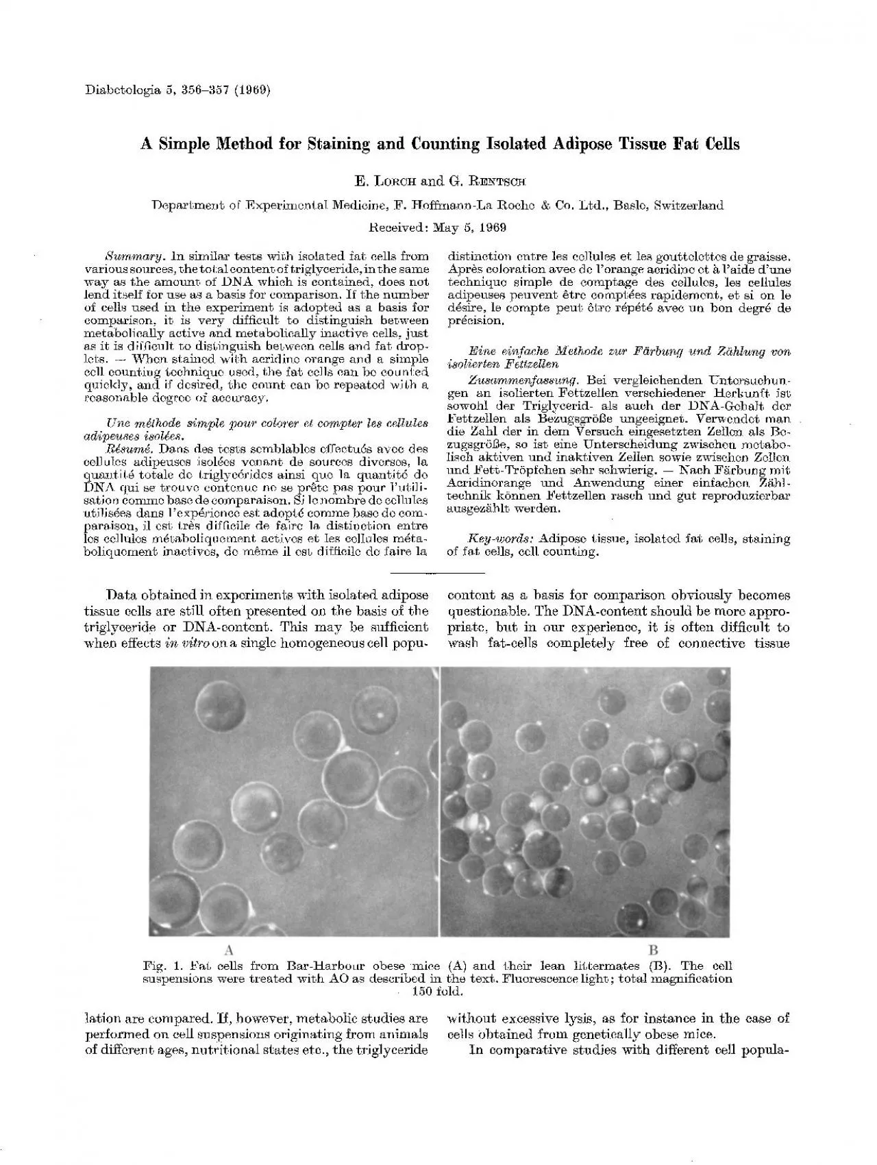
LoRc and G RENrSCH A Simple Method for Staining and Counting 357 tions number of cells seems to be the most reasonable reference basis at present We tried therefore to find a simple counting ID: 939553

cell cells counting suspension cells cell suspension counting fat fluorescence slide trough orange drop microscope chem metabolism nuclei show

Share:

Link:

Embed:

Download Presentation from below link

Download Pdf The PPT/PDF document "and Counting Adipose Tissue Co Viay 5 1..." is the property of its rightful owner. Permission is granted to download and print the materials on this web site for personal, non-commercial use only, and to display it on your personal computer provided you do not modify the materials and that you retain all copyright notices contained in the materials. By downloading content from our website, you accept the terms of this agreement.


Presentation Transcript

and Counting Adipose Tissue & Co. !Viay 5, 1969 LoRc~{ and G. REN~rSCH: A Simple Method for Staining and Counting 357 tions, number of cells seems to be the most reasonable reference basis at present. We tried, therefore, to find a simple counting technique that would allow results to be reproduced at any time. In counting fat cells, difficulties are en- countered since one has to disth~guish between living cells, fat droplets and dead cells. Using the fluorescent dye Acridine Orange (AO), the differentiation becomes very easy, because fluorescence of different wavelengths appears after staining a cell population with (AO) 1. The nuclei of normal, living cells show a green fluo- rescence, whereas the nuclei of dying or dead cells develop a fluorescence ranging from orange to red. Fat droplets show a pale green fluorescence. rectangular frame (1  1 era) was cut out of ad- hesive tape (of about 0.2 mm thickness), and was glued to a siliconized microscope slide forming a ':trough" for the reception of the cell suspension. With the tape frame facing downwards, a cell suspension (5 ~zl) pre- pared according to RODB~, 2 and containhlg appro- ximately 105 cells per ml, was applied from underneath into the middle of the trough. This application was made with a plastic-tipped pipette (Oxford-sam- pler or Eppendorf-mieropipette) held vertically with the tip pointing upwards, in order to prevent cell loss due to flotation within the pipette tip. The slide was then turned over, and 5 ~1 of a solution of 1 mg of AO-HC1 in 10 ml of Hanks-solution was added to the cell suspension. The AO sample used was purifi

ed chro- matographically 3 from commercial material (Fluka AG, Chemische Fabrik, St. Gallen, Switzerland). A silieonized cover slide with a scratched-in millimeter net was put on top of the trough, the millimeter net facing the drop and thus permitting both the cells and the scratched scale to be in focus simultaneously. After 3--10 rain standing at room temperature, the stained cell-suspension was observed in a Zeiss fluor- escence microscope at 50-fold magnification. Technical data of microscopy were: UV lamp: Osram tIBO 200, high pressure mercury lamp ; blue filter BG 23 ; barrier filter: Zeiss 53, transmitting light of wave length greater than 530 nm. Since the cells floathlg to the surface of the drop did not seem to be distributed statistically, all the cells of the drop were counted. Using the same original cell suspension, 6 consecutive countings performed in this manner gave, for example, the following counts: 298, 295, 306, 289, 301,292 cells. authors would like to express their appreciation to Miss ANDY LIPPOK fer her skflful technical assistance. 3. M. : 2. Auflage. Leipzig: Akad, Verlagsgesellschaft Geest und Portig KG, i959. RODBELL, M.: Metabolism of Isolated Fat Cells. I. Effects of Hormones on Glucose Metabolism and Lipo- lysis. J. biol. Chem. 239, 375 380 (1964). ZAN~:~, V. : Uber den Nachweis definierter, reversibler Assoziate (,,reversible Polymerisate") des Acridin- orange durch Absorptions- und Fluoreszenzmessungen in w~i~riger L6sung. Z. physik. Chem. 199, 225--258 (1952). Dr. E. Lo~ct{ Dr. G. RESTSe~{ Abtlg. fiir experimentelle Medizin F. Hoffmann-La l%oebe & Co. CH-4000 Basel